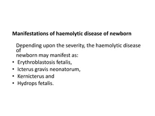
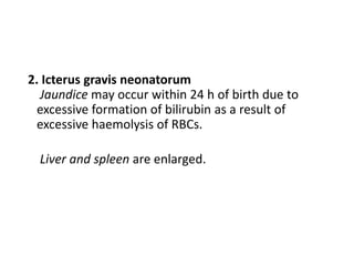
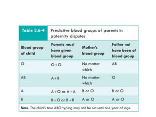
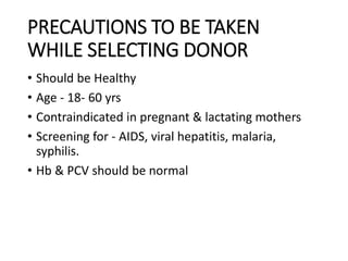
Call Girls Cuttack Just Call 9907093804 Top Class Call Girl Service Available
DOC-20230928-WA0010..pptx
1. Physiology Of Blood groups
and Its applied aspects
By - Dr. Imran Khan H
B.Sc ( Psychology) , M.B.B.S
1 st Year PG ( MD Physiology) KIMS,Hubballi.
2. OBJECTIVES
BLOOD GROUPS
History
Introduction
Classical ABO blood grouping system
• Agglutinogens
• Agglutinins
• Types of ABO blood groups
• Population distribution of ABO
blood groups
• Inheritance of ABO blood groups
• Determination of ABO blood groups
Rhesus (Rh) blood grouping
system
• Rh antigens
• Rh antibodies
• Inheritance of Rh antigens
• Haemolytic disease of
newborn
Clinical applications of blood
grouping
BLOOD TRANSFUSION
3. History of Blood Transfusion
• 1628
• British physician William Harvey discovers the circulation
of blood. The first known blood transfusion is attempted
soon afterward.
• 1658
• Microscopist Jan Swammerdam observes and describes
red blood cells.
4. • 1818
• British obstetrician James Blundell performs the first
successful transfusion of human blood to a patient for the
treatment of postpartum hemorrhage.
• 1873-1880
• U.S. physicians attempt transfusing milk from cows, goats
and humans.
5. • 1665
• The first recorded successful blood transfusion occurs in
England: Physician Richard Lower keeps dog alive by
transfusing blood from other dogs
• 1667
• Jean-Baptiste Denis in France and Richard Lower and
Edmund King in England separately report successful
transfusions from sheep to humans.
6. • 1884
• Saline infusion replaces milk as a “blood substitute” due
to the increased frequency of adverse reactions to milk.
• 1901
• Karl Landsteiner, an Austrian physician, discovers the first
three human blood groups.
7. • It was not until the year 1900, when Karl Landsteiner at the
University of Vienna, discovered why some blood transfusions
were successful while others could be deadly.
• Landsteiner discovered the ABO blood group system by mixing
the red cells and serum of each of his staff. He demonstrated that
the serum of some people agglutinated the red cells of other.
From these early experiments, he identified three types, called A,
B and C (C was later to be re-named O for the German “Ohne”,
meaning “without”, or “Zero”, “null” in English).
• The fourth less frequent blood group AB, was discovered a year
later. In 1930, Landsteiner received the Nobel Prize in physiology
and medicine for his work
8. • The gene that determines human ABO blood type
is located on chromosome 9 (9q34.1) and is called
ABO glycosyltransferase.
• The ABO locus has three main allelic forms: A, B,
and O, as mentioned above and each of them is
responsible for the production of its glycoprotein.
• It is therefore the combination of alleles that are
inherited from parents that determines which
glycoproteins (antigens) are found on persons’
blood cells and thereby their ABO blood type
9. INTRODUCTION
one of the classes into which people or their blood can be separated on the basis
of the presence or absence of specific antigens in their blood called also blood
type.
Agglutinogens -refer to the antigens present on the cell
membranes of RBCs. A variety of antigens are present on
the cell membrane, but only a few of them are of practical
significance.
Agglutinins -refer to the antibodies against the agglutinogens. These are present in the
plasma.
Agglutination of RBCs can be caused by the antigens present on their cell membranes
in the presence of suitable
agglutinins (antibodies). That is why, these antigens are
called agglutinogens.
10. BLOOD GROUPING SYSTEM
Depending upon the type of agglutinogen present or absent
on the red cell membranes, various blood grouping systems
are known, which can be classified as:
Major blood group systems are based on the presence of
agglutinogens which are widely prevalent in the population
and are known to cause worst transfusion reactions. These
include:
• The classical ABO blood grouping system and
• Rhesus (Rh), (CDE) blood grouping system
11. Minor blood group systems are based on the
presence of
agglutinogens which are found only in small
proportion of
the population and occasionally produce mild
transfusion
reactions. These include:
• M and N blood grouping and
• P blood group system
12. LANDSTEINER'S LAW
Karl Landsteiner in 1900, framed a law in relation to
agglutinogens and agglutinins, which states that:
If an agglutinogen is present on the red cell membrane of
an individual, the corresponding agglutinin must be absent
in the plasma and
If an agglutinogen is absent from the cell membrane of
RBCs of an individual, the corresponding agglutinin must
be present in the plasma.
13. It is important to note that
• The Landsteiner law is applicable to ABO blood
group system only.
• The law is not applicable to other blood group
systems - because there are no naturally occurring
agglutinins in these systems.
14. CLASSICAL ABO BLOOD GROUPING
SYSTEM
The classical ABO blood grouping system is based on the
presence of A and B agglutinogens on the cell membrane
of RBCs.
A and B agglutinogens are complex oligosaccharides
differing in their terminal sugars.
The A and B antigens present on the membranes
of RBCs are also present in many other tissues like
salivary glands, pancreas, kidney, liver, lungs and testis;
and also in body fluids like saliva, semen and amniotic
fluid.
The antigens on RBCs membrane are glycolipids,
while in the tissues and body fluids they are soluble
glycoproteins
15. ANTI-A AND ANTI-B AGGLUTININS
• Anti-A agglutinin and anti-B agglutinin refer to the
antibody, i.e. which reacts with or acts on the antigen A
and antigen B, respectively.
• There are two types of α agglutinins: the α1 and α proper.
• The α and β agglutinins are globulins of IgM type and
cannot cross the placenta.
• The α and β agglutinins act best at low temperature
(5–20°C) and are therefore also called as cold antibodies.
16. TYPES OF ABO BLOOD GROUPS
Depending upon the presence or absence of A and B
agglutinogens and α and β agglutinins, there are four
types of blood groups:
Blood group A is characterized by:
• Presence of A agglutinogen and absence of B
agglutinogen on the cell membrane of RBCs.
• Presence of anti-B agglutinin and absence of anti-A
agglutinin from the plasma.
17. Blood group B is characterized by:
• Presence of B agglutinogen and absence of A agglutinogen
on the cell membrane of RBCs and
• Presence of anti-A agglutinin and absence of anti-B
agglutinin from the plasma.
Blood group AB is characterized by:
• Presence of both A and B agglutinogens on the cell
membrane of RBCs and
• Absence of both anti-A and anti-B or agglutinins from
the plasma
18. Blood group O is characterized by:
• Absence of both A and B agglutinogens on the cell
membrane of RBCs and
• Presence of both anti-A and anti-B agglutinins in
the
plasma.
21. INHERITANCE OF ABO BLOOD GROUPS
• Agglutinogens A and B or the non-antigenic
substances
which determine the blood groups are genetically
inherited
as Mendelian dominant in the classical Mendelian
pattern.
The ABO phenotypes and possible genotypes are as
under:
22. APPERANCE OF ANTIGENS &
ANTIBODIES
• Agglutinogens A and B first appear in the sixth week of
fetal life. Their concentration at birth is one-fifth of adult
level and it progressively rises during puberty and
adolescence.
•
Anti-A (or a) and Anti-B (or b) agglutinins (specific
blood group antibodies) are absent at birth, but they appear
10–15 days after birth and reach a maximum concentration
by the age of 10 years.
23. MECHANISM
The probable mechanism of appearance of α and β
agglutinins is described.
• Antigens very similar to A and B antigens are
commonly present in the intestinal bacteria and
foods.
• When the newborn is exposed to these antigens,
these are absorbed into the blood and stimulate the
formation of antibodies against the antigens
recognized as non-self (i.e. not present in the own
body) by the immune system.
26. • Determination of blood groups—the RBCs showing
agglutination with antisera are:
• 1-of blood group ‘A’ with
antisera A;
• 2-of blood group B with antisera B;
• 3- of blood
group ‘AB’ with antisera A and B (both) and
• 4-of blood group
‘O’ with none.
27. Rh BLO0D GROUPING SYSTEM
• Rh Antigens - called Rh as these were first
discovered in RBC of rhesus monkey.
• Discovered by Landsteiner & Weiner in 1940.
• 3 types of Rh antigen, C,D & E,
• D IS COMMONEST & causes severe transfusion
reaction.
• " Rh antigens are integral membrane proteins & not
found in other tissues.
28. Rh Antibodies
• There are no natural antibodies of Rh antigens, while in
ABO system of blood grouping α or β antibodies are
always present naturally if the appropriate antigen is
absent .
• Rh antibodies (also called anti-D) are produced only
when an Rh -ve individual is transfused with Rh +ve blood
or when a Rh -ve mother gives birth to Rh +ve baby (Rh
+ve RBCs of foetus enter into the maternal circulation),
Rh antibodies are of IgG type and can cross the placenta.
Since these react best at body temperature so are also
called warm antibodies
29. • Once produced, the Rh antibodies persist in the
blood
for years and can produce serious reactions during
the second transfusion
30. INHERITANCE OF Rh ANTIGENS
• The Rh antigen (D antigen) is inherited as dominant
gene D. When gene D is absent from a chromosome, its
place is occupied by the alternate form (allelomorph)
called ‘d’. Rh gene is inherited from both the father and
the mother.
• Rh +ve individual may have two genotypes. DD (homozygous) or Dd
(heterozygous) of 85% Rh +ve individuals
about 35% have DD genotype and 50% have Dd genotype.
• The genotype of Rh -ve individual is dd.
• Therefore, the genotype (gene composition) of offspring
will be:
– DD when gene D is carried by both sperm and ovum
– Dd when one gamete carries D and other d and – dd, when both
the gametes carry gene d.
31.
32. HAEMOLYTIC DISEASE OF NEWBORN
Haemolytic disease of newborn occurs as a result of
incompatibility of Rh blood groups between the mother and
the fetus.
•
Mechanism of haemolytic disease of newborn in
Rh incompatibility
1. Entrance of Rh +ve fetal RBCs into Rh -ve mother’s circulation
during first pregnancy.
At the time of delivery, the fetal RBCs enter maternal circulation
because of severance of umbilical cord. Before delivery, usually
the foetal and maternal circulation do not mix.
Since the Rh +ve RBCs enter maternal circulation during delivery,
so the first child
is usually normal.
33. • 2. Production of Rh antibodies (anti-D) in mother.
During postpartum period, i.e. within a month after
delivery, the mother develops Rh antibodies in her
blood.
As mentioned earlier, the Rh antibodies are of IgG
type and are able to cross the placental barrier.
Once formed the Rh antibodies persist for a long
period in mother’s blood.
34. • 3. Rh incompatibility reaction during second
pregnancy.
When the Rh -ve mother in the second pregnancy
also bears a Rh +ve child, the Rh antibodies present
in the mother’s blood enter the fetal circulation by
crossing the placental barrier and cause
agglutination of fetal RBCs leading to haemolytic
disease of newborn.
35. Manifestations of haemolytic disease of newborn
Depending upon the severity, the haemolytic disease
of
newborn may manifest as:
• Erythroblastosis fetalis,
• Icterus gravis neonatorum,
• Kernicterus and
• Hydrops fetalis.
36. 1. Erythroblastosis fetalis is characterized by:
Erythroblastosis, i.e. appearance of large number
of erythroblasts in the peripheral blood.
• Anaemia occurs due to excessive haemolysis of
RBCs by Rh antibodies. Infant may even die of
severe anaemia.
37. 2. Icterus gravis neonatorum
Jaundice may occur within 24 h of birth due to
excessive formation of bilirubin as a result of
excessive haemolysis of RBCs.
Liver and spleen are enlarged.
38. 3. Kernicterus. It is a neurological syndrome occurring
in newborns with severe haemolysis.
The excessive bilirubin
formed may enter the brain tissue as the blood–brain
barrier is not well developed in infants and cause
damage.
The bilirubin mostly affects the basal ganglia producing
disturbance of motor activities. It usually develops
when serum bilirubin level exceeds 18 mg/dL.
39.
40. • 4. Hydrops fetalis, i.e. the fetus is grossly
oedematous. It occurs when haemolysis is very
severe. Usually, there occurs intrauterine death of
fetus or if born prematurely or even at
term, the infant dies within a few hours.
41. Prevention and treatment
Prevention of haemolytic disease of newborn. The
haemolytic disease in the newborn during second
pregnancy can be prevented by injecting single dose of
Rh antibodies (anti-D) in the form of Rh-
immunoglobulin to mother soon
after child birth (1st pregnancy). These antibodies will
destroy the Rh +ve RBCs of the fetus which have gained
access to maternal circulation. In this way active
antibodies will not be formed by the mother.
42. Treatment of haemolytic disease of the newborn is
replacement of baby’s Rh +ve blood with Rh -ve
blood exchange transfusion.
43. CLINICAL APPLICATIONS OF
BLOOD GROUPS.
• In blood transfusion.
• In Preventing Hemolytic Disease.
• In Paternity Disputes.
• In Medicolegal Cases.
• In knowing Susceptibility to Diseases.
47. DONORS & RECIPIENT
• Donor - person who donate the blood
• Recipient - person who receives the blood.
• Universal donor –O Rh Negative.
• Universal recipient - AB Rh positive
48. PRECAUTIONS TO BE TAKEN
WHILE SELECTING DONOR
• Should be Healthy
• Age - 18- 60 yrs
• Contraindicated in pregnant & lactating mothers
• Screening for - AIDS, viral hepatitis, malaria,
syphilis.
• Hb & PCV should be normal
49. PRECAUTIONS DURING BLOOD
TRANSFUSION
Absolute indication .
Cross matching
-Major - Donor's RBC + Recipient plasma
-Minor -Donor's plasma+ Recipient RBC
• Rh +ve blood should never be transfused to Rh –ve
person.
• Donor's blood should always be screened for
diseases.
50. PRECAUTIONS DURING BLOOD
TRANSFUSION
• Blood bag/bottle should be checked.
• Blood transfusion should be given at slow rate.
• Proper Aseptic measures.
• Careful watch on recipient condition - for first 10-
15min.
• " Should stop if any reaction
54. Forms of autologous blood
transfusion
• Three main techniques for autologous transfusion
are predeposit autologous donation (PAD), acute
normovolaemic haemodilution (ANH), and
perioperative cell salvage (PCS).
55. STORAGE OF BLOOD FOR
TRANSFUSION
• One unit 420 ml mixed with 120 ml ACD (Acid
citrate dextrose)
Contents of ACD mixture are:
– Acid citrate (monohydrous), 0.48 g,
– Trisodium citrate, 1.32 g,
– Dextrose 1.47 g and
– Distilled water 100 mL.
56. • Dextrose (glucose) present in ACD mixture provides
energy
for maintenance of sodium–potassium pump
activity.
• Anticoagulant activity is provided by the citrates
present in the ACD mixture which also decreases
the pH of blood
57. IMPORTANT FACTS ABOUT
BLOOD TRANFUSION
• One can safely donate 1 unit of blood every 6
month.
• Blood can be stored for 21 days with above
conditions
• WBC & platelet virtually absent after 24 hrs of
storage.
• After transfusion 80% RBC survive for 24 hrs &
destroyed at a rate of 1% per day.
58. Bombay Blood Group
• It is the rarest type of Blood group, only 4 per
million of the world population have this blood
group .
• Also called the HH group.
• It is the Rarest Blood Group first discovered in
Mumbai (then Bombay)
• in the year 1952 by Dr. Y.M Bhide and that’s how it
got its name.
59. • How was it discovered?
•
This incident happened when a patient was admitted
to the KEM hospital and required blood transfusion.
During the regular blood diagnosis, the red cells in the
blood were grouped like that of O group and hence
blood with blood group O was transfused to the
patient. The patient eventually developed a hemolytic
transfusion reaction and the blood transfusion was
stopped.
60. • All the blood groups primarily contain a protein called
the H antigen.
• The antigens A and B are made from the H antigen. So,
when someone has the A blood group, it means that
they have the antigen of type ‘A’ and antibody of type
‘B’ in their blood. Similarly, a B blood group person has
antigen of type ‘ B’ and antibody of type ‘A’ in his/her
blood.
• Blood group AB has both type A and B antigens in their
blood but no antibodies, whereas ‘O’ blood group has
‘A’ and ‘B’ antibodies and no antigens.
61. • The HH or Bombay blood group do not produce this
H antigen, subsequently not producing either A or
B antigen. They only have the ‘H’ antibody which
none of the other blood groups has due to which
blood transfusion for the Bombay blood with any
other group is not possible.
62. • In Bombay, it could be present in 1 per 10,000 of the local
residents.
• In India, there are about 179 people known to have the
Bombay phenotype blood group.
• Interestingly in the year 2010 in Bangalore, a 42-year-old
father Mr. Annappa was found to have this rare blood group
when he wanted to donate blood to his ailing son. That
made the total count of people to 180 in India.
• This blood group is commonly mistaken as ‘o’ and might not
be identified if proper blood grouping or testing practices
are not followed.
63. • Mr. Annappa’s blood group initially showed up as ‘O’
with no reaction to Anti-A and Anti – B antibodies.
Repeated testing was done to perform the reverse
grouping and confirm the Bombay blood type.
• People of this blood type can receive only their blood
type. In most diagnostic centres where blood tests are
done, only the presence and absence of AB and Rh is
tested in the blood. Since Bombay blood group do not
possess the A or B antigens, it is usually comprehended
as the ‘O’ blood type.
64. • It is only after a specific test for H Antigen in the
blood is performed which can make a distinction
between Bombay blood group and ‘O’ blood group.
Blood transfusions for the Bombay blood group
with any other blood type may lead to a fatal
hemolytic transfusion reaction. To be on the safer
side, if one is of the blood type ‘O’, make sure it is
not the Bombay blood type through proper testing
at a standard diagnostic centre.
66. • Karl Landsteiner (14
June 1868- 26 June 1943
) aged 75.
• Known for Research of
blood group system,
discovery of Rh factor,
discovery of poliovirus
• AwardsNobel Prize in
Physiology or
Medicine (1930)
• Dr. Y.M Bhende
• (27 Sep-1911 – 2 Dec -
1998 )
• In 1952, Dr Bhende
along with his co-
authors published a
paper in The Lancet
proposing a new blood
group –The Bombay
blood group.
67. References
• Medical Physiology for Undergraduate Students,
1/e
Indu Khurana
• Blood Groups and Red Cell Antigens-Laura
Dean, MD
• Guyton and Hall Textbook of Medical Physiology
14th Edition.
• Ganong's Review of Medical Physiology-Twenty sixth
Edition