CAD CAM DENTURES IN PROSTHODONTICS : Dental advancements
1-cardiovascular disorders
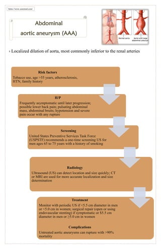
1. › Localized dilation of aorta, most commonly inferior to the renal arteries
Abdominal
aortic aneurysm (AAA)
Risk factors
Tobacco use, age >55 years, atherosclerosis,
HTN, family history
H/P
Frequently asymptomatic until later progression;
possible lower back pain; pulsating abdominal
mass, abdominal bruits; hypotension and severe
pain occur with any rupture
Screening
United States Preventive Services Task Force
(USPSTF) recommends a one-time screening US for
men ages 65 to 75 years with a history of smoking
Radiology
Ultrasound (US) can detect location and size quickly; CT
or MRI are used for more accurate localization and size
determination
Treatment
Monitor with periodic US if <5.5 cm diameter in men
or <5.0 cm in women; surgical repair (open or using
endovascular stenting) if symptomatic or $5.5 cm
diameter in men or ≥5.0 cm in women
Complications
Untreated aortic aneurysms can rupture with >90%
mortality
https://www.gsmmed.com/
2. ›Acute pericarditis
•Acute inflammation of the pericardial sac accompanied by pericardial effusion
•Caused by viral infection, tuberculosis, systemic lupus erythematosus (SLE), uremia,
neoplasm, drug toxicity (e.g., isoniazid, hydralazine), post-MI inflammation (Dressler
syndrome), radiation, recent heart surgery
Treatment
Treat underlying cause; pericardiocentesis for large effusions; nonsteroidal anti-
inflammatory drugs (NSAIDs) for pain and inflammation; colchicine may be
useful for preventing recurrence owing to viral or idiopathic causes
Radiology
CXR is helpful in ruling out
other systemic causes;
effusion frequently seen on
echocardiogram
ECG
Global ST elevation, PR
depression
H/P
Anterior chest pain with inspiration (i.e., pleuritic chest pain), dyspnea, cough; pain lessens
with leaning forward; fever, friction rub (best heard when leaning forward); pulsus paradoxus
(i.e., fall in systolic blood pressure >10 mm Hg with inspiration) occurs because increased
physiologic right ventricle (RV) filling during inspiration combined with pathologic left
ventricle (LV) compression by pericardial effusion causes impaired LV filling, decreased
stroke volume, and decreased inspiratory systolic blood pressure infection
Complications
Chronic constrictive
pericarditis if
untreated
https://www.gsmmed.com
/
3. Acute
Rheumatic fever
H/P
Migratory arthritis, hot and swollen joints,
fever, subcutaneous nodules on extensor
surfaces, Sydenham chorea (i.e.,
purposeless involuntary movement),
erythema marginatum (i.e., painless rash)
Labs
Increased erythrocyte sedimentation rate
(ESR), C-reactive protein (CRP), and
white blood cell (WBC) count; 90% of
patients have antistreptococcal antibodies
ECG
Increased PR
interval
Complications
Progressive valve
damage if untreated
Treatment
NSAIDs for joint inflammation; use
corticosteroids, if carditis is severe;
β-lactam (penicillin family) antibiotic for
infection
https://www.gsmmed.com
• Uncommon sequela of untreated group A
streptococcus infection
• Streptococcus infection can provoke autoantibodies
that attack joints and heart valves (mitral > aortic >
tricuspid).
• Incidence is low in the United States because of
antibiotic treatment.
• The term “rheumatic heart disease” describes both
the acute carditis (pericarditis, myocarditis,
valvulitis) and chronic valvular damage.
Diagnosis
Made using Jones
criteria
4. jator“Angina pectoris”
www.gsmmed.com
› Etiology
a. Temporary myocardial ischemia during
exertion that causes chest pain
b. Most commonly caused by CAD; also
occurs secondary to arterial vasospasm
(Prinzmetal angina) and valvular disease
c. Gastroesophageal reflux disease (GERD)
and esophageal spasm can mimic symptoms.
H/P
substernal chest pain that may radiate to left shoulder, arm, jaw, or back
Labs
stress testing or nuclear studies used for diagnosis
a. Important in assessment of chest pain
b. Seeks to increase cardiac workload to assess myocardial ischemia
c. Accomplished either through exercise or pharmacologic testing
Treatment
sublingual nitroglycerin and cessation of intense activity until completion
of workup; full workup (including stress testing or nuclear studies) for
cause is needed to define long-term treatment
5. › Intimal tear leads to blood entering
media, causing formation of false lumen
›Stanford classification—Stanford A aortic
dissection involves ascending aorta; Stanford
B is distal to left subclavian artery
› AORTIC
DISSECTION
Risk factors
• HTN, coarctation of the aorta, syphilis, Ehlers-Danlos
syndrome, Marfan syndrome, trauma (rare)
H/P
• Acute, “ripping” chest pain, syncope; decreased peripheral
pulses, normal or increased blood pressure
ECG
• Normal or LVH
Radiology
• Widening of aorta and superior mediastinum on CXR; CT with
contrast, echocardiogram, MRI, magnetic resonance
angiography (MRA), or angiography good for definite diagnosis
Treatment
• Stabilize blood pressure (e.g., b-blocker, nitroprusside) if
unstable; Stanford A dissections need emergency surgery;
Stanford B dissections can be treated medically unless
rupture or occlusion develops
Complications
• Possible MI, renal insufficiency, ischemic colitis, stroke,
or paraplegia
https://www.gsmmed.com
6. › Abnormal communications between arteries and veins
› Congenital or acquired
› Large AVM can cause local ischemia and increase the risk of thrombus formation.
H/P
Palpable, warm, pulsating masses, if superficial;
painful if mass compresses adjacent structures
Treatment
Surgical removal or sclerosis, if
symptomatic, or if located in brain or bowel
“Arteriovenous
malformations (AVM)”
https://www.gsmmed.com/
7. “Atherosclerosis”
www.gsmmed.com
›Gradual narrowing of arteries caused by endothelial
dysfunction, progressive formation of plaques (which
consist of lipids and smooth muscle), and the
associated inflammatory response.
›Plaques can calcify, rupture, and thrombose, which
leads to further narrowing of arteries and progressive
occlusion of blood flow.
H/P
•asymptomatic for most of disease progression; later sequelae include angina, claudication,
progressive hypertension (HTN), retinal changes, extra heart sounds, myocardial infarction
(MI), and stroke
Labs
•stress testing, echocardiography, nuclear studies, or angiography can be used to detect coronary
ischemia
•a. Exercise stress test—patient exercises on an aerobic fitness machine at increasingly strenuous
workloads; heart rate and ECG are constantly monitored; test is continued until patient achieves
85% of predicted maximal heart rate (predicted maximal heart rate = 220 - age) or patient develops
angina or signs of ischemia as seen on ECG; ischemic heart disease is diagnosed with signs of
reproducible angina or obvious signs of ischemia at low workloads
•b. Nuclear exercise test—thallium-201 or technetium-99m-sestamibi is injected during exercise
testing, and scintigraphy (e.g., planar or single positron emission computed tomography [SPECT])
is performed to assess myocardial perfusion; used in cases of suspected ischemic heart disease in
which results of regular exercise stress testing are equivocal
•c. Exercise stress test with echocardiography—exercise stress testing performed in conjunction
with echocardiography to increase sensitivity of detecting myocardial ischemia
•d. Pharmacologic stress testing—administration of cardiac inotrope (e.g., dobutamine) in place of
exercise to increase myocardial demand; frequently performed in conjunction with SPECT or
performed in patients for whom comorbidities interfere with the ability to perform exercise
•e. Positron emission tomography (PET) myocardial imaging—injection of positron- emitting
isotopes with subsequent three-dimensional detection imaging to evaluate heart for perfusion
defects and tissue viability
•f. Coronary angiography—gold standard for identifying CAD but more invasive than other
techniques
Treatment
•management is primarily intended to minimize risk factors (e.g., tobacco use, HTN,
hyperglycemia, hypercholesterolemia); diet low in fats and cholesterol and high in antioxidants
(e.g., vitamins E and C, β-carotene) is helpful in preventing disease
8. https://www.gsmmed.com/
› Lack of coordinated atrial contractions with independent sporadic ventricular contractions
› Caused by rapid, disorderly firing from a second atrial focus
• Pulmonary disease, CAD,
HTN, anemia, valvular
disease, pericarditis,
hyperthyroidism,
rheumatic heart disease
(RHD), sepsis, alcohol
use
Risk factors
• Possibly
asymptomatic;
shortness of breath,
chest pain,
palpitations,
irregularly irregular
pulse
H/P
• No discernible P
waves, irregular
QRS rate
ECG
• Anticoagulation; rate control via calcium
channel blockers, b-blockers, or digoxin;
electric or chemical (i.e., class IA, IC, or III
antiarrhythmics) cardioversion if presenting
within initial 2 days; cardioversion can be
performed in delayed presentation if absence
of thrombi is confirmed by transesophageal
echocardiogram; if presenting after 2 days or
if thrombus is seen on echocardiogram, then
anticoagulate and wait 3 to 4 weeks before
cardioversion; AV nodal ablation can be
considered for recurrent cases
Treatment
• Increased risk of
MI, heart failure;
poor atrial
contraction
• causes blood
stasis, which leads
to mural thrombi
formation and a
risk of
embolization
Complications
Atrial fibrillation (Afib)
0
https://www.gsmmed.com/
9. https://www.gsmmed.com/
› Caused by rapid firing of an ectopic focus in the atria
• CAD, congestive heart
failure (CHF), chronic
obstructive pulmonary
disease (COPD), valvular
disease, pericarditis
Risk factors
• possibly asymptomatic;
palpitations, syncope
H/P
• regular
tachycardia >150
bpm with
occasionally set
ratio of P waves
to QRS; sawtooth
pattern of P
waves
ECG
• rate control with calcium channel
blockers, b-blockers; electrical or
chemical (class IA, IC, or III
antiarrhythmics) cardioversion if
unable to be controlled with
medication; catheter ablation to
remove ectopic focus may be possible
in some casesan be considered for
recurrent cases
Treatment • may degenerate
into Afib
Complications
Atrial flutter (Aflutter)
0
https://www.gsmmed.com/
10. “Atrial septal defect”
(ASD)
›Opening in atrial septum allowing movement of blood between atria
›Initially, blood flow is left-to-right across defect.
H/P
Possibly asymptomatic; large defects can cause
cyanosis, heart failure symptoms, dyspnea,
fatigue, or failure to thrive; strong impulse at
lower left sternal border, wide fixed split S2,
systolic ejection murmur at upper left sternal
border
ECG
Right axis deviation
Radiology
Echocardiogram shows blood flow between
atria, dilated RV, and large heart; CXR shows
increased pulmonary vascular markings
caused by pulmonary HTN
Treatment
Small defects do not need repair but require
antibiotic prophylaxis before surgery or dental
work; surgical closure for symptomatic infants
or when pulmonary blood flow is twice that of
systemic blood flow
Complications
Untreated ASD leads to right-to-left
shunt (i.e., Eisenmenger syndrome),
RV dysfunction, pulmonary HTN,
arrhythmias
www.gsmmed.com
11. › Bradycardia
› HR < 60 bpm
› Caused by increased vagal tone or nodal disease
› Predisposition to development of ectopic beats
https://www.gsmmed.com
/
14. “Cardiac output (CO)”
1. Heart rate (HR)
a. Number of cardiac contractions per unit time; commonly expressed as beats per
minute (bpm)
b. If HR is too high (normal = 60 to 100 bpm), then diastolic filling is decreased.
2. Stroke volume (SV)
a. SV is the change in blood volume from immediately before initiation of
contraction to completion of contraction (i.e., SV = end diastolic volume to end
systolic volume).
b. It is determined by contractility (i.e., SV = [end diastolic volume] - [end
systolic volume]), preload (amount of myocardial stretch at end of diastole), and
afterload (resistance ventricles must overcome to empty their contents).
c. SV increases with catecholamine release, an increase in intracellular Ca, a
decrease in extracellular Na, digoxin use, anxiety, and exercise.
d. SV decreases with b-blockers, heart failure, acidosis, and hypoxia.
3. Fick principle
CO = SV × HR =
a. Rate of O2 use can be determined by comparing O2 content in expired air to that
in inhaled air; arterial and venous O2 content can be measured directly from the
corresponding vasculature.
b. CO increases during exercise, initially by increasing SV and later by increasing HR.
4. Mean arterial pressure = CO × total peripheral resistance (TPR) = diastolic arterial pressure
+ 1/3 pulse pressure
5. Pulse pressure = systolic arterial pressure - diastolic arterial pressure
www.gsmmed.com
("#$% '( )2 +,%)
[(#"$%"/#0 )2 1'2$%2$) × (4%2'+, )2 1'2$%2$)]
15. https://www.gsmmed.com/
› Large pericardial effusion causes compression of heart and greatly
decreased CO; can result from progressive, acute pericarditis, chest
trauma, LV rupture following MI, or dissecting aortic aneurysm
› High mortality
• Dyspnea, tachycardia,
tachypnea; JVD, pulsus
paradoxus
H/P
• Low voltage, sinus
tachycardia; electrical
alternans is relatively
specific but not sensitive
ECG
• Enlarged cardiac
silhouette on CXR; large
effusion seen on
echocardiogram
Radiology
• Immediate
pericardiocentesis
Treatment
Cardiac tamponade
0 j
https://www.gsmmed.com/
16. ›Chronic constrictive pericarditis
› Sequela of chronic untreated pericardial irritation
› Diffuse thickening of pericardium with possible
calcifications leads to decreased diastolic filling and
decreased CO.
› Most commonly caused by radiation or heart
surgery
https://www.gsmmed.com/
H/P
Symptoms consistent with heart failure (JVD, dyspnea on exertion, orthopnea,
peripheral edema), increasing JVD with inspiration (Kussmaul sign); Afib
common
Labs
Cardiac catheterization shows equal pressure in all chambers
Radiology
Possible pericardial calcifications on CXR; echocardiogram, computed
tomography (CT), and magnetic resonance imaging (MRI) show pericardial
thickening
Treatment
NSAIDs, colchicine, corticosteroids; surgical excision of pericardium (high
mortality)
17. ›Deep vein thrombosis (DVT)
› Development of thrombosis in large vein; most common
in lower extremity
› Location, in order of decreasing frequency: calf,
femoral, popliteal, and iliac veins
› Can cause inflammation of affected vein (i.e.,
thrombophlebitis)
https://www.gsmmed.com/
Risk factors
Prolonged inactivity (travel, immobilization), heart failure, hypercoagulable
states, neoplasm, pregnancy, oral contraceptive pill (OCP) use, tobacco use,
vascular trauma
H/P
Possibly asymptomatic; deep leg pain, swelling, warmth
Labs
D-dimer will be elevated with DVT formation, but test is more useful in using
a normal result to rule out DVT
Radiology
Compressive venous US is used for detection
Treatment
Leg elevation; low molecular weight heparin or unfractionated heparin
initially, warfarin for long-term management; inferior vena cava (IVC) filter
should be placed in a patient with contraindications to anticoagulation
Complications
Clot can embolize to lungs (i.e., pulmonary embolus) with 40% mortality; chronic
DVT can cause chronic venous insufficiency
18. “Dyslipidemia”
› Abnormal serum cholesterol levels (high low-density lipoprotein [LDL] and/or low high-density lipoprotein [HDL]) that
are associated with increased risk of ischemic heart disease
› Can result from a congenital disorder (less common) or an acquired condition (most common)
› Normal cholesterol physiology
a. Cholesterols and triglycerides are carried by lipoproteins.
b. Increased LDL leads to increased CAD risk; increased HDL is protective.
c. Increased LDL and decreased HDL result from a diet high in fatty foods, tobacco use, obesity, alcohol use, diabetes
mellitus (DM), and certain medications (e.g., oral contraceptive pills [OCP], diuretics).
H/P
usually asymptomatic; extremely high triglycerides and LDL lead to xanthomas (i.e., lipid
deposits in tendons), xanthelasmas (i.e., lipid deposits in eyelids), and cholesterol emboli in
retina (visible on funduscopic examination); symptoms are more severe and appear earlier
in life in primary disorders compared with acquired conditions
Labs
increased total cholesterol and LDL; possible decreased HDL; total cholesterol may be >300 to
600 mg/dL in primary disorders; screening for hyperlipidemia is performed in men >35 years
of age and women >45 years of age (younger if patient has other risk factors for CAD)
Treatment
focuses on prevention of cardiovascular disease and includes tobacco cessation, exercise, and dietary
restrictions (e.g., low fat, low cholesterol); guidelines published in 2013 by the American College of
Cardiology (ACC) and American Heart Association (AHA) recommend starting moderate- or high-intensity
statin therapy in patients who meet specific criteria, without specific target values for LDL, HDL, or other
lipid parameters; the guidelines do not specifically recommend other cholesterol-lowering medications due
to lack of sufficient evidence, although these other medications are often used in patients who do not tolerate
moderate- or high-intensity statin therapy
www.gsmmed.com
19. www.gsmmed.com
1. Measures flow of electrical impulses
through the heart to provide
information regarding cardiac function
2. Reviewing an ECG (a consistent
order of analysis is useful for picking
up abnormalities)
a. Check calibration on tracing
b. Rhythm (regular, irregular,
pathognomonic signs?)
c. Rate (normal, tachycardia,
bradycardia?)
d. Intervals (PR, QRS, ST)
e. QRS axis (normal, deviated?)
f. P wave (normal, abnormal?)
g. QRS complex (normal, hypertrophy,
widened, infarction?)
h. ST segment and T wave (normal,
depressed, elevated, inverted?)
3. Morphology of action potentials
varies with location in the heart (see
Figure 1-4).
20. › Malformation of atrioventricular valves, atrial septum, and/or ventricular septum during
fetal development causes a variety of valvular and septal defects.
› Found in 20% of children with Down syndrome
Has ASD, VSD, and a single atrioventricular canal.
Has ASD and minor atrioventricular valve abnormalities.
Left axis deviation
Echocardiogram or cardiac
Incomplete form resembles presentation for ASD; complete form
causes heart failure symptoms, pneumonitis; murmurs consistent
with particular defect
Surgical correction
https://www.gsmmed.com
Complete defect
Incomplete defect
ECG
H/P
Treatment
Radiology
“Endocardial cushion
defect”
21. › Endocarditis
• Bacterial infection of endocardium (i.e., inner lining of heart), with or without valve involvement
• More common in patients with congenital heart defects, intravenous drug abuse, or prosthetic valves
• Patients with SLE may present in a similar manner with noninfective endocarditis (Libman-Sacks endocarditis).
• Both acute (sudden presentation) and subacute (insidious progression) forms
a. Acute endocarditis is caused by Staphylococcus aureus, Streptococcus pneumoniae, Streptococcus pyogenes,
Neisseria gonorrhoeae
b. Subacute endocarditis caused by viridans streptococci, Enterococcus, fungi, and Staphylococcus epidermidis
• Duke criteria are used as guide for making diagnosis.
https://www.gsmmed.com
/
22. Treatment
Long-term (4 to 6 weeks) IV antibiotics (initially broad spectrum, then bug specific); b-lactam plus
an aminoglycoside is the most commonly used regimen (adjusted for resistance and particular
pathogen); antibiotic prophylaxis before surgery or dental work if valves are damaged; valve
replacement may be necessary for severely damaged valves
H/P
Fever (very high in acute form), chills, night sweats, fatigue, arthralgias; possible new murmur;
small, tender nodules on finger and toe pads (i.e., Osler nodes); peripheral petechiae (i.e.,
Janeway lesions), subungual petechiae (i.e.,
splinter hemorrhages), retinal hemorrhages (i.e., Roth spots)
Labs
Serial blood cultures will grow same pathogen; increased ESR; increased CRP
Radiology
Echocardiogram (preferably transesophageal echocardiogram [TEE]) may show
vegetations on valves; CXR may reveal congestion consistent with septic emboli
and right-sided heart failure
Complications
Severe damage to endocardium and valves, septic embolization, or abscess formation, if
untreated
23. ›Eosinophilic granulomatosis with polyangiitis
(Churg-Strauss syndrome)
› Inflammation of small or medium arteries
https://www.gsmmed.com/
H/P
Asthmatic symptoms, fatigue, malaise mononeuropathy (pain,
paresthesia, or weakness); erythematous or papular rash
Labs
Increased serum eosinophils, increased ESR, p-ANCA; lung
biopsy may show eosinophilic granulomas
Treatment
Corticosteroids, immunosuppressive agents
24. “Fetal circulation”
1) Gas exchange occurs in
uteroplacental circulation.
2) Fetal Hgb has greater O2
affinity than adult Hgb and
pulls O2 from maternal
blood.
3) Umbilical arteries carry
deoxygenated blood to
placenta; umbilical veins
carry oxygenated blood
from placenta to portal
system.
4) Changes occurring after birth
a. Lung expansion causes increased pulmonary blood flow, leading to an
increase in relative blood oxygenation.
b. A decreasing serum level of prostaglandin E2 results in ductus arteriosus
closure; umbilical cord clamping results in end of placental circulation and
an increase in systemic vascular resistance.
c. This increased vascular resistance, in turn, induces ductus venosus closure
and umbilical artery and vein constriction.
d. Left atrial pressure increases (because of increased pulmonary blood flow)
and umbilical circulation decreases, causing a decrease in IVC pressure.
e. Decrease in IVC and right atrial pressures leads to foramen ovale closure.
www.gsmmed.com
25. › Heart block
›Impaired myocardial conduction that occurs when electrical impulses encounter
tissue that is electronically inexcitable, resulting in an arrhythmia
https://www.gsmmed.com/
First degree
a. Caused by increased vagal tone or functional conduction impairment
b. H/P = asymptomatic
c. ECG = PR >0.2 sec
d. Treatment = none necessary
Second degree—Mobitz I (Wenckebach)
a. Caused by intranodal or His bundle conduction defect, drug effects
(e.g., b- blockers, digoxin, calcium channel blockers), or increased
vagal tone
b. H/P = asymptomatic
c. ECG = progressive PR lengthening until skipped QRS; PR
progression, then resets and begins again
d. Treatment = adjust doses of medications associated with heart
block; treatment usually not necessary unless symptomatic
bradycardia is present (pacemaker indicated)
Second degree—Mobitz II
a. Caused by an infranodal conduction problem (bundle of His, Purkinje fibers)
b. H/P = usually asymptomatic
c. ECG = randomly skipped QRS without changes in PR interval
d. Treatment = ventricular pacemaker
e. Complications = can progress to third-degree heart block
Complete or third-degree heart block
a. Cause is absence of conduction between atria and ventricles
b. H/P = syncope, dizziness, hypotension
c. ECG = no relationship between P waves and QRS
d. Treatment = avoid medications affecting atrioventricular (AV) conduction;
ventricular pacemaker
26. https://www.gsmmed.com/
› IgA immune complex–mediated vasculitis affecting arterioles, capillaries, and venules
› More frequently in children than adults
• Biopsy of purpura
demonstrates IgA
deposition; similar
findings in renal biopsy
Labs
• Recent upper respiratory infection;
palpable purpura on the buttocks and
lower extremities, abdominal pain and
GI bleeding, polyarticular arthritis/
arthralgias, hematuria
H/P • Frequently self-limited; use
corticosteroids for severe GI
symptoms
Treatment
Henoch-Schönlein purpura 0
https://www.gsmmed.com/
27. ›Blood pressure ≥180/120 mm Hg (nonpregnant patient) without symptoms and
without evidence of end-organ damage.
› HYPERTENSIVE
URGENCY
Hypertensive
emergency
(malignant
HTN)
• BP ≥180/120 mm Hg with evidence of end-organ damage (e.g., progressive renal failure,
pulmonary edema, aortic dissection, encephalopathy, papilledema)
H/P
• By definition, hypertensive urgency is asymptomatic; no signs of end-organ
damage
Treatment
• For hypertensive emergency, the goal blood pressure varies by the systemic effects seen; drugs
used may include IV nitroprusside, nitroglycerin, labetalol, nicardipine; once blood pressure is
controlled, convert to oral drugs for further blood pressure reduction and maintenance therapy
https://www.gsmmed.com/
28. › Kawasaki disease
› Necrotizing inflammation of large, medium, and small vessels
› Most commonly seen in young children
› Coronary vasculitis develops in 25% of patients, leading to
possible aneurysm, MI, or sudden death.
Labs
Possible autoantibodies to endothelial cells
https://www.gsmmed.com/
30. › Multifocal atrial tachycardia (MAT)
› Caused by several ectopic foci in the atria that
discharge automatic impulses (multiple
pacemakers), resulting in tachycardia
https://www.gsmmed.com
/
31. “Myocardial infarction (MI)”
Risk factors
increased age, HTN,
hypercholesterolemia, family
history of CAD, DM, and
tobacco use; males > females;
postmenopausal females >
premenopausal females
H/P
chest pain (“elephant on chest”)
in distribution similar to
episodes of angina; possible
shortness of breath, diaphoresis,
nausea, and vomiting;
examination findings can
include tachycardia, decreased
blood pressure, pulmonary
rales, new S4, and new systolic
murmur
ECG
ST elevation and T-wave
changes; possible new
arrhythmia, left bundle branch
block (LBBB), or Q-wave
changes
Labs
serial cardiac enzymes
a. Changes in enzymes in the initial 24 hr after MI are helpful
for making a diagnosis of acute infarction, so enzymes are
measured every 8 hr in the first 24 hr after presentation (three
sets total).
b. Creatine kinase myocardial fraction (CK-MB) increases 2
to 12 hr post-MI, peaks in 12 to 40 hr, and decreases in 24 to
72 hr.
c. Lactase dehydrogenase (LDH) increases in 6 to 24 hr and
peaks in 3 to 6 days (rarely used for diagnosis).
d. Troponin-I increases in 2 to 3 hr, peaks in 6 hr, and
gradually decreases over 7 days.
Complications
infarct extension, arrhythmias, myocardial
dysfunction, papillary muscle necrosis,
wall rupture, aneurysm, mural thrombus,
pericarditis, Dressler syndrome (fever,
pericarditis, and increased erythrocyte
sedimentation rate [ESR] 2 to 4 weeks
post-MI)
Treatment
a. Acutely, give IV morphine, supplemental O2, nitroglycerin, aspirin, heparin (unfractionated heparin
for patients undergoing percutaneous coronary intervention [PCI], low molecular weight heparin for
patients not managed with PCI), β-blocker, a statin, and antiplatelet therapy (clopidogrel or ticagrelor).
b. For ST-elevation MI (STEMI), perform PCI if possible; patients undergoing PCI should also receive
a GP IIb/IIIa inhibitor (abciximab, tirofiban, or eptifibatide). If PCI is not available within 12 hours of
presentation, consider fibrinolysis with tPA.
c. Administer potassium and magnesium to keep levels >4 mEq/L and >2 mEq/L; if patient is
hypotensive, stop nitroglycerin and give intravenous (IV) fluids; give amiodarone for patients with
ventricular tachycardia (Vtach).
d. If emergent cardiac catheterization was not performed, perform cardiac catheterization to measure
vessel patency and consider possible PTCA or CABG if significant stenosis is found.
e. Long-term treatment 5 risk reduction medications should include low-dose acetylsalicylic acid (ASA)
or clopidogrel, a β-blocker, an angiotensin-converting enzyme inhibitor (ACE-I), an aldosterone
antagonist, and a statin (HMG-CoA reductase inhibitor); exercise, smoking cessation, and dietary
modifications are also important for risk reduction.
www.gsmmed.com
›Tissue death resulting from ischemia caused by occlusion of coronary vessels or vasospasm; often
secondary to thrombus formation following plaque rupture
32. ›Inflammatory reaction in heart limited to cardiac muscle involvement
›Most commonly caused by infection (e.g., viruses [Coxsackie virus, parvovirus B-19,
HHV-6, adenovirus, echovirus, Epstein Barr virus (EBV), cytomegalovirus (CMV),
influenza virus], bacteria, rickettsiae, fungi, parasites)
›occasionally caused by drug toxicity (e.g., doxorubicin, chloroquine, penicillins,
sulfonamides, cocaine, radiation), toxins, or endocrine abnormalities
Myocarditis
H/P
Patient may report history of recent upper
respiratory infection; pleuritic chest pain, dyspnea,
S3 or S4 heart sound, possible diastolic murmur,
possible friction rub
ECG
ST- and T-wave changes, conduction abnormalities
Radiology
Possible cardiomegaly on CXR; echocardiogram
useful in assessing heart function
Labs
Difficult to diagnose because of variations in laboratory
findings; viral titers and serology may help suggest a
particular infectious agent; myocardial biopsy frequently
shows myocyte inflammation with primarily monocytes and
macrophages and focal areas of necrosis
Treatment
Treat infection; stop offending medications; avoid
exertional activity; treat heart failure symptoms as for
acute exacerbation of heart failure
https://www.gsmmed.com/
33. “Paroxysmal supraventricular tachycardia
(PSVT)”
› Tachycardia (HR >100 bpm) arising in atria or AV junction
› Occurs mostly in young patients with healthy hearts
› Cause frequently is reentry anomaly:
a. AV nodal reentry—presence of both slow and fast conduction pathways in AV node; conduction proceeds quickly
through fast pathway and progresses up slow pathway in retrograde fashion; conduction loop is created, resulting
in reentrant tachycardia
b. AV reentry as found in Wolff-Parkinson-White (WPW) syndrome—similar to AV nodal reentry, but instead of fast
and slow pathways existing in the AV node, a separate accessory conduction pathway exists between the atria and
ventricles that returns a conduction impulse to the AV node to set up a reentry loop; ECG shows a delta wave (i.e.,
slurred upstroke of the QRS) and shortened PR
H/P
sudden tachycardia; possible chest pain, shortness of breath, palpitations, syncope
ECG
waves hidden in T waves; 150 to 250 bpm HR; normal QRS
Treatment
carotid massage, Valsalva maneuver, or IV adenosine may halt an acute arrhythmia, but cardioversion or
calcium channel blocker is required in cases of hemodynamic instability; pharmacologic therapy (e.g., b-
blocker or calcium channel blocker for AV nodal reentrant tachycardia and type IA or IC antiarrhythmic for
WPW syndrome) or catheter ablation of accessory conduction pathways is frequently used for long-term
control in symptomatic patients
www.gsmmed.com
34. ›Patent ductus arteriosus (PDA)
›Failure of ductus arteriosus to close after birth.
›Left-to-right shunt (aorta to pulmonary artery)
https://www.gsmmed.com/
Risk factors
Prematurity, high altitude, first-trimester maternal rubella, maternal
prostaglandin administration; females > males
H/P
Possibly asymptomatic; heart failure symptoms, dyspnea; wide pulse pressure,
continuous “machinery” murmur at second left intercostal space, loud S2,
bounding pulses
ECG
Possible LVH
Radiology
Possible cardiomegaly on CXR; echocardiogram shows large left atrium (LA)
and LV; angiography confirms diagnosis
Treatment
Indomethacin induces closure; surgical closure if unresponsive
35. ›Peripheral vascular disease (PVD)
› Occlusion of peripheral blood supply secondary
to atherosclerosis
https://www.gsmmed.com/
Risk factors
HTN, DM, CAD, smoking
H/P
leg pain with activity that improves with rest (i.e., intermittent claudication),
resting leg pain in severe disease; dry skin, skin ulcers, decreased hair growth
in affected area; male erectile dysfunction with aortoiliac disease
Labs
Ankle-brachial index (ABI) is ratio of systolic blood pressure at ankle to that
at brachial artery; ABI ≤0.9 indicates vascular insufficiency at ankle; ABI <0.4
indicates severe disease (frequently seen with resting pain)
Radiology
US is useful for locating stenosis and variations in blood pressure; CT or MR
angiography or traditional angiography will map narrowing in the arterial
distribution of interest
Treatment
Exercise (increases collateral circulation); instruction in foot examination (early
detection of ulcers from vascular insufficiency); treatment of underlying diseases;
ASA, pentoxifylline, or cilostazol to help to slow occlusion; percutaneous transluminal
angioplasty (PTA) indicated for failed nonoperative treatment, significant disability
caused by claudication, or predictable benefit and improvement in prognosis; bypass
grafting if incapacitating claudication, resting pain, or necrotic foot lesions develop;
prolonged ischemia may require limb amputation
36. “Persistent truncus arteriosus”
•Likely LVH,
RVH
ECG
•Cyanosis after birth; dyspnea,
fatigue, failure to thrive; heart
failure symptoms soon develop;
harsh systolic murmur at lower
left sternal border, loud S1 and
S2, bounding pulses
H/P •Angiography or
echocardiogram used for
diagnosis; CXR may show
boot-shaped heart, no
pulmonary artery, and large
aorta arching to right side
Radiology
• Surgical
correction
Treatment
www.gsmmed.com
› Failure of aorta and pulmonary artery to separate
during development results in a single vessel that
supplies systemic and pulmonary circulation
37. › Polyarteritis nodosa
› Inflammation of small or medium arteries leads to ischemia.
› Affects kidneys, heart, gastrointestinal (GI) tract, muscles, nerves, joints; spares the lungs
https://www.gsmmed.com
/
38. “Premature ventricular contraction (PVC)”
•usually asymptomatic;
possible palpitations,
syncope
H/P
•early and wide QRS
without preceding P
wave followed by brief
pause in conduction
ECG
•none if patient is
healthy; b-blockers in
patients with CAD
Treatment
•associated with increased
risk of sudden death in
patients with CAD
Complications
www.gsmmed.com
› Caused by ectopic beats from a ventricular origin
› Common, frequently benign; can also be caused by
hypoxia, abnormal serum electrolyte levels,
hyperthyroidism, caffeine use
39. Risk Factors
H/P
Treatment
Complications
›Cause is idiopathic
›Accounts for >95% all cases of HTN
› Diagnosed when systolic blood pressure ≥140
mm Hg and/or diastolic blood pressure ≥90 mm
Hg, as measured in three readings taken at three
separate appointments
Family history of HTN, high-salt diet
(especially if salt sensitive), tobacco use,
obesity, increased age; blacks > whites
Asymptomatic until progression, then headache may be the
only symptom until complications develop; blood pressure
≥140/90 mm Hg; arteriovenous nicking (i.e., apparent
retinal-vein narrowing secondary to arterial wall
thickening), cotton-wool spots, or retinal hemorrhages (i.e.,
flame hemorrhages) on funduscopic examination; loud S2,
possible S4
›Do not start medications until three consecutive high
readings have been recorded
a. Initially, prescribe weight loss, exercise, salt
restriction, smoking cessation, and alcohol reduction.
b. A thiazide diuretic, calcium channel blocker, ACE-I,
or ARB is typically the first drug prescribed unless
comorbid condition indicates otherwise
Untreated or poorly treated disease increases risk
Of CAD, stroke, aortic aneurysm, aortic dissection,
CHF, kidney disease, and ophthalmologic disease
Primary
(Essential) HTN
www.gsmmed.com
40. ›HTN due to an identifiable cause (see Table 1-10)
›Some causes can be reversible, whereas others are progressive.
www.gsmmed.com
42. • Inflammation of aortic arch and its branches
• Can cause cerebrovascular and myocardial ischemia
“Takayasu arteritis“
Risk factors
Asian heritage, women 10 to 40
years of age
H/P
Malaise, vertigo, syncope; fever,
decreased carotid and limb pulses
https://www.gsmmed.com/
Labs
Biopsy of affected vessel shows plasma cells and
lymphocytes in media and adventitia, giant cells,
and vascular fibrosis
Radiology
Arteriography may detect abnormal vessels and stenoses;
CT or MRI is useful for detecting vessel wall abnormalities
Treatment
Corticosteroids, immunosuppressive agents; bypass grafting of obstructed vessels
43. › Commonly caused by subacute granulomatous inflammation of the
external carotid and vertebral arteries
› Half of patients also have polymyalgia rheumatica.
› TEMPORAL
(GIANT CELL)
ARTERITIS
Risk factors
• Women > men, 50 years of age and older
H/P
• New onset of headache (unilateral or bilateral) with scalp
pain, temporal region tenderness, jaw claudication,
transient or permanent monocular blindness, weight loss,
myalgias, arthralgias, fever; funduscopic examination
should be performed to address vision loss (may show
thrombosis of ophthalmic or ciliary arteries)
Labs
• Increased ESR; temporal artery biopsy shows
inflammation in vessel media and lymphocytes, plasma
cells, or giant cells in vessel adventitia
Radiology
• US may show stenosis or occlusion of temporal or
occipital arteries
Treatment
• Prednisone for 1 to 2 months followed by taper; low-dose
ASA to reduce risk of vision loss or stroke from vessel
occlusion; vitamin D and calcium supplementation to
reduce risk of osteoporosis from prolonged high-dose
corticosteroid use; ophthalmology follow-up
https://www.gsmmed.com
44. › Tetralogy of Fallot
• VSD, RVH, overriding aorta, RV outflow obstruction
Treatment
Prostaglandin E to maintain PDA; O2, propranolol, IV fluids, morphine, knee-to-
chest positioning during cyanotic episodes; surgical correction
Risk factors
Down syndrome, cri-du-chat
syndrome, trisomy 13 and 18
ECG
Right axis deviation
H/P
Early cyanosis, dyspnea, fatigue; children squat for relief during hypoxemic episodes; systolic
ejection murmur at left sternal border, RV lift, single S2
Radiology
Echocardiogram or cardiac catheterization
used for diagnosis; boot- shaped heart seen on
CXR
https://www.gsmmed.com
/
45. › Total anomalous
pulmonary venous return
› Pulmonary veins fail to empty into the left atrium and instead
empty into the systemic venous circulation (most commonly the
left brachiocephalic vein); incompatible with life unless the
foramen ovale or ductus arteriosus remains patent
https://www.gsmmed.com
/
46. “Transposition of the great vessels”
•Apert syndrome, Down
syndrome, cri-du-chat
syndrome, trisomy 13
or 18
Risk factors
•Cyanosis after birth;
cyanosis worsens as
PDA closes; loud S2
H/P
•Narrow heart base,
abnormal pulmonary
markings on CXR;
echocardiogram used for
diagnosis
Radiology
•Keep PDA open with
prostaglandin E; balloon
atrial septostomy to
widen VSD; prompt
surgical correction
Treatment
www.gsmmed.com
› Parallel pulmonary and systemic circulations; aorta
connected to RV; pulmonary artery connected to LV
› Cause is poorly understood but is likely linked to cardiac
septal development in the truncus arteriosus.
›Incompatible with life (fetus is stillborn) unless
comorbid PDA or VSD
47. › Tricuspid atresia
› Failure of tricuspid valve to form,
preventing blood flow from the right atrium
to the right ventricle; usually accompanied by
ASD, VSD, and right ventricular hypoplasia
https://www.gsmmed.com
/
48. “Unstable
angina”
› Worsening angina that occurs at rest .
› Frequently caused by plaque rupture, hemorrhage, or thrombosis in coronary arteries .
› One-third of patients have an MI within 3 years.
› Any patient suspected of having an MI must have a workup in a hospital setting with an ECG and serial cardiac enzymes.
H/P
angina with worse pain and increased frequency than in prior episodes; symptoms occur
at rest; less responsive to prior treatment regimens
ECG
ST depression, T-wave flattening or inversion
Treatment
seeks to relieve cause of ischemia and decrease myocardial O2 demand
a. Pharmacotherapy = IV morphine, supplemental O2, nitroglycerin, aspirin, b-blockers (to reduce cardiac
workload), a statin (preferably before percutaneous coronary intervention [PCI]); if no PCI planned, use
clopidogrel or ticagrelor for antiplatelet therapy; if PCI, use glycoprotein (GP) IIb/IIIa inhibitor (abciximab,
tirofiban, or eptifibatide) for antiplatelet therapy; anticoagulate with unfractionated heparin (if PCI planned)
or low molecular weight heparin (if no PCI planned) to help prevent further thrombus formation; administer
potassium and magnesium to keep K⁺ levels >4 mEq/L and Mg⁺2 levels >2 mEq/L
b. Percutaneous transluminal coronary angioplasty (PTCA)
(1) Suggested in cases that are nonresponsive to medications
(2) Catheter inserted through femoral or brachial artery and maneuvered through heart to stenotic vessel
(3) Balloon on catheter inflated to dilate stenosis
(4) Catheters can also be used for atherectomy (i.e., plaque is shaved by burr on catheter) or stent placement
(i.e., intravascular support structure).
c. Coronary artery bypass graft (CABG)
(1) Considered for left main stenosis >50%, three-vessel disease, or history of CAD and DM
(2) Donor vessel grafted to coronary artery to bypass obstruction
(3) Saphenous vein and internal mammary artery are most commonly used.
www.gsmmed.com
49. Incompetent venous valves that cause
elongation, dilation, and tortuosity of veins
H/P
Usually asymptomatic; pain and fatigue
that lessens with leg elevation; possible
visible or palpable veins, increased local
pigmentation, edema, or ulceration
Treatment
Exercise, compression hosiery, leg
elevation; surgical removal or injection
sclerotherapy for cosmetic improvement or
symptomatic varicosities
www.gsmmed.com
50. › Ventricular fibrillation (Vfib)
› Lack of ordered ventricular contraction leads to no CO and is rapidly fatal.
› Frequently occurs after severe MI, post-Vtach
https://www.gsmmed.com
/
51. › Opening in ventricular septum allowing
shunting of blood .
› Most common congenital heart defect
Ventricular
septal defect (VSD)
H/P
Asymptomatic if small; frequent
respiratory infections, failure to thrive,
dyspnea, shortness of breath, heart failure
symptoms with larger defects; pansystolic
murmur at lower left sternal border, loud
pulmonic S2, systolic thrill
Radiology
Echocardiogram
shows shunt
ECG
Left ventricular hypertrophy,
right ventricular hypertrophy;
frequently normal
Complications
If untreated, Eisenmenger syndrome
develops (irreversible); increased risk of
endocarditis ease
Treatment
Clinically, follow small defects; diuretics
or ACE-I are useful for decreasing fluid
volume and vascular resistance in patients
with large shunts; repair large defects soon
(before Eisenmenger syndrome develops)
https://www.gsmmed.com
52. › Ventricular
tachycardia (Vtach)
Risk factors
CAD, history of MI
H/P
possibly asymptomatic if brief; palpitations, syncope, hypotension
ECG
series of regular, wide QRS complexes independent of P waves
Treatment
electrical cardioversion followed by antiarrhythmic medications (class IA, IB, II, or
III); for recurrent Vtach, internal defibrillator may be necessary (senses ventricular
arrhythmia and automatically releases electric pulse to restore normal rhythm)
Complications
sustained Vtach can quickly deteriorate into Vfib if not corrected
› Series of 3+ PVCs with HR 160 to 240 bpm
https://www.gsmmed.com