SlideShare a Scribd company logo
1 of 14
Download to read offline
1
Supraorbital
margins
Zygomatic
arch
 The soft tissues covering the cranial vault from the scalp
 It is hair bearing area of the skull. The forehead, though not covered with
hair, is included in the scalp.
Anterior Supraorbital margins
Posterior External occipital protuberance
Laterally on each
side
Superior temporal lines and Zygomatic arch
Superior temporal
External occipital
protuberance
SCALP
Extent of scalp
2
S - Skin
C - Connective tissue (superficial fascia)
A - Aponeurosis
L - Loose Areolar Connective Tissue (subaponeurotic layer)
P - Pericranium
Skin  The skin of scalp is characterized by plenty of hair
follicles, sebaceous glands and sweat glands.
 Firmly attached to the epicranial aponeurosis through
dense fascia
 Abundance sebaceous glands Sebaceous cyst are
common
Connective
tissue
 Binds skin to subjacent aponeurosis
 The superficial fascia is characterized by dense
connective tissue. It contains the blood vessels and
nerves of the scalp. The fibrous strands in the superficial
fascia are fixed to the walls of the blood vessels. This
The musculoaponeurotic layer consists of two parts:
i. Epicranial aponeurosis (galea aponeurotica)
ii. ii. Muscular part composed of frontal and
occipital bellies of the occipitofrontalis
muscle.
Structure
3
factor prevents retraction of the blood vessels, when
injured. Hence, scalp wounds bleed profusely. The
bleeding in the scalp is controlled by application of direct
pressure of the digit or by placing the surgical sutures.
 Inflammation cause little swelling but are much painful
Aponuerosis
and muscle
 The galea aponeurotica covers the vault like a helmet.
 EPICRANIAL APONUEROSIS It is freely movable on the
pericranium along with the overlying and adherent scalp
and fascia.
 On each side it is attached to the superior temporal lines.
Anteriorly ,it receives the insertion of the frontalis.
Posteriorly ,receives insertion of the occipital bellies and
attached to the external occipital protuberance.
OCCIPITOFRONTALIS
LOOSE
AEREOLAR
TISSUE
 The subaponeurotic layer (fourth layer) forms a potential
space filled with loose areolar tissue beneath the
aponeurotic layer.
 The emissary veins, which communicate the veins of the
scalp with the intracranial venous sinuses, pass through
this space. This space is closed on all sides except
anteriorly, where it extends into the upper eyelid It is
known as the dangerous area of scalp.
 It Extends anteriorly into the eyelids.
 Posteriorly to the highest and superior nuchal lines and
on each side to the superior temporal lines.
PERICRANIUM  Loosely attached to the surface of the bones,but is firmly
adherent to the sutures where the sutural ligaments bind
the pericranium to the endocranium.
4
5
 Arteries supplying the scalp are branches of either the external carotid
artery or the ophthalmic artery which is a branch of the internal carotid
artery.
 External carotid arteries
o Occipital arteries
o Posterior auricular arteries
o Superficial temporal arteries
 Internal carotid arteries
o Supratrochlear arteries
o Supraorbital arteries
1. The following arteries supply the anterior quadrant:
i. Supratrochlear
ii. Supraorbital
The above two arteries are the branches of the ophthalmic artery
(a branch of internal carotid artery).
iii. Superficial temporal artery is the terminal branch of external carotid
artery.
2. The following arteries supply the posterior quadrant of scalp
i. Posterior auricular
ii. Occipital artery
The above two arteries are branches of external carotid artery.
Arterial Supply of the Scalp
6
 Veins draining the scalp follow a pattern similar to the arteries.
a. The supratrochlear and the supraorbital vein unite at the medial angle
of eye to form angular vein.
b. The superficial temporal vein joins the maxillary vein to form
retromandibular vein.
c. The posterior division of retromandibular vein unites with the posterior
auricular vein to form external jugular vein.
d. The occipital vein drains into the suboccipital venous plexus. Which lies
beneath the floor of the upper part of the posterior triangle, the plexus in
turn drains into the vertebral veins or internal jugular vein.
 Of the deep parts of the scalp
o Via emissary veins that communicates with the dural sinuses
o Emissary vein connect the extracranial veins with the intracranial venous
sinuses to equalize the pressure, the veins are valveless.
o Parietal emissary vein, which passes through parietal foramen and
communicates with the superior sagittal sinus.
o Mastoid emissary vein, which passes through mastoid foramen and
communicates with the sigmoid sinus.
Venous Drainage of the Scalp
7
 The lymphatics in the occipital region initially drain to occipital nodes which
drain into upper deep cervical nodes.
 lymphatics from the upper part of the scalp drain in two directions
o Posterior to the vertex of the head they drain to mastoid nodes.
o Anterior to the vertex of the head they drain to pre-auricular and
parotid nodes.
Lymphatic Drainage
8
 Two main sources
o Cranial nerves :- (anterior to ear & vertex) by trigeminal nerves
o Cervical nerves:- (posterior to ear and vertex) by C2 &C3 nerves
Anterior to the ears and the
vertex
Posterior to the ears and the vertex
Sensory nerves
 Supratrochlear nerve
 Supraorbital nerve
 Zygomaticotemporal
nerve
 Auriculotemporal nerve
By branches of all four
divisions of the trigeminal
nerve
Sensory nerves
 Great auricular nerve
 Lesser occipital nerve
 Greater occipital nerve
 Third occipital nerve
By branches of all four divisions of the
spinal cutaneous nerves (C2 and C3)
Motor nerves
 Temporal branch of
facial nerve
Motor nerves
 Posterior auricular branch of
facial nerve
Nerve Supply
9
 Skin of the scalp is the commonest site in the body for sebaceous cysts as it
is rich in sebaceous glands being the hairiest
 Cutaneous tissue of the second layer is divided into a number of loculi
containing fat in which infection sends to become localised and causes
severe pain.
 Wounds into the highly vascular second layer tend to bleed freely as the
walls of the blood vessels are attached to the fibrous septa so that they
cannot contract and retract to stop the haemorrhage
 A wound which cuts through the third layer gapes but the edges of a
wound which does not penetrate the aponeurosis remain together This is
because the aponeurosis is under tension because of its muscular
component and retracts only when divided
 The emissary veins which traverse the fourth layer I.e. the subaponeurotic
space connect the veins of the second layer with the intracranial venous
sinuses. A superficial infection of the scalp can spread via this system to
produce venous sinus thrombosis or meningitis
 The subaponeurontic space affords an easy plane of cleavage Blood or pus
collecting in this layer tracks freely under the first three layers but cannot
pass into either the superficial temporal region or the occipital region
because of the attachment of the occipitofrontalis muscle. Blood can,
however, spread into the upper eyelid causing a 'black eye that may form a
few hours after a severe head injury. This layer is known as the 'dangerous
layer as infection tends to spread readily within it.
 The pericranium is easily stripped up by collection of pus or blood beneath
it. The affected bone in outlined as the sutural membrane limits the spread
of collected fluid. This is particularly well seen in birth injuries involving the
skull and causing a cephalhematoma.
 Caput succedaneum is the formation of swelling in the newborn skull due to
stagnation of fluid in the scalp layers. This is a temporary swelling since it
Clinical Application
10
results from the venous congestion of the scalp due to compression through
the birth canal. The swelling of caput succedaneum is diffuse because it is
not restricted to any particular bone. It disappears within 24 hours after
delivery.
 In newborn babies, there is slow accumulation of blood in this space, when
there is intracranial hemorrhage due to fracture of the bone of vault and
associated dural tear. The leakage of blood outside the cranium in the
potentially large subaponeurotic space delays the symptoms of cerebral
compression. Hence, the slow accumulation of blood in the fourth layer is
known as Safety valve hematoma.
11
 The temple is the area between the temporal lines and the zygomatic arch
which must be studied in the view of the skull or norma lateralis .
 The temporal fossa is a shallow depression bounded above by the superior
temporal line on the frontal and parietal bones and below by the
zygomatic arch.
 The bones taking part in the formation of the fossa are the squamous part
of temporal (T), the greater wing of the sphenoid (S) and lower part of the
frontal (F) and perietal (P) bones. These bones articulate at an H-shaped
suture called the pterion
 The superior and inferior temporal lines are the duplication of the temporal
line on the parietal bone. The superior temporal line marks the upper limit
of the norma lateralis.
Lower part of the
Frontal
Greater Wing of the
Sphenoid
Squamous part of
temporal
Lower part of the
perietal
TEMPORAL FOSSA
12
Bounded by
It contents consist of:
13
There are six layers in this region.
 The first and second layers are the same as in the scalp I,e. skin and
cutaneous tissue
 The third layer is a thin expansion of the epicranial aponeurosis of
the scalp from which the auricularis anterior and superior muscles
arise
 The fourth layer is the temporal fascia (Fig 2.7) which is attached
above to the superior temporal line and below to the zygomatic arch
It is a rugged membrane. The superior temporal vessels and the
auriculotemporal nerve lie upon it, and it is perforated by the middle
temporal artery and vein .
 The fifth layer is the temporalis muscle . which arises from the whole
of temporal fossa. The large fan- shaped muscle converges towards
the coronoid process mandible .
Layers in temporal region
14
 The sixth or the last layer is the same as in the scalp i.e. pericranium.
Youtube - https://youtu.be/1b40B8sgrho
Facebook – De Nto

More Related Content

What's hot

What's hot (20)

Anterior triangle of the neck
Anterior triangle of the neckAnterior triangle of the neck
Anterior triangle of the neck
 
Posterior triangle of the neck
Posterior triangle of the neckPosterior triangle of the neck
Posterior triangle of the neck
 
Pharyngeal arches
Pharyngeal archesPharyngeal arches
Pharyngeal arches
 
Scalp and face
Scalp and faceScalp and face
Scalp and face
 
Scalp
ScalpScalp
Scalp
 
Anatomy of temporal region
Anatomy of temporal regionAnatomy of temporal region
Anatomy of temporal region
 
DEEP CERVICAL FASCIA.pptx
DEEP CERVICAL FASCIA.pptxDEEP CERVICAL FASCIA.pptx
DEEP CERVICAL FASCIA.pptx
 
Suboccipital triangle
Suboccipital triangleSuboccipital triangle
Suboccipital triangle
 
3rd week of human development
3rd week of human development3rd week of human development
3rd week of human development
 
Suboccipital triangle
Suboccipital triangleSuboccipital triangle
Suboccipital triangle
 
Scalp Anatomy
Scalp AnatomyScalp Anatomy
Scalp Anatomy
 
Anatomy of the neck; skin, fascia and muscles
Anatomy of the neck;  skin, fascia and musclesAnatomy of the neck;  skin, fascia and muscles
Anatomy of the neck; skin, fascia and muscles
 
Pectoral region
Pectoral regionPectoral region
Pectoral region
 
Parotid region and facial nerve
Parotid region and facial nerveParotid region and facial nerve
Parotid region and facial nerve
 
Scalp, temple and face
Scalp, temple and faceScalp, temple and face
Scalp, temple and face
 
cervical fascia
cervical fasciacervical fascia
cervical fascia
 
8. lateral and back of the leg
8. lateral and back of the leg8. lateral and back of the leg
8. lateral and back of the leg
 
Breast (Mammary gland)
Breast (Mammary gland)Breast (Mammary gland)
Breast (Mammary gland)
 
Anatomy of the Scalp
Anatomy of the ScalpAnatomy of the Scalp
Anatomy of the Scalp
 
Posterior triangle of the neck
Posterior triangle of the neckPosterior triangle of the neck
Posterior triangle of the neck
 

Similar to Scalp Anatomy

Scalp the anatomy and its applied aspects.pptx
Scalp the anatomy and its applied aspects.pptxScalp the anatomy and its applied aspects.pptx
Scalp the anatomy and its applied aspects.pptxBudhichandra Singh
 
anatomy of Scalp full detail
anatomy of Scalp full detailanatomy of Scalp full detail
anatomy of Scalp full detailGANESH DOI
 
Cranial cavity and dural venous sinuses .pptx
Cranial cavity and dural venous sinuses .pptxCranial cavity and dural venous sinuses .pptx
Cranial cavity and dural venous sinuses .pptxBudhichandra Singh
 
Scalp Anatomy by Dr Zobayer Mahmud Khan
Scalp Anatomy by Dr Zobayer Mahmud KhanScalp Anatomy by Dr Zobayer Mahmud Khan
Scalp Anatomy by Dr Zobayer Mahmud KhanZobayer Mahmud
 
Osteology of head and face
Osteology of head and faceOsteology of head and face
Osteology of head and facesweetdil
 
Topographic anatomy of the brain and brain part of the skull
Topographic anatomy of the brain and brain part of the skullTopographic anatomy of the brain and brain part of the skull
Topographic anatomy of the brain and brain part of the skullRustam Sultonov
 
Anatomy of the Eye (whole).pptx
Anatomy of the Eye (whole).pptxAnatomy of the Eye (whole).pptx
Anatomy of the Eye (whole).pptxdratulkranand
 
HEAD AND NECK 23.pdf
HEAD AND NECK 23.pdfHEAD AND NECK 23.pdf
HEAD AND NECK 23.pdfcradto
 
ANATOMY OF EXTERNAL AUDITORY CANAL
ANATOMY OF EXTERNAL AUDITORY CANALANATOMY OF EXTERNAL AUDITORY CANAL
ANATOMY OF EXTERNAL AUDITORY CANALKanu Saha
 

Similar to Scalp Anatomy (20)

Scalp
ScalpScalp
Scalp
 
1 The scalp.pptx
1 The scalp.pptx1 The scalp.pptx
1 The scalp.pptx
 
The Scalp.pptx
The Scalp.pptxThe Scalp.pptx
The Scalp.pptx
 
Scalp the anatomy and its applied aspects.pptx
Scalp the anatomy and its applied aspects.pptxScalp the anatomy and its applied aspects.pptx
Scalp the anatomy and its applied aspects.pptx
 
the scalp by dr. haydar muneer salih
the scalp by dr. haydar muneer salihthe scalp by dr. haydar muneer salih
the scalp by dr. haydar muneer salih
 
GENERAL ANATOMY.pptx
GENERAL ANATOMY.pptxGENERAL ANATOMY.pptx
GENERAL ANATOMY.pptx
 
anatomy of Scalp full detail
anatomy of Scalp full detailanatomy of Scalp full detail
anatomy of Scalp full detail
 
Cranial cavity and dural venous sinuses .pptx
Cranial cavity and dural venous sinuses .pptxCranial cavity and dural venous sinuses .pptx
Cranial cavity and dural venous sinuses .pptx
 
Scalp Anatomy by Dr Zobayer Mahmud Khan
Scalp Anatomy by Dr Zobayer Mahmud KhanScalp Anatomy by Dr Zobayer Mahmud Khan
Scalp Anatomy by Dr Zobayer Mahmud Khan
 
Osteology of head and face
Osteology of head and faceOsteology of head and face
Osteology of head and face
 
Scalp (2)
Scalp (2)Scalp (2)
Scalp (2)
 
Scalp[1]
Scalp[1]Scalp[1]
Scalp[1]
 
Topographic anatomy of the brain and brain part of the skull
Topographic anatomy of the brain and brain part of the skullTopographic anatomy of the brain and brain part of the skull
Topographic anatomy of the brain and brain part of the skull
 
The Scalp
The ScalpThe Scalp
The Scalp
 
Anatomy of the Eye (whole).pptx
Anatomy of the Eye (whole).pptxAnatomy of the Eye (whole).pptx
Anatomy of the Eye (whole).pptx
 
Face and neck
Face and neckFace and neck
Face and neck
 
HEAD AND NECK 23.pdf
HEAD AND NECK 23.pdfHEAD AND NECK 23.pdf
HEAD AND NECK 23.pdf
 
Face and scalp
Face and scalpFace and scalp
Face and scalp
 
الاسراء 2015
الاسراء 2015الاسراء 2015
الاسراء 2015
 
ANATOMY OF EXTERNAL AUDITORY CANAL
ANATOMY OF EXTERNAL AUDITORY CANALANATOMY OF EXTERNAL AUDITORY CANAL
ANATOMY OF EXTERNAL AUDITORY CANAL
 

Recently uploaded

18-04-UA_REPORT_MEDIALITERAСY_INDEX-DM_23-1-final-eng.pdf
18-04-UA_REPORT_MEDIALITERAСY_INDEX-DM_23-1-final-eng.pdf18-04-UA_REPORT_MEDIALITERAСY_INDEX-DM_23-1-final-eng.pdf
18-04-UA_REPORT_MEDIALITERAСY_INDEX-DM_23-1-final-eng.pdfssuser54595a
 
_Math 4-Q4 Week 5.pptx Steps in Collecting Data
_Math 4-Q4 Week 5.pptx Steps in Collecting Data_Math 4-Q4 Week 5.pptx Steps in Collecting Data
_Math 4-Q4 Week 5.pptx Steps in Collecting DataJhengPantaleon
 
internship ppt on smartinternz platform as salesforce developer
internship ppt on smartinternz platform as salesforce developerinternship ppt on smartinternz platform as salesforce developer
internship ppt on smartinternz platform as salesforce developerunnathinaik
 
How to Make a Pirate ship Primary Education.pptx
How to Make a Pirate ship Primary Education.pptxHow to Make a Pirate ship Primary Education.pptx
How to Make a Pirate ship Primary Education.pptxmanuelaromero2013
 
Kisan Call Centre - To harness potential of ICT in Agriculture by answer farm...
Kisan Call Centre - To harness potential of ICT in Agriculture by answer farm...Kisan Call Centre - To harness potential of ICT in Agriculture by answer farm...
Kisan Call Centre - To harness potential of ICT in Agriculture by answer farm...Krashi Coaching
 
Painted Grey Ware.pptx, PGW Culture of India
Painted Grey Ware.pptx, PGW Culture of IndiaPainted Grey Ware.pptx, PGW Culture of India
Painted Grey Ware.pptx, PGW Culture of IndiaVirag Sontakke
 
Computed Fields and api Depends in the Odoo 17
Computed Fields and api Depends in the Odoo 17Computed Fields and api Depends in the Odoo 17
Computed Fields and api Depends in the Odoo 17Celine George
 
Pharmacognosy Flower 3. Compositae 2023.pdf
Pharmacognosy Flower 3. Compositae 2023.pdfPharmacognosy Flower 3. Compositae 2023.pdf
Pharmacognosy Flower 3. Compositae 2023.pdfMahmoud M. Sallam
 
BASLIQ CURRENT LOOKBOOK LOOKBOOK(1) (1).pdf
BASLIQ CURRENT LOOKBOOK  LOOKBOOK(1) (1).pdfBASLIQ CURRENT LOOKBOOK  LOOKBOOK(1) (1).pdf
BASLIQ CURRENT LOOKBOOK LOOKBOOK(1) (1).pdfSoniaTolstoy
 
Introduction to ArtificiaI Intelligence in Higher Education
Introduction to ArtificiaI Intelligence in Higher EducationIntroduction to ArtificiaI Intelligence in Higher Education
Introduction to ArtificiaI Intelligence in Higher Educationpboyjonauth
 
call girls in Kamla Market (DELHI) 🔝 >༒9953330565🔝 genuine Escort Service 🔝✔️✔️
call girls in Kamla Market (DELHI) 🔝 >༒9953330565🔝 genuine Escort Service 🔝✔️✔️call girls in Kamla Market (DELHI) 🔝 >༒9953330565🔝 genuine Escort Service 🔝✔️✔️
call girls in Kamla Market (DELHI) 🔝 >༒9953330565🔝 genuine Escort Service 🔝✔️✔️9953056974 Low Rate Call Girls In Saket, Delhi NCR
 
Proudly South Africa powerpoint Thorisha.pptx
Proudly South Africa powerpoint Thorisha.pptxProudly South Africa powerpoint Thorisha.pptx
Proudly South Africa powerpoint Thorisha.pptxthorishapillay1
 
Organic Name Reactions for the students and aspirants of Chemistry12th.pptx
Organic Name Reactions  for the students and aspirants of Chemistry12th.pptxOrganic Name Reactions  for the students and aspirants of Chemistry12th.pptx
Organic Name Reactions for the students and aspirants of Chemistry12th.pptxVS Mahajan Coaching Centre
 
ENGLISH5 QUARTER4 MODULE1 WEEK1-3 How Visual and Multimedia Elements.pptx
ENGLISH5 QUARTER4 MODULE1 WEEK1-3 How Visual and Multimedia Elements.pptxENGLISH5 QUARTER4 MODULE1 WEEK1-3 How Visual and Multimedia Elements.pptx
ENGLISH5 QUARTER4 MODULE1 WEEK1-3 How Visual and Multimedia Elements.pptxAnaBeatriceAblay2
 
Class 11 Legal Studies Ch-1 Concept of State .pdf
Class 11 Legal Studies Ch-1 Concept of State .pdfClass 11 Legal Studies Ch-1 Concept of State .pdf
Class 11 Legal Studies Ch-1 Concept of State .pdfakmcokerachita
 
POINT- BIOCHEMISTRY SEM 2 ENZYMES UNIT 5.pptx
POINT- BIOCHEMISTRY SEM 2 ENZYMES UNIT 5.pptxPOINT- BIOCHEMISTRY SEM 2 ENZYMES UNIT 5.pptx
POINT- BIOCHEMISTRY SEM 2 ENZYMES UNIT 5.pptxSayali Powar
 
Enzyme, Pharmaceutical Aids, Miscellaneous Last Part of Chapter no 5th.pdf
Enzyme, Pharmaceutical Aids, Miscellaneous Last Part of Chapter no 5th.pdfEnzyme, Pharmaceutical Aids, Miscellaneous Last Part of Chapter no 5th.pdf
Enzyme, Pharmaceutical Aids, Miscellaneous Last Part of Chapter no 5th.pdfSumit Tiwari
 
Introduction to AI in Higher Education_draft.pptx
Introduction to AI in Higher Education_draft.pptxIntroduction to AI in Higher Education_draft.pptx
Introduction to AI in Higher Education_draft.pptxpboyjonauth
 

Recently uploaded (20)

18-04-UA_REPORT_MEDIALITERAСY_INDEX-DM_23-1-final-eng.pdf
18-04-UA_REPORT_MEDIALITERAСY_INDEX-DM_23-1-final-eng.pdf18-04-UA_REPORT_MEDIALITERAСY_INDEX-DM_23-1-final-eng.pdf
18-04-UA_REPORT_MEDIALITERAСY_INDEX-DM_23-1-final-eng.pdf
 
_Math 4-Q4 Week 5.pptx Steps in Collecting Data
_Math 4-Q4 Week 5.pptx Steps in Collecting Data_Math 4-Q4 Week 5.pptx Steps in Collecting Data
_Math 4-Q4 Week 5.pptx Steps in Collecting Data
 
internship ppt on smartinternz platform as salesforce developer
internship ppt on smartinternz platform as salesforce developerinternship ppt on smartinternz platform as salesforce developer
internship ppt on smartinternz platform as salesforce developer
 
How to Make a Pirate ship Primary Education.pptx
How to Make a Pirate ship Primary Education.pptxHow to Make a Pirate ship Primary Education.pptx
How to Make a Pirate ship Primary Education.pptx
 
Model Call Girl in Tilak Nagar Delhi reach out to us at 🔝9953056974🔝
Model Call Girl in Tilak Nagar Delhi reach out to us at 🔝9953056974🔝Model Call Girl in Tilak Nagar Delhi reach out to us at 🔝9953056974🔝
Model Call Girl in Tilak Nagar Delhi reach out to us at 🔝9953056974🔝
 
Kisan Call Centre - To harness potential of ICT in Agriculture by answer farm...
Kisan Call Centre - To harness potential of ICT in Agriculture by answer farm...Kisan Call Centre - To harness potential of ICT in Agriculture by answer farm...
Kisan Call Centre - To harness potential of ICT in Agriculture by answer farm...
 
Painted Grey Ware.pptx, PGW Culture of India
Painted Grey Ware.pptx, PGW Culture of IndiaPainted Grey Ware.pptx, PGW Culture of India
Painted Grey Ware.pptx, PGW Culture of India
 
Staff of Color (SOC) Retention Efforts DDSD
Staff of Color (SOC) Retention Efforts DDSDStaff of Color (SOC) Retention Efforts DDSD
Staff of Color (SOC) Retention Efforts DDSD
 
Computed Fields and api Depends in the Odoo 17
Computed Fields and api Depends in the Odoo 17Computed Fields and api Depends in the Odoo 17
Computed Fields and api Depends in the Odoo 17
 
Pharmacognosy Flower 3. Compositae 2023.pdf
Pharmacognosy Flower 3. Compositae 2023.pdfPharmacognosy Flower 3. Compositae 2023.pdf
Pharmacognosy Flower 3. Compositae 2023.pdf
 
BASLIQ CURRENT LOOKBOOK LOOKBOOK(1) (1).pdf
BASLIQ CURRENT LOOKBOOK  LOOKBOOK(1) (1).pdfBASLIQ CURRENT LOOKBOOK  LOOKBOOK(1) (1).pdf
BASLIQ CURRENT LOOKBOOK LOOKBOOK(1) (1).pdf
 
Introduction to ArtificiaI Intelligence in Higher Education
Introduction to ArtificiaI Intelligence in Higher EducationIntroduction to ArtificiaI Intelligence in Higher Education
Introduction to ArtificiaI Intelligence in Higher Education
 
call girls in Kamla Market (DELHI) 🔝 >༒9953330565🔝 genuine Escort Service 🔝✔️✔️
call girls in Kamla Market (DELHI) 🔝 >༒9953330565🔝 genuine Escort Service 🔝✔️✔️call girls in Kamla Market (DELHI) 🔝 >༒9953330565🔝 genuine Escort Service 🔝✔️✔️
call girls in Kamla Market (DELHI) 🔝 >༒9953330565🔝 genuine Escort Service 🔝✔️✔️
 
Proudly South Africa powerpoint Thorisha.pptx
Proudly South Africa powerpoint Thorisha.pptxProudly South Africa powerpoint Thorisha.pptx
Proudly South Africa powerpoint Thorisha.pptx
 
Organic Name Reactions for the students and aspirants of Chemistry12th.pptx
Organic Name Reactions  for the students and aspirants of Chemistry12th.pptxOrganic Name Reactions  for the students and aspirants of Chemistry12th.pptx
Organic Name Reactions for the students and aspirants of Chemistry12th.pptx
 
ENGLISH5 QUARTER4 MODULE1 WEEK1-3 How Visual and Multimedia Elements.pptx
ENGLISH5 QUARTER4 MODULE1 WEEK1-3 How Visual and Multimedia Elements.pptxENGLISH5 QUARTER4 MODULE1 WEEK1-3 How Visual and Multimedia Elements.pptx
ENGLISH5 QUARTER4 MODULE1 WEEK1-3 How Visual and Multimedia Elements.pptx
 
Class 11 Legal Studies Ch-1 Concept of State .pdf
Class 11 Legal Studies Ch-1 Concept of State .pdfClass 11 Legal Studies Ch-1 Concept of State .pdf
Class 11 Legal Studies Ch-1 Concept of State .pdf
 
POINT- BIOCHEMISTRY SEM 2 ENZYMES UNIT 5.pptx
POINT- BIOCHEMISTRY SEM 2 ENZYMES UNIT 5.pptxPOINT- BIOCHEMISTRY SEM 2 ENZYMES UNIT 5.pptx
POINT- BIOCHEMISTRY SEM 2 ENZYMES UNIT 5.pptx
 
Enzyme, Pharmaceutical Aids, Miscellaneous Last Part of Chapter no 5th.pdf
Enzyme, Pharmaceutical Aids, Miscellaneous Last Part of Chapter no 5th.pdfEnzyme, Pharmaceutical Aids, Miscellaneous Last Part of Chapter no 5th.pdf
Enzyme, Pharmaceutical Aids, Miscellaneous Last Part of Chapter no 5th.pdf
 
Introduction to AI in Higher Education_draft.pptx
Introduction to AI in Higher Education_draft.pptxIntroduction to AI in Higher Education_draft.pptx
Introduction to AI in Higher Education_draft.pptx
 

Scalp Anatomy

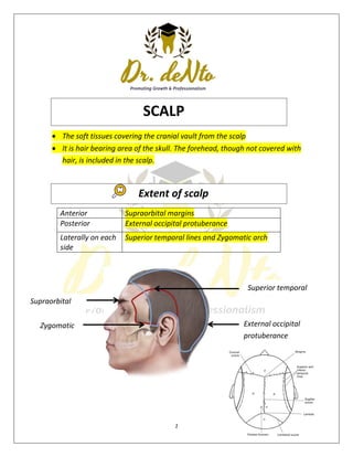
  • 1. 1 Supraorbital margins Zygomatic arch  The soft tissues covering the cranial vault from the scalp  It is hair bearing area of the skull. The forehead, though not covered with hair, is included in the scalp. Anterior Supraorbital margins Posterior External occipital protuberance Laterally on each side Superior temporal lines and Zygomatic arch Superior temporal External occipital protuberance SCALP Extent of scalp
  • 2. 2 S - Skin C - Connective tissue (superficial fascia) A - Aponeurosis L - Loose Areolar Connective Tissue (subaponeurotic layer) P - Pericranium Skin  The skin of scalp is characterized by plenty of hair follicles, sebaceous glands and sweat glands.  Firmly attached to the epicranial aponeurosis through dense fascia  Abundance sebaceous glands Sebaceous cyst are common Connective tissue  Binds skin to subjacent aponeurosis  The superficial fascia is characterized by dense connective tissue. It contains the blood vessels and nerves of the scalp. The fibrous strands in the superficial fascia are fixed to the walls of the blood vessels. This The musculoaponeurotic layer consists of two parts: i. Epicranial aponeurosis (galea aponeurotica) ii. ii. Muscular part composed of frontal and occipital bellies of the occipitofrontalis muscle. Structure
  • 3. 3 factor prevents retraction of the blood vessels, when injured. Hence, scalp wounds bleed profusely. The bleeding in the scalp is controlled by application of direct pressure of the digit or by placing the surgical sutures.  Inflammation cause little swelling but are much painful Aponuerosis and muscle  The galea aponeurotica covers the vault like a helmet.  EPICRANIAL APONUEROSIS It is freely movable on the pericranium along with the overlying and adherent scalp and fascia.  On each side it is attached to the superior temporal lines. Anteriorly ,it receives the insertion of the frontalis. Posteriorly ,receives insertion of the occipital bellies and attached to the external occipital protuberance. OCCIPITOFRONTALIS LOOSE AEREOLAR TISSUE  The subaponeurotic layer (fourth layer) forms a potential space filled with loose areolar tissue beneath the aponeurotic layer.  The emissary veins, which communicate the veins of the scalp with the intracranial venous sinuses, pass through this space. This space is closed on all sides except anteriorly, where it extends into the upper eyelid It is known as the dangerous area of scalp.  It Extends anteriorly into the eyelids.  Posteriorly to the highest and superior nuchal lines and on each side to the superior temporal lines. PERICRANIUM  Loosely attached to the surface of the bones,but is firmly adherent to the sutures where the sutural ligaments bind the pericranium to the endocranium.
  • 4. 4
  • 5. 5  Arteries supplying the scalp are branches of either the external carotid artery or the ophthalmic artery which is a branch of the internal carotid artery.  External carotid arteries o Occipital arteries o Posterior auricular arteries o Superficial temporal arteries  Internal carotid arteries o Supratrochlear arteries o Supraorbital arteries 1. The following arteries supply the anterior quadrant: i. Supratrochlear ii. Supraorbital The above two arteries are the branches of the ophthalmic artery (a branch of internal carotid artery). iii. Superficial temporal artery is the terminal branch of external carotid artery. 2. The following arteries supply the posterior quadrant of scalp i. Posterior auricular ii. Occipital artery The above two arteries are branches of external carotid artery. Arterial Supply of the Scalp
  • 6. 6  Veins draining the scalp follow a pattern similar to the arteries. a. The supratrochlear and the supraorbital vein unite at the medial angle of eye to form angular vein. b. The superficial temporal vein joins the maxillary vein to form retromandibular vein. c. The posterior division of retromandibular vein unites with the posterior auricular vein to form external jugular vein. d. The occipital vein drains into the suboccipital venous plexus. Which lies beneath the floor of the upper part of the posterior triangle, the plexus in turn drains into the vertebral veins or internal jugular vein.  Of the deep parts of the scalp o Via emissary veins that communicates with the dural sinuses o Emissary vein connect the extracranial veins with the intracranial venous sinuses to equalize the pressure, the veins are valveless. o Parietal emissary vein, which passes through parietal foramen and communicates with the superior sagittal sinus. o Mastoid emissary vein, which passes through mastoid foramen and communicates with the sigmoid sinus. Venous Drainage of the Scalp
  • 7. 7  The lymphatics in the occipital region initially drain to occipital nodes which drain into upper deep cervical nodes.  lymphatics from the upper part of the scalp drain in two directions o Posterior to the vertex of the head they drain to mastoid nodes. o Anterior to the vertex of the head they drain to pre-auricular and parotid nodes. Lymphatic Drainage
  • 8. 8  Two main sources o Cranial nerves :- (anterior to ear & vertex) by trigeminal nerves o Cervical nerves:- (posterior to ear and vertex) by C2 &C3 nerves Anterior to the ears and the vertex Posterior to the ears and the vertex Sensory nerves  Supratrochlear nerve  Supraorbital nerve  Zygomaticotemporal nerve  Auriculotemporal nerve By branches of all four divisions of the trigeminal nerve Sensory nerves  Great auricular nerve  Lesser occipital nerve  Greater occipital nerve  Third occipital nerve By branches of all four divisions of the spinal cutaneous nerves (C2 and C3) Motor nerves  Temporal branch of facial nerve Motor nerves  Posterior auricular branch of facial nerve Nerve Supply
  • 9. 9  Skin of the scalp is the commonest site in the body for sebaceous cysts as it is rich in sebaceous glands being the hairiest  Cutaneous tissue of the second layer is divided into a number of loculi containing fat in which infection sends to become localised and causes severe pain.  Wounds into the highly vascular second layer tend to bleed freely as the walls of the blood vessels are attached to the fibrous septa so that they cannot contract and retract to stop the haemorrhage  A wound which cuts through the third layer gapes but the edges of a wound which does not penetrate the aponeurosis remain together This is because the aponeurosis is under tension because of its muscular component and retracts only when divided  The emissary veins which traverse the fourth layer I.e. the subaponeurotic space connect the veins of the second layer with the intracranial venous sinuses. A superficial infection of the scalp can spread via this system to produce venous sinus thrombosis or meningitis  The subaponeurontic space affords an easy plane of cleavage Blood or pus collecting in this layer tracks freely under the first three layers but cannot pass into either the superficial temporal region or the occipital region because of the attachment of the occipitofrontalis muscle. Blood can, however, spread into the upper eyelid causing a 'black eye that may form a few hours after a severe head injury. This layer is known as the 'dangerous layer as infection tends to spread readily within it.  The pericranium is easily stripped up by collection of pus or blood beneath it. The affected bone in outlined as the sutural membrane limits the spread of collected fluid. This is particularly well seen in birth injuries involving the skull and causing a cephalhematoma.  Caput succedaneum is the formation of swelling in the newborn skull due to stagnation of fluid in the scalp layers. This is a temporary swelling since it Clinical Application
  • 10. 10 results from the venous congestion of the scalp due to compression through the birth canal. The swelling of caput succedaneum is diffuse because it is not restricted to any particular bone. It disappears within 24 hours after delivery.  In newborn babies, there is slow accumulation of blood in this space, when there is intracranial hemorrhage due to fracture of the bone of vault and associated dural tear. The leakage of blood outside the cranium in the potentially large subaponeurotic space delays the symptoms of cerebral compression. Hence, the slow accumulation of blood in the fourth layer is known as Safety valve hematoma.
  • 11. 11  The temple is the area between the temporal lines and the zygomatic arch which must be studied in the view of the skull or norma lateralis .  The temporal fossa is a shallow depression bounded above by the superior temporal line on the frontal and parietal bones and below by the zygomatic arch.  The bones taking part in the formation of the fossa are the squamous part of temporal (T), the greater wing of the sphenoid (S) and lower part of the frontal (F) and perietal (P) bones. These bones articulate at an H-shaped suture called the pterion  The superior and inferior temporal lines are the duplication of the temporal line on the parietal bone. The superior temporal line marks the upper limit of the norma lateralis. Lower part of the Frontal Greater Wing of the Sphenoid Squamous part of temporal Lower part of the perietal TEMPORAL FOSSA
  • 13. 13 There are six layers in this region.  The first and second layers are the same as in the scalp I,e. skin and cutaneous tissue  The third layer is a thin expansion of the epicranial aponeurosis of the scalp from which the auricularis anterior and superior muscles arise  The fourth layer is the temporal fascia (Fig 2.7) which is attached above to the superior temporal line and below to the zygomatic arch It is a rugged membrane. The superior temporal vessels and the auriculotemporal nerve lie upon it, and it is perforated by the middle temporal artery and vein .  The fifth layer is the temporalis muscle . which arises from the whole of temporal fossa. The large fan- shaped muscle converges towards the coronoid process mandible . Layers in temporal region
  • 14. 14  The sixth or the last layer is the same as in the scalp i.e. pericranium. Youtube - https://youtu.be/1b40B8sgrho Facebook – De Nto