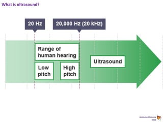
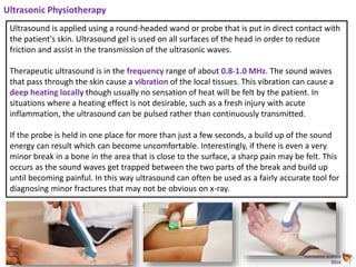
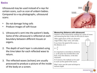
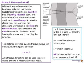
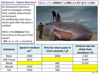
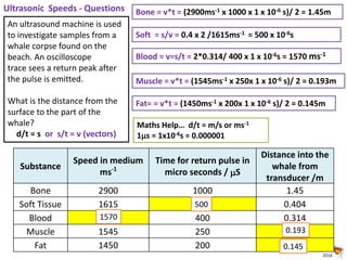
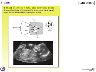
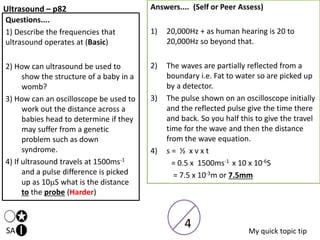
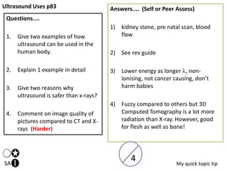
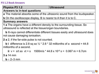
Medical Physics
P3 Physics
Mr D Powell
Animated Science
2016
Connection
• Connect your learning to the
content of the lesson
• Share the process by which the
learning will actually take place
• Explore the outcomes of the
learning, emphasising why this will
be beneficial for the learner
Demonstration
• Use formative feedback – Assessment for
Learning
• Vary the groupings within the classroom
for the purpose of learning – individual;
pair; group/team; friendship; teacher
selected; single sex; mixed sex
• Offer different ways for the students to
demonstrate their understanding
• Allow the students to “show off” their
learning
Activation
• Construct problem-solving
challenges for the students
• Use a multi-sensory approach – VAK
• Promote a language of learning to
enable the students to talk about
their progress or obstacles to it
• Learning as an active process, so the
students aren’t passive receptors
Consolidation
• Structure active reflection on the lesson
content and the process of learning
• Seek transfer between “subjects”
• Review the learning from this lesson and
preview the learning for the next
• Promote ways in which the students will
remember
• A “news broadcast” approach to learning
P3 1.1 X-Rays – What are they and
how are they used in medicine –
p208
INSPIRE
C
I can simply recall some examples
of what X-rays are used for as a list
of ideas.
I can compare a normal X-ray to
a CT-scan
I can explain in detail how an X-
ray is produced in a detailed flow
chart of steps.
I can discuss in detail the Ionising effect of X-rays low kV and high kV
and sievert as a unit of energy delivery per kg of tissue.
A
B
CFS Science
P3 1.1 X-Rays (p208)
Hierarchical Progress Criteria
A*
+ link to sieverts +CGP p81
questions
A/B + all in detail
C + Q1-5 in basic form
2) In your book create a detailed flow
chart/diagram to show/explain how an X-ray
is taken
5) CCDs.....(why are they useful?)
1) List some examples of what X-rays are used
for with some basic details/ ideas for their
uses....
3) What are the health issues for X-Rays?
4) Explain the use of a contrast medium....
6) CT Scans are.... (explain in detail)
Animated Science
2016
Schematic – ignore labels!
Animated Science
2016
X-Ray Process
X-rays are useful in applications such as medical imaging of
bone fractures and dental problems.
1. They are directed at the part of the body under
investigation.
2. They are transmitted through the body - except in areas
where they are absorbed by dense structures like bone.
3. In older X-ray machines, white photographic film is
placed behind the patient.
4. X-rays pass through the patient’s body and into the
photographic film.
5. The film turns black where X-rays hit it. Bones absorb
(stop) X-rays, so the film stays white where the bones
are. Fractures show up as dark areas in the image of the
bones on the film.
NB: This method
can be used for
dental treatment,
as decayed teeth
will absorb X-rays
less strongly than
healthy teeth.
Task..
Review this
information and
then explain it to
another student
Animated Science
2016
Ionising effect of X-rays
The ionising properties of X-rays mean that
they can damage the DNA in cells. The ionising
effect of X-rays is more damaging to actively
dividing cells in the body.
Low doses of X-rays may cause cancer -
whereas high doses may kill cancerous cells.
Cancer treatment
Cancer is a disease in which cells divide
uncontrollably because of changes in their
DNA, forming tumours.
One method of treating cancer is to direct high
energy X-rays at the tumours. This causes so
much damage to the cancerous cells that they
die. This treatment is called radiotherapy.
Animated Science
2016
Precautions when using X-rays
Patients are limited to the number of X-rays they are
allowed to have so their bodies are not exposed to too
much radiation.
X-ray machines also produce relatively low energy X-rays,
which reduce the risk of them damaging human tissue.
Hospital staff are also at risk from repeated exposure to
low levels of X-rays. Shielded walls containing lead are
built into all X-ray rooms to protect people outside the
room. They have warning signs to show when the room is
in use so that people do not enter.
Only trained specialist staff - called radiographers - are allowed to use X-ray
machines. They routinely leave the room, or stand behind a screen containing
lead, whenever X-ray machines are in use.
In situations where radiographers cannot stand behind a screen, they wear lead
aprons which act as a protective layer of clothing.
Animated Science
2016
Complex Equipment...
CCDs...A CCD is a charge-coupled
device. Modern X-ray machines use CCDs
instead of photographic film. The images are
formed electronically, allowing them to be
recorded and stored more easily than the images
from photographic film.
CT scans...Traditional X-ray imaging gives a two-
dimensional (2D) view of the body from one
angle. This can result in detail being obscured by
other structures in the body. Computerised
tomography (CT) scans involve taking a range of
X-ray images from various positions.
These are processed by a computer to build
a three-dimensional (3D) image. This image can
be manipulated in order to see the structures
within the body at different layers and from
different points of view. This lets a doctor gain a
much greater insight into what is wrong with a
patient.
Animated Science
2016
Common Radiation Units...
Quantity Name Symbol Unit Year System
Exposure
(X)
röntgen R
esu/0.0012
93 g of air
1928 non-SI
Absorbed
dose (D)
rad rad 100 erg·g−1 1953 non-SI
gray Gy J·kg−1 1974 SI
Activity (A)
curie Ci 3.7×10¹⁰ s-1 1953 non-SI
becquerel Bq s−1 1974 SI
Dose
equivalent
(H)
röntgen
equivalent
man
rem 100 erg·g−1 1971 non-SI
sievert Sv J·kg−1 1977 SI
GCSE requires Bq but Sv may appear in questions!
X Rays in Medicine p81 CGP
SA
Questions....
1) Describe all you can about an X-
Ray as a wave. (Basic)
2) Give an example of how a
human X-ray works.
3) How can an X-ray be detected by
a computer.
4) What is a CT Scan.
5) How can you use them to treat
cancer (Harder)
5
My quick topic tip
Answers.... (Self or Peer Assess)
1) High freq, short wavelength, EM radiation
or waves that travel at 3 x 10⁸ms-1 in a
vacuum
2) Passes through soft tissue but not bone so
a photograph is created (goes dark on
tissue and stays white on bone)
3) From tiny charge coupled devices or CCD
silicon chips in an array
4) Computerised Axiel Tomography. A 3D
detector picks up X-rays to form a 3D
image
5) x-rays ionise or damage tissue and can be
directed onto tumours in the body.
Animated Science
2016
P3 1.1. Book Answers
Physics P3 1.1 X-rays
Answers to in-text questions
a A crack is a gap that X-rays can pass through.
b To keep the light out without stopping the X-rays.
c They would damage or kill living cells or cause cancer.
Summary answers
1 a penetrate b absorb c damage
2 a To make an image of the patient’s bones on the fi lm.
b To stop light from affecting the fi lm.
c To prevent damage by the X-rays to the parts of the body not being X-
rayed. High doses can kill living cells and low doses can cause cell mutation
and cancerous growth.
3 Advantage: A CT scan distinguishes between different types of soft tissue;
an ordinary X-ray machine does not. (Or a CT scanner can give a three-
dimensional image whereas an ordinary X-ray image is two-dimensional.)
Disadvantage: The radiation dose from a CT scan is much greater than
from an ordinary X-ray imaging machine. (Or CT scanners are much more
expensive to buy and operate than ordinary X-ray machines.)
P3 1.2 Ultrasound – what is it and how
is it used in a hospital?
INSPIRE
C
Be able to recall simple examples of
Ultrasound in some detail and one in a lot
of detail.
Complete calculations to work out the
distance from a transducer to an organ
boundary /Be able to compare an MRI to an
Ultrasound in detail
Be able to explain how an A scan ultrasound
scan works in terms of reflection and what
they are used for in hospitals.
Explain how the Piezoelectric effect works to another student or teacher
A
B
CFS Science
Animated Science
2016
What is ultrasound?
Animated Science
2016
Ultrasonic Physiotherapy
Ultrasound is applied using a round-headed wand or probe that is put in direct contact with
the patient's skin. Ultrasound gel is used on all surfaces of the head in order to reduce
friction and assist in the transmission of the ultrasonic waves.
Therapeutic ultrasound is in the frequency range of about 0.8-1.0 MHz. The sound waves
that pass through the skin cause a vibration of the local tissues. This vibration can cause a
deep heating locally though usually no sensation of heat will be felt by the patient. In
situations where a heating effect is not desirable, such as a fresh injury with acute
inflammation, the ultrasound can be pulsed rather than continuously transmitted.
If the probe is held in one place for more than just a few seconds, a build up of the sound
energy can result which can become uncomfortable. Interestingly, if there is even a very
minor break in a bone in the area that is close to the surface, a sharp pain may be felt. This
occurs as the sound waves get trapped between the two parts of the break and build up
until becoming painful. In this way ultrasound can often be used as a fairly accurate tool for
diagnosing minor fractures that may not be obvious on x-ray.
Animated Science
2016
Ultrasonic Jewellery Cleaners
An ultrasonic jewellery cleaner is an electronic device designed to
remove dirt from rings, necklaces and other items of jewellery.
These devices are readily found on home shopping channels or on
the internet.
To begin the cleaning process, the cleaning tank is filled with
warm water or a cleaning solution. Cleaning solutions can consist
of non-ionic surfactants, detergents or ammonia. Once on, the
machine's motor produces ultrasonic energy, which is transmitted
with vibrating energy waves. On average, ultrasonic jewellery
cleaners emit at least 40,000 sound waves per second.
1. The vibrating motion of the ultrasonic waves creates
microscopic bubbles in the water or cleaning solution in a
process called cavitation
2. millions of tiny bubbles knock into one another and into the
items resting in the cleaning tank.
3. The cavitation process gently knocks dirt off the jewellery.
4. The motion is very effective at penetrating the tiny crevices in
jewellery that traditional cleaning cloths and topical cleaners
cannot easily reach.
Animated Science
2016
Ultrasonic Washing Machine
LG recently put on sale a washing machine that doesn't
require detergent to clean lightly soiled clothes.
The machine is fitted with electrodes on the side of the
tub that electrolyse the water, and an ultrasonic wave
generator at the base of the machine.
1. The ultrasonic waves form tiny air bubbles, help
loosen grime and grit on clothes in a purely
mechanical action.
2. Electrolyzing the water produces active oxygen, or
forms of oxygen such as hydrogen peroxide and
ozone, and hypochlorous acid, a mild bleaching
agent. Hypochlorous acid kills bacteria while active
oxygen dissolves such dirt as the residue of body
sweat.
3. LG claims users can half the cost of doing normal
laundry. Reducing the amount of detergent sent into
waste water streams is also environmentally friendly.
Animated Science
2016
Breaking down kidney stones
A high powered ultrasound wave is used to break
down kidney stones and other stones in the
body.
The stones vibrate until they shake themselves
apart and are then easily passed out of the body
via the urethra.
Animated Science
2016
Basics
Ultrasound may be used instead of x-rays for
certain scans, such as scan of unborn babies.
Compared to x-ray photographs, ultrasound
scans:
• Do not damage living cells
• Produce images of soft tissue
1. Ultrasound is sent into the patient's body.
Some of the ultrasound is reflected at each
boundary between different tissues or
organs.
2. The depth of each layer is calculated using
the time taken for each reflected wave to
return.
3. The reflected waves (echoes) are usually
processed to produce a picture of the inside
of the body on a screen.
Animated Science
2016
Advantages / Disadvantages
Animated Science
2016
Ultrasonic How does it work?
When ultrasound waves reach a
boundary between two media
(substances) with different densities,
they are partly reflected back. The
remainder of the ultrasound waves
continue to pass through. A detector
placed near the source of the
ultrasound waves is able to detect the
reflected waves. It can measure the
time between an ultrasound wave
leaving the source and it reaching the
detector.
The distance travelled by an ultrasound wave can
be calculated using this equation:
s = v × t
An ultrasound machine can be used to detect
cracks or flaws in materials such as metal.
s = distance in metres, m
(often d is used for GCSE P1
unit but s for P3)
v = speed in metres per
second, m/s
t = time in seconds, s
NB: but remember this is an
echo so you must half it!
Animated Science
2016
Ultrasonic Crack Detection - Example
Step 1: Work out the time between the pulse being emitted and detected
In this case the time is given as 120 µs
Step 2: Convert µs into s
1 s = 1 000 000 µs
120 µs = 120 ÷ 1 000 000 = 0.00012 s (1.2 × 10 -4 s)
Step 3: Put the values into the formula s = v x t
Total distance = 6320ms-1 × 0.00012s = 0.758 m
Step 4 Echos?
s (to the crack) = 0.758 ÷ 2 = 0.379 m
An ultrasound machine is used to check
for cracks in aluminium. An oscilloscope
trace sees a return peak 120 µs after the
pulse is emitted. The speed at which
sound travels through aluminium is 6,320
m/s.
What is the distance from the surface to
the crack?
Animated Science
2016
Ultrasound – Topical Questions
An ultrasound machine is
used to investigate samples
from a whale corpse found
on the beach.
An oscilloscope trace sees a
return peak after the pulse is
emitted.
What is the distance from
the surface to the part of the
whale?
d/t = s or s/t = v (vectors)
Substance
Speed in medium
ms-1
Time for return pulse in
micro seconds / S
Distance into the
whale from
transducer /m
Bone 2900 1000 1.45
Soft Tissue 1615 0.404
Blood 400 0.314
Muscle 1545 250
Fat 1450 200
Bone = v*t = (2900ms-1 x 1000 x 1 x 10-6 s)/ 2 = 1.45m
Animated Science
2016
Ultrasonic Speeds - Questions
An ultrasound machine is used
to investigate samples from a
whale corpse found on the
beach. An oscilloscope
trace sees a return peak after
the pulse is emitted.
What is the distance from the
surface to the part of the
whale?
d/t = s or s/t = v (vectors)
Substance
Speed in medium
ms-1
Time for return pulse in
micro seconds / S
Distance into the
whale from
transducer /m
Bone 2900 1000 1.45
Soft Tissue 1615 0.404
Blood 400 0.314
Muscle 1545 250
Fat 1450 200
Bone = v*t = (2900ms-1 x 1000 x 1 x 10-6 s)/ 2 = 1.45m
500
0.193
0.145
1570
Soft = s/v = 0.4 x 2 /1615ms-1 = 500 x 10-6s
Blood = v=s/t = 2*0.314/ 400 x 1 x 10-6s = 1570 ms-1
Muscle = v*t = (1545ms-1 x 250x 1 x 10-6 s)/ 2 = 0.193m
Fat= = v*t = (1450ms-1 x 200x 1 x 10-6 s)/ 2 = 0.145m
Maths Help… d/t = m/s or ms-1
1s = 1x10⁻⁶s = 0.000001
Animated Science
2016
This ultrasound is designed to look the structure
of the eye.
• A is the partial reflection as the waves enter
the cornea,
• B is a reflection of the pulse at the lens,
• C is due to the reflection off the back of the
eye. Further “blips” are due to the waves
reflecting off bone in the skull.
Exam Questions
The distance to the back of the eye scanned in this
way was known to be 48mm. What would be the
distance from the eye lens to the front of the model
eye using this scale?
48mm = 3.9 sq
7.8 squares = 96mm or 1 sq = 12.3
1.1 square = 12.3mm *1.1 = 13.54mm
14mm = distance
or simply 48mm * (1.1sq /3.9sq) = 14mm
Smaller pulses are due to..
• Extra distance in tissue
results in more signal
absorption
• Smaller fraction of signal
reflected at second surface
• The pulse will be more
spread over time
• The signal is diffracted
Animated Science
2016
The screen shows an ultrasound pulse which
takes 32 millionths (32S or 32 x 10-6S) to travel
across each square of the grid.
Exam Questions
1) How many internal boundaries are present in this sample...
Two (excluding the initial pulse external boundary and far side boundary)
2) How long does each pulse take to travel from the body surface to the nearest
internal body
difference is 2.8 sq (there and back)
t = ½ * 2.8 * 32 millionths of a second = 44.8S.
3) If the wavespeed is 1500ms-1. What is the distance from the body surface to the
nearest tissue boundary?
v = s/t so vt = s 1500ms-1 * 44.8 x 10-6 s = 0.067m = 6.7cm
Animated Science
2016
Extra DetailsThe Eye – real world ideas....
Animated Science
2016
Extended Thinking...
Summary
Animated Science
2016
Extended ideas on Speed
Animated Science
2016
Extra DetailsB - Scans
Animated Science
2016
Piezoelectric Effect – Extra Thinking
Ultrasound – p82
SA
Questions....
1) Describe the frequencies that
ultrasound operates at (Basic)
2) How can ultrasound be used to
show the structure of a baby in a
womb?
3) How can an oscilloscope be used to
work out the distance across a
babies head to determine if they
may suffer from a genetic
problem such as down
syndrome.
4) If ultrasound travels at 1500ms-1
and a pulse difference is picked
up as 10S what is the distance
to the probe (Harder)
Answers.... (Self or Peer Assess)
1) 20,000Hz + as human hearing is 20 to
20,000Hz so beyond that.
2) The waves are partially reflected from a
boundary i.e. Fat to water so are picked up
by a detector.
3) The pulse shown on an oscilloscope initially
and the reflected pulse give the time there
and back. So you half this to give the travel
time for the wave and then the distance
from the wave equation.
4) s = ½ x v x t
= 0.5 x 1500ms-1 x 10 x 10-6S
= 7.5 x 10⁻³m or 7.5mm
My quick topic tip
4
Ultrasound Uses p83
SA
Questions....
1. Give two examples of how
ultrasound can be used in the
human body.
2. Explain 1 example in detail
3. Give two reasons why
ultrasound is safer than x-rays?
4. Comment on image quality of
pictures compared to CT and X-
rays (Harder)
Answers.... (Self or Peer Assess)
1) kidney stone, pre natal scan, blood
flow
2) See rev guide
3) Lower energy as longer , non-
ionising, not cancer causing, don’t
harm babies
4) Fuzzy compared to others but 3D
Computed Tomography is a lot more
radiation than X-ray. However, good
for flesh as well as bone!
My quick topic tip
4
Transparent concrete
LiTraCon in the US has now embedded optical fibres within poured concrete blocks,
creating (partially) transparent concrete that’s just as strong as its non-
transparent brother.
Animated Science
2016
P3 1.2 Book Answers
Physics P3 1.2 Ultrasound
Answers to in-text questions
a The material absorbs some of the ultrasonic sound from the loudspeaker.
b On the oscilloscope display, B is nearer to A than it is to C.
Summary answers
1 a The organs have a different density to the surrounding tissue. So
ultrasound is reflected at the tissue/organ boundaries.
b X-rays cannot differentiate different tissues easily and ultrasound does
not cause damaging ionisation.
2 a 2, if the far-side pulse is not counted.
b i difference is 2.8 sq so ½ * 2.8 * 32 millionths of a second = 44.8
millionths of a second.
ii v = s/t so vt = s 1500m/s * 44.8 x 10⁻⁶ s = 0.067 m = 6.7cm
3 a 14 mm
b ± 2–3 mm
P3 1.3 Refractive Index
• What is it?
• What does it mean?
• How does it have an effect on light?
INSPIRE
C
Be able to explain simply how a light ray
slows down or refracts/changes direction
in a prisms. (Laser Video/ White light
video & Prac) (Whiteboard)
+conduct a highly precise practical
where “n” is consistent
Conduct a practical to establish the
refractive index “n” of a prism. A
measure of optical density. (use Sin)
+ work out “n” graphically AND / OR critical angle of a semi-circular
prism
A
B
CFS Science
weak
reflected
ray
r
i
n
sin
sin

Animated Science
2016
weak
reflected
ray
NB: When light travels from an optically denser medium to a less
dense medium, rays are bent away from the normal. The
incident substance has a larger refractive index than the other
substance
Refractive Index Theory…
i
• Light enters a prism
• It slows down
• Turns towards the normal
• This process is called refraction
• We can measure the incident angle (i)
• We can measure the refracted angle (r)
• We find the ratio of the sine function of each
gives us a consistent value for a material.
• This is the refractive index of a substance
n
r
i

)sin(
)sin(
Example Material n
Vacuum 1
Air 1.00029
Carbon dioxide 1.0005
Diamond 2.419
Cubic Zirconia 2.15
Water ice 1.31
Prism ?
cornea (human) 1.3375
Animated Science
2016
Sine – Maths Support
Sine, in mathematics, is a trig function of
an angle.
• The sine of an angle is defined in the context
of a right triangle
• for the specified angle, it is a simple ratio of
sides
"Opposite" is opposite to the angle θ
"Adjacent" is adjacent (next to) to the angle θ
"Hypotenuse" is the long diagonal one










hyp
opp
hyp
opp
1
sin
sin
 














9.36
6.0sin
5
3
sin
1
1
Animated Science
2016
Sine – Calculator Support 2nd function!
Animated Science
2016
y = mx + c – Graphical Method
)sin()sin(
...
sin(
)sin(
rni
cmxy
but
n
r
i



Angle of
refraction, r
(degrees)
Angle of
incidence, i
(degrees)
sin r sin i n= Sin(i) / sin(r)
6.6 10 0.11 0.17 1.51
13 20 0.22 0.34 1.52
19 30 0.33 0.50 1.54
25 40 0.42 0.64 1.52
31 50 0.52 0.77 1.49
36 60 0.59 0.87 1.47
39 70 0.63 0.94 1.49
y = 1.4703x + 0.0119
0.00
0.10
0.20
0.30
0.40
0.50
0.60
0.70
0.80
0.90
1.00
0.00 0.20 0.40 0.60 0.80
Sini
Sin r
Finding the Refractive Index of a Prism (Graphical
Method)
Use the generic formula to plot
the graph, then the gradient or
slope is the refractive index of
the prism!
Animated Science
2016
Sine – Maths Extension!
Sine, in mathematics, is a trig function of
an angle. The sine of an angle is defined in the
context of a right triangle: for the specified
angle, it is the ratio of the length of the side
that is opposite that angle to the length of
the longest side of the triangle (i.e., the
hypotenuse).
Extension
In AS Maths / Physics you will learn that
we can plot the Sin of an angle i.e.
sin(x) as a function to get a curve!
Animated Science
2016
Sine – Maths Support
Sine, in mathematics, is a trig function of
an angle.
• The sine of an angle is defined in the context
of a right triangle
• for the specified angle, it is a simple ratio of
sides
"Opposite" is opposite to the angle θ
"Adjacent" is adjacent (next to) to the angle θ
"Hypotenuse" is the long diagonal one










hyp
opp
hyp
opp
1
sin
sin
 














9.36
6.0sin
5
3
sin
1
1
Refractive Index Plenary Questions – CGP p84
Questions....
1) What causes refraction?
2) Write down the formula for refractive index.
3) If a light ray enters some jelly at an angle of
42 from air (nair = 1) and is refracted to an
angle of 35  what is nJelly = ??? (Harder
Maths)
4) Why does a diamond sparkle so bright!
5) Draw an accurate diagram of refraction in a
rectangular block of glass of an incident ray
of monochromatic red light at an angle of
about 30 to the normal. (challenge)
Answers.... (Self or Peer Assess)
1) When a light wave slows or
speeds up (optical density)
2) Ref index n = sin i / sin r
3) nJelly = sin 42 /sin 35 = 1.18
4) It has a very high refractive
index and splits the light up to
make a spectrum!
5) See revision guide, but must
be pretty good – no marks for
wobbly lines!
Animated Science
2016
P3 1.3 Book Answers
Physics P3 1.3 Refractive index
Answers to in-text questions
a 1.54
b The angles were not measured precisely enough.
Summary answers
1 a less
b more
c less
2 a sin 40 ÷ sin 26 = 1.47
b The angle of refraction was measured incorrectly and the
measurement should have been smaller.
3 a 25.5°
b 70.1°
Animated Science
2016
P3 1.3/4 Homework - Spectacle Lens Answers
1. 500 nm to 1000 nm
2. No, the range is not suitable, as it does not cover the
full range of visible light wavelengths.
3. Yes, because the refractive index of polycarbonate at
1.584 is higher than the next best ophthalmic glass at
1.523
4. 1.569–1.570
5. To improve the validity of results because the lens
manufacturer may be biased
6. The variables are continuous.
7. The temperature of the material.
8. Presentation makes comparison of the refractive index
easier in the table than a graph.
Task: Use the answers
to self assess your
homework.
Write your score out
of /8
Then give yourself a *
& I
P3 1.4 The Endoscope – how can we use a
special property of “refraction” to look
inside the body!
INSPIRE
C
Explain simply with use of 3 precise
diagrams the idea of the “critical
angle”
Complete critical angle
calculations for a wider range
of materials.
Explain how this idea can be
applied to a human body (video /
water demo)
Extend your thinking to explain how a coherent image is formed from the
many fibres in an endoscope?
A
B
CFS Science
Animated Science
2016
More Uses of Optical Fibres….
1. Illuminating models or road signs using only one bulb
2. Endoscopy - seeing down inside a patient’s body
3. Communications – sending information along a light beam. Useful for
telephone, television, radio, computer networks, stereo links, control in
aircraft
4. Security fencing – very difficult to bypass
5. Fibre optic lamp
Advantages of fibre optics over copper wire
1. Cheap – glass is made from silica, the basic constituent of sand
2. Light in weight – useful in aircraft
3. Light beam can carry a huge amount of information
Such fibres can be made to carry information such as TV channels or telephone
conversations. Other applications of fibre optics include its use in medicine to
see inside the human body and in road signs where one light bulb and a set of
fibres is used to illuminate different parts of the sign thus saving electrical
energy. A further recent application is in security fences. The metal strands of
the fence contain a piece of fibre optic material down which a beam of light
passes. If the strand is cut the light beam is interrupted and an alarm sounds. It
is thought that this type of system is impossible to bypass.
Animated Science
2016
As the angle of incidence
increases towards the critical
angle ( glass = 420 ) the refracted
ray gets weaker and the reflected
ray gets stronger.
weak reflected
ray
NB: When light travels from an optically denser medium to a less dense medium,
rays are bent away from the normal. The incident substance has a larger refractive
index than the other substance
TIR
Animated Science
2016
Critical angle depends upon the
refractive indices of the media
nprism
C
Critical Angle?
C
n
n
n
n
prism
prism
prism
prism
prism
prism
air
prism
sin
1
sin
1
sin
90sin
sin
sin




















prism
prism
prism
n
C
n
C
C
n
1
sin
1
sin
sin
1
1
Filter
Colour
Critical
Angle 
Refractive
Index
None
(white)
46 1.39
Red
Green
Blue
Animated Science
2016
Critical angle depends upon the
refractive indices of the media
nprism
C
Critical Angle?
C
n
n
n
n
prism
prism
prism
prism
prism
prism
air
prism
sin
1
sin
1
sin
90sin
sin
sin




















prism
prism
prism
n
C
n
C
C
n
1
sin
1
sin
sin
1
1
Filter
Colour
Critical
Angle 
Refractive
Index
None
(white)
46 1.39
Red 46.5 1.378
Green 46 1.39
Blue 45.5 1.40
Animated Science
2016
1
2
C
AIR
WATER
For Water
33.1
1
sin C
C = 48.80
For Crown Glass
50.1
1
sin C C = 41.80
Critical Angle Example?








 
prismn
C
1
sin 1
Animated Science
2016
1
1
sin
n
C 
24.44
Diamonds Sparkle the most!
 Diamond has a refractive index of 2.417.
 This means that colours are spread out more.
 TIR occurs many times inside the diamond before
emerging.
Can you work out the
critical angle?
Animated Science
2016
Material n
Vacuum 1 (per definition)
Air @ STP 1.000277
Gases @ 0 °C and 1 atm
Air 1.000293
Carbon dioxide 1.00045
Liquids @ 20 °C
Water 1.3330
Solids @ room temperature
Diamond 2.419
Cubic Zirconia 2.15
Strontium titanate 2.41
Amber 1.55
Water ice 1.31
cornea (human) 1.3375
Extra Examples…
Animated Science
2016
A coherent bundle of optical fibres in which
the relative spatial coordinates of each fibre
are the same at the two ends of the bundle.
Such a bundle are used for the transmission
of images. Endoscope
A non-coherent fibre bundle, as you would
expect, does not have this precise matrix
alignment since they need only transmit
light for illumination purposes. They are
cheaper to produce. Christmas Tree.
Applications of Fibre Optics…
Total Internal Reflection - p90
SA
Questions....
1. Explain the key features of TIR
with an optical fibre.
2. Give an example of the use of this
in industry?
3. What is the formula for refractive
index to find the critical angle.
4. Work out the refractive index for
diamond if c = 24 (Harder)
5. Explain in detail how an
endoscope works?
Answers.... (Self or Peer Assess)
1) Sheath of plastic around a core of
different refractive index “n”, light at
critical angle is TIR along the core.
2) Connections for data or internet, fibre
optic Christmas tree, key hole surgery
3) 1/sin(c) = refractive index
4) 1/sin(24) = 2.46
5) Light is passed down an optical fibre to
light up the part of the patient. A
separate ordered bundle of fibres is
then placed next to it to receive the
light.
My quick topic tip?
Animated Science
2016
P3 1.4 Book Answers
Physics P3 1.4 The endoscope
Answers to in-text questions
a 90°
b 1.47
Summary answers
1 a refraction
b total internal reflection
c partial reflection
d reflection, refraction
b Any two advantages:
1. The endoscope uses light which is non-ionising, (unlike X-rays).
2. Movement of the fragments can be seen with an endoscope.
3. Fragments may be hidden by other fragments on an X-ray picture.
3 a 48.8°
b 1.49
Animated Science
2016
i
Refraction – Summary / Revision
Main idea is... That the angle of incidence increases the
angle of refraction will also increase.
The theory of this is that light is slowed down by a prism
due to an increase in optical density in comparison with air.
c = f so the theory is that if  reduces then so will c as the
frequency or colour is a constant through the prism.
NB: When light travels from an optically denser medium to
a less dense medium, rays are bent away from the normal.
If the incident substance has a larger refractive index than
the other substance i.e. Air -> prism. This reverses on the
way out and bends towards the normal.
• Light enters a prism
• It slows down
• Turns towards the normal
• This process is called refraction
• We can measure the incident
angle (i)
• We can measure the refracted
angle (r)
• We find the ratio of the sine
function of each gives us a
consistent value for a material.
• This is the refractive index of a
substance
Example Material n
Vacuum 1
Air 1.00029
Carbon dioxide 1.0005
Diamond 2.419
Cubic Zirconia 2.15
Water ice 1.31
Prism ?
cornea (human) 1.3375
Filter Colour
Critical Angle

Refractive
Index
Wavelength 
/ x 10-6
m
None (white) 46 1.39 -
Red 46.5 1.378 0.62
Green 46 1.39 0.53
Blue 45.5 1.40 0.41
• Light enters a prism at the critical angle. “c”
• The angle of refraction becomes 90
• Ratio sin formula simplifies as sin 90 = 1
• The refractive index “n” can be worked out from 1
angle instead of 2.
• If the light has a different wavelength then it refracts
or slows down differently in the prism. Blue refracts
the most so the critical angle is lower in the same
prism.
C
nprism
sin
1

n
r
i

)sin(
)sin(
Animated Science
2016
Think – what causes this?
A convex lens... (converging)
• Produces a magnified real image
on a screen
• Has a focal point (or focal length)
P3 1.5/1.6 Lenses – how can we
scale an image on a camera with a
lens or system of lenses.
INSPIRE
B/C
Draw/explain a simple diagram to show
how an image can be focused by a convex
or concave lens and magnified.
+include a series of steps in your own words
(resourced from the revision guide / book / PPT
explaining how you constructed your diagrams.
Including details on virtual images. & examples
+draw the diagrams with a precise
scale (i.e. onto graph paper)
Try the 15 questions on “curved mirrors” for ray diagram practice
S
A
CFS Science
P3 1.5/1.6 Lenses p216 – 219
Hierarchical Progress Criteria
S + Q5 for homework – in detail
A + Q3/4 precise in pencil including guide.
B + Q2 in full detail
C/D Q1 – ruler pencil + basics of Q2
1) Use the images here to
complete the ray diagrams
for both converging and
diverging situations.
2) Go on to detail the key
features of each lens in
detail in the space
provided.
3) Use a textbook or CGP
revision guide and graph
paper to draw & explain
accurate step by step
diagrams for both types of
lens (not the same as the
ones on the sheet!. You
can pick you own scale and
focal lengths. Use a ruler
and pencil for this.
4) Explain the concept of
magnification (in your
book/on the sheet)
5) Homework: do research at
home and answer
spectacle lenses sheet
P3 1.5/1.6 Lenses p216 – 219
Hierarchical Progress Criteria
S +Q5 for homework – in detail
A + Q3/4 precise in pencil including guide.
B + Q2 in full detail
C Q1 – ruler pencil + basics of Q2
1) Use the images here to
complete the ray
diagrams for both
converging and
diverging situations.
2) Go on to detail the key
features of each lens in
detail in the space
provided.
3) Use a textbook or CGP
revision guide and
graph paper to draw &
explain accurate step
by step diagrams for
both types of lens. You
can pick you own scale
and focal lengths. Use a
ruler and pencil for this.
4) Explain the concept of
magnification (in your
book/on the sheet)
5) Homework: do
research at home and
go on to explain a use
for the lenses in detail
Convex – Converging
• Convex lenses are thicker in the middle.
• A convex lens is a converging lens as the rays
converge
• When parallel rays of light pass through a
convex lens the refracted rays converge at
one point called the principal focus.
• The distance between the principal focus and
the centre of the lens is called the focal
length.
Concave - Diverging
• are thinner at the middle.
• Rays of light that pass through the lens are
spread out (they diverge).
• A concave lens is a diverging lens spreading the
light rays
• When parallel rays of light pass through a
concave lens the refracted rays diverge so that
they appear to come from one point called
the principal focus.
• The distance between the principal focus and
the centre of the lens is called the focal length.
• The image is virtual and diminished (smaller)
Animated Science
2016
Convex Lenses
• Convex lenses are thicker in
the middle.
• A convex lens is a converging
lens as the rays converge
• When parallel rays of light pass
through a convex lens the
refracted rays converge at one
point called the principal
focus.
• The distance between the
principal focus and the centre
of the lens is called the focal
length.
Use of Convex Lenses – The Camera
A camera consists of three main parts;
1. The body which is light tight and contains
all the mechanical parts.
2. The lens which is a convex (converging)
lens).
3. The film or a charged couple device in
the case of a digital camera.
Animated Science
2016
Concave Lenses
• are thinner at the middle.
• Rays of light that pass through the lens
are spread out (they diverge).
• A concave lens is a diverging lens
spreading the light rays
• When parallel rays of light pass through
a concave lens the refracted rays
diverge so that they appear to come
from one point called the principal
focus.
• The distance between the principal
focus and the centre of the lens is
called the focal length.
• The image
is virtual and diminished (smaller)
Convex – Converging
• Convex lenses are thicker at the middle.
• A convex lens is a converging lens as the rays converge
• When parallel rays of light pass through a convex lens the refracted rays
converge at one point called the principal focus.
• The distance between the principal focus and the centre of the lens is
called the focal length.
Concave - Diverging
• are thinner at the middle.
• Rays of light that pass through the lens are spread out (they diverge).
• A concave lens is a diverging lens spreading the light rays
• When parallel rays of light pass through a concave lens the refracted rays
diverge so that they appear to come from one point called the principal
focus.
• The distance between the principal focus and the centre of the lens is called
the focal length.
• The image is virtual and diminished (smaller)
Animated Science
2016
Use of Convex Lenses – The Camera
The rays of light from the person are converged
by the convex lens forming an image on the film
or charged couple device in the case of a digital
camera.
• The angle at which the light enters the lens
depends on the distance of the object from
the lens.
• If the object is close to the lens the light rays
enter at a sharper angled.
• As the lens can only bend the light to a
certain agree the image needs to be
focussed in order to form on the film.
• This is achieved by moving the lens away
from the film.
• The image formed is said to be real because
the rays of lighted from the object pass
through the film and inverted (upside down)
Animated Science
2016
Examples for Convex and Concave
Animated Science
2016
Magnification
• A magnifying glass is a convex lens which produces a
magnified (larger) image of an object.
• A magnifying glass produces an upright, magnified
virtual image.
• The virtual image produced is on the same side of
the lens as the object.
• For a magnified image to be observed the distance
between the object and the lens must be shorter
than the focal length of the lens.
Worked example
An object appears to be
50 cm high when viewed
through a magnifying
glass.
The object is actually
only 20 cm high.
What is the
magnification.
M = 50 ÷ 20
M = 2.5
The image is 2.5 times
larger than the object.
Animated Science
2016
Bitesize Revision – Tricky! distance of
object
Size Orientation Type Position
Further than
2F
Diminished Inverted Real
Between 2F
and F
2F Same size Inverted Real 2F
Between 2F
and F
Magnified Inverted Real
Further than
2F
F
No image because the emerging rays are parallel to
the axis
Closer than
F
Magnified Upright Virtual
Same side
as object
A Converging lenses can be
investigated by changing the
distance between the object and
the lens. Using the setup in the
diagram the object can be moved
closer or further away from the lens.
The object distance is measured
in focal lengths: F, 2F (twice the
focal length), and so on. The image
distance can also be measured in
focal lengths.
The image..
• Is inverted and real - unless the
object is at F or closer
• gets larger as the object gets
closer
• gets further away as the object
gets closer - unless the object is
at F or closer
http://www.bbc.co.uk/schools/gcsebitesize/science/triple_aqa/medical_applications_physics/lenses/revision/4/
3
3
Lenses and Images p85
Questions....
1. Draw a converging lens with a
principal focus point?
2. Draw the same for diverging lens
but with virtual point? (Harder)
3. Write down the three rules for a
converging lens (in short form).
4. Write down the three rules for a
diverging lens. (in short form).
5. What do we mean by “inverted”
Answers.... (Self or Peer Assess)
1) See rev guide – must used ruler!
2) See rev guide – must used ruler!
3) See rev guide
4) See rev guide
5) Upside down
?
Lenses p86
SA
Questions....
1. When a converging lens is used
what happens to the image of a
flower pointed upwards?
2. If you use a converging lens and
double the distance of the
object from the lens, what
happens to it?
3. When using a diverging lens
(concave) describe the image
formed? (Harder)
Answers.... (Self or Peer Assess)
1) It inverts.
2) Stays inverted by x2 the height.
3) Virtual and correct way up.
My quick topic tip
?
Magnification and Power
SA
Questions....
1. What type of lens do you need in
a magnifying glass?
2. Where must the object be to
enlarge it and why?
3. What is the magnification
formula?
4. What is the power of a lens?
5. What is the power of a
converging lens of focal length
0.1m (Harder)
Answers.... (Self or Peer Assess)
1) Converging or convex.
2) Closer than the focal point so the image
appears at the focal point bigger (see rev
guide)
3) Mag = image height / object height
4) Power (D) = 1 / focal length (m) or P =
1/f
5) Power (D) = 1/0.1m = 10 D
My quick topic tip
5
Animated Science
2016
Fundus….
Animated Science
2016
Instructions: Close one eye and focus the other on the appropriate letter
(R for right or L for left).
Place your eye a distance from the screen approximately equal to 3× the
distance between the R and the L.
Move your eye towards or away from the screen until you see the other
letter disappear.
For example, close your right eye, look at the "L" with your left eye, and
the "R" will disappear.
Blind Spot? Hint Try drawing a R
& L in your book!
Animated Science
2016
P3 1.5 Book Answers
Physics P3 1.5 Lenses
Answers to in-text questions
a A 5 cm focal length lens.
b Inverted.
c To make it appear much larger, so any flaws can be seen.
Summary answers
1 a converging, real
b diverging, virtual
2 a Upright, enlarged and virtual.
b i Inverted, magnified and real.
ii The slide must be moved towards the screen.
3 a The image is real, inverted and enlarged. Magnification = 3.
b The image would be smaller than it was and would still be upside down.
Animated Science
2016
P3 1.6 Book Answers
Physics P3 1.6 Using lenses
Answers to in-text questions
Summary answers
b f = 1.8 cm
P3 1.7/8 The Eye – how is it
structured and how does it
work and what is lens “power”
INSPIRE
C
Discuss the idea of a blind spot
through a practical.
Compare short (myopia) and long sighted
(hyperopia) vision problems and explain
how you can solve them.
Label the structure & understand the
function of a human eye (pre lesson work). &
work out the Power of a lens from a practical
+Link to ideas of laser eye surgery to correct (personal research)
A
B
CFS Science
f
D
1

The Human Eye
Task: Label your diagram, then add detail to each item for the
function where appropriate…
P3 1.7/8 The Eye p220-223
Hierarchical Progress Criteria
S +Q4 homework – explained in detail
A
+Q3 with correct adjustments to the
diagrams
B + Q2 all correct for table
C Q1 – full details
1. Work out the missing values
from the practical in the table
for Lens A and B to identify
the focal lengths of the lens,
and explain which is “fatter”.
(Also complete a practical of
your own if you have time).
2. Link to previous work on
lenses and use the diagrams
on this sheet to explain how
long or short sightedness
causes a problem and explain
how it can be corrected (in
detail – redrawing the
corrected diagrams)
3. Homework: Use a 2D printed
diagram of the eye from p220
to label the structure and
function of the eye.
4. Extension: do research at
home and go on to explain
what is Astigmatism and
what can be done about it?
Lens A
v (screen to
lens) /m
u lens to
screen /m
1/v 1/u
focal length
f /m
Power
D = 1/f
0.268 0.173 3.73 5.78 0.11 9.5
0.185 0.355
0.132 1.35
Lens B
0.895 0.075
1.134 0.073
0.698 0.076
Own Lens
f
D
1

P3 1.7/8 The Eye p220-223
Hierarchical Progress Criteria
S +Q4 homework – explained in detail
A
+Q3 with correct adjustments to the
diagrams
B + Q2 all correct for table
C Q1 – full details
1. Work out the missing values
from the practical in the table
for Lens A and B to identify
the focal lengths of the lens,
and explain which is “fatter”.
(Also complete a practical of
your own if you have time).
2. Link to previous work on
lenses and use the diagrams
on this sheet to explain how
long or short sightedness
causes a problem and explain
how it can be corrected (in
detail – redrawing the
corrected diagrams)
3. Homework: Use a 2D printed
diagram of the eye from p220
to label the structure and
function of the eye.
4. Extension: do research at
home and go on to explain
what is Astigmatism and
what can be done about it?
Lens A
v (screen to
lens) /m
u lens to
screen /m
1/v 1/u
focal length
f /m
Power
D = 1/f
0.268 0.173 3.73 5.78 0.11 9.5
0.185 0.355 5.41 2.82 0.12 8.2
0.132 1.35 7.58 0.74 0.12 8.3
Lens B
0.895 0.075 1.12 13.33 0.07 14.5
1.134 0.073 0.88 13.70 0.07 14.6
0.698 0.076 1.43 13.16 0.07 14.6
f
D
1

Animated Science
2016
Focus length of a Convex Lens
Animated Science
2016
Focusing Power – Practical…
Mr Powell - Medium Lens (Converging)
v (screen to lens)
/m
u lens to screen
/m
1/v 1/u focal length f /m
Power
D = 1/f
0.268 0.173 3.73 5.78 0.11 9.5
0.185 0.355 5.41 2.82 0.12 8.2
0.132 1.35 7.58 0.74 0.12 8.3
Mr Powell Small Lens (Converging) - fatter!
v (screen to lens)
/m
u lens to screen
/m
1/v 1/u focal length f /m
Power or D =
1/f
0.895 0.075 1.12 13.33 0.07 14.5
1.134 0.073 0.88 13.70 0.07 14.6
0.698 0.076 1.43 13.16 0.07 14.6
https://www.youtube.com/watch?v=LdZKsPH6M9k – focal length practical
Sight Issues
Name
Short
sightedness
Long
Sightedness
Issue
Eye ball may
be too long
focus is
before the
retina
Eye ball is too
short, focus is
beyond the
retina
Correcti
ng Lens
Type
concave
(diverging)
spectacle
lens.
Convex
(converging)
spectacle lens.
What
does
this do
Lengthens
the focal
distance to
make image
sharp
Shortens the
focal distance
to make image
sharp
Short sight
Long sight
Sight Issues
Name
Short
sightedness
Long
Sightedness
Issue
Correct
ing
Lens
Type
What
does
this do
Short sight
Long sight
Animated Science
2016
Long Sight.. (fuzzy short distance)
Issue....
A person who is long sighted can focus
clearly on distant objects but cannot focus
on near objects.
This is because the eyeball is too short.
Light from near objects is focussed at a
point behind the retina resulting in a
blurred image.
Correction...
This defect can be corrected by wearing a
convex (converging) spectacle lens.
The rays of light from a near object are
converged before entering the eye so that
the cornea and eye lens can direct the
focal point onto the retina.
Animated Science
2016
Short Sight (fuzzy long distance)
Issue....
A person who is short sighted can focus
clearly on near objects but cannot focus
on distant objects.
This is because the eyeball is too long.
Light from distant objects is focussed at a
point in front of the retina resulting in a
blurred image.
Correction...
This defect can be corrected by wearing a
concave (diverging) spectacle lens.
The rays of light from a near object are
diverged before entering the eye so that
the cornea and eye lens can direct the
focal point onto the retina.
Animated Science
2016
Correcting Poor Eyesight… Short sight
correction...
This defect can be
corrected by wearing a
concave (diverging or
concave) spectacle
lens.
Task: Sketch how the light is focused incorrectly, then draw a
lens to show the correction and label
Long Sight correction
This defect can be
corrected by wearing a
convex (converging or
convex) spectacle lens.
Correcting vision – p89
SA
Questions....
1. Explain the issue with Myopia
or near/ short sightedness
2. Do the same for Hyperopia
(long sightedness)
3. How do we correct each one of
the above. (Harder)
Answers.... (Self or Peer Assess)
1) See images below....
3) Correct long with converging lens,
opposite for short.
Learn the
diagrams
and focal
points
My quick topic tip
3
http://lookafteryoureyes.org/eye-conditions/short-and-long-sight/
Animated Science
2016
P3 1.7 Book Answers
Physics P3 1.7 The eye
Answers to in-text questions
a Ciliary muscles – change the thickness of the eye lens; cornea – protects
the front of the eyes and helps to focus light; eye lens – focuses light on to
the retina; iris – controls the amount of light entering the eye; pupil – allows
light to pass through the eye lens; retina – a layer of light-sensitive cells on
which the image is formed; suspensory ligaments – attach the eye lens to
the ciliary muscles.
b To let as much light in as possible in darkness.
c +20 D
Summary answers
1 a cornea
b iris
c lens, retina
2 a +2.0 D
b −2.5 D
3 a Each eye lens becomes thinner.
b The power of each eye lens decreases.
Animated Science
2016
P3 1.7 Book Answers
Physics P3 1.8 More about the eye
Answers to in-text questions
a short-sighted
b The image would not be in focus and would
be blurred.
Summary answers
1 a Short sight.
b A diverging lens.
2 a The lens is a converging lens with a focal
length of 50 cm.
b The sight defect is long sight. It may be
caused by an eyeball that is too short or an
eye lens that is not strong enough.
3 The lens with the higher refractive index
would be flatter.
Animated Science
2016
Lenses Homework/ Review Questions (HT)
8. A camera was used to take photographs of an object . Complete the ray
diagram to show how the lens produces an image of the object. (4 Marks)
9. The focal length of the lens is 5 cm. A student looking through the lens
sees the image of a pin. Complete the ray diagram below to show how
the image of the pin is formed. (3 marks)
NB: answer diagrams in pencil only!
15
1. What does a convex lens do to light? (2 marks)
2. Where must the object be to enlarge it? (1 mark)
3. What is the magnification formula? (1 mark)
4. What is the power of a lens? (1 mark)
5. What is the power of a converging lens of focal
length 0.1m? (1 mark)
6. What does a cilliary muscle do? ( 1 mark)
7. Where is the aqueous humour in the eye? (1
mark)
Animated Science
2016
Lenses Homework/ Review Questions (HT)
8. A camera was used to take photographs of an object . Complete the ray
diagram to show how the lens produces an image of the object. (4 Marks)
9. The focal length of the lens is 5 cm. A student looking through the lens
sees the image of a pin. Complete the ray diagram below to show how
the image of the pin is formed. (3 marks)
NB: answer diagrams in pencil only!
15
1. Refract and converge the light
to a focal point (2 marks)
2. Closer than the focal point so
the image appears at the focal
point bigger (see rev guide) (1
mark)
3. Mag = image height / object
height (1 mark)
4. Power (D) = 1 / focal length (m)
or P = 1/f (1 mark)
5. Power (D) = 1/0.1m = 10 D (1
mark)
6. They adjust the thickness of the
lens and are attached to the
suspensory ligaments (1 mark)
7. Behind the cornea but in front
of the lens to give structure to
the front of the eye. (1 mark)
Animated Science
2016
Ray Diagrams Practice Question
The focal length of the lens is 5 cm. A student looking
through the lens sees the image of a pin. Complete the ray
diagram below to show how the image of the pin is
formed. (3 marks)
3 Marks...
1. Line horizontal from
pin head to lens (arrow
to lens)
2. Line through focal
point to previous line
and followed through
the lens
3. Virtual object drawn at
intersection with
arrow up.
Animated Science
2016
Ray Diagrams Practice Question
A camera was used to take photographs of an object .
Complete the ray diagram to show how the lens produces
an image of the object. (4 Marks)
Any two correct construction lines:
if more than 2 construction lines treat as
a list (max 2)
• line passing straight through centre
of lens (& out other side)
• line travelling parallel to principal
axis & then being refracted through
principal focus (on RHS)
• line travelling through principal
focus (on LHS) & then being
refracted to be parallel to principal
axis (on RHS)
-------------- &
Inverted image drawn (with arrow) in
correct location (1 mark)
--------------------- &
one arrowhead from object to image on
any construction ray (1 mark)
conflicting arrowheads negate this mark
Animated Science
2016

P3 Medical Physics

  • 1.
  • 2.
    Animated Science 2016 Connection • Connectyour learning to the content of the lesson • Share the process by which the learning will actually take place • Explore the outcomes of the learning, emphasising why this will be beneficial for the learner Demonstration • Use formative feedback – Assessment for Learning • Vary the groupings within the classroom for the purpose of learning – individual; pair; group/team; friendship; teacher selected; single sex; mixed sex • Offer different ways for the students to demonstrate their understanding • Allow the students to “show off” their learning Activation • Construct problem-solving challenges for the students • Use a multi-sensory approach – VAK • Promote a language of learning to enable the students to talk about their progress or obstacles to it • Learning as an active process, so the students aren’t passive receptors Consolidation • Structure active reflection on the lesson content and the process of learning • Seek transfer between “subjects” • Review the learning from this lesson and preview the learning for the next • Promote ways in which the students will remember • A “news broadcast” approach to learning
  • 4.
    P3 1.1 X-Rays– What are they and how are they used in medicine – p208 INSPIRE C I can simply recall some examples of what X-rays are used for as a list of ideas. I can compare a normal X-ray to a CT-scan I can explain in detail how an X- ray is produced in a detailed flow chart of steps. I can discuss in detail the Ionising effect of X-rays low kV and high kV and sievert as a unit of energy delivery per kg of tissue. A B CFS Science
  • 5.
    P3 1.1 X-Rays(p208) Hierarchical Progress Criteria A* + link to sieverts +CGP p81 questions A/B + all in detail C + Q1-5 in basic form 2) In your book create a detailed flow chart/diagram to show/explain how an X-ray is taken 5) CCDs.....(why are they useful?) 1) List some examples of what X-rays are used for with some basic details/ ideas for their uses.... 3) What are the health issues for X-Rays? 4) Explain the use of a contrast medium.... 6) CT Scans are.... (explain in detail)
  • 6.
  • 7.
    Animated Science 2016 X-Ray Process X-raysare useful in applications such as medical imaging of bone fractures and dental problems. 1. They are directed at the part of the body under investigation. 2. They are transmitted through the body - except in areas where they are absorbed by dense structures like bone. 3. In older X-ray machines, white photographic film is placed behind the patient. 4. X-rays pass through the patient’s body and into the photographic film. 5. The film turns black where X-rays hit it. Bones absorb (stop) X-rays, so the film stays white where the bones are. Fractures show up as dark areas in the image of the bones on the film. NB: This method can be used for dental treatment, as decayed teeth will absorb X-rays less strongly than healthy teeth. Task.. Review this information and then explain it to another student
  • 8.
    Animated Science 2016 Ionising effectof X-rays The ionising properties of X-rays mean that they can damage the DNA in cells. The ionising effect of X-rays is more damaging to actively dividing cells in the body. Low doses of X-rays may cause cancer - whereas high doses may kill cancerous cells. Cancer treatment Cancer is a disease in which cells divide uncontrollably because of changes in their DNA, forming tumours. One method of treating cancer is to direct high energy X-rays at the tumours. This causes so much damage to the cancerous cells that they die. This treatment is called radiotherapy.
  • 9.
    Animated Science 2016 Precautions whenusing X-rays Patients are limited to the number of X-rays they are allowed to have so their bodies are not exposed to too much radiation. X-ray machines also produce relatively low energy X-rays, which reduce the risk of them damaging human tissue. Hospital staff are also at risk from repeated exposure to low levels of X-rays. Shielded walls containing lead are built into all X-ray rooms to protect people outside the room. They have warning signs to show when the room is in use so that people do not enter. Only trained specialist staff - called radiographers - are allowed to use X-ray machines. They routinely leave the room, or stand behind a screen containing lead, whenever X-ray machines are in use. In situations where radiographers cannot stand behind a screen, they wear lead aprons which act as a protective layer of clothing.
  • 10.
    Animated Science 2016 Complex Equipment... CCDs...ACCD is a charge-coupled device. Modern X-ray machines use CCDs instead of photographic film. The images are formed electronically, allowing them to be recorded and stored more easily than the images from photographic film. CT scans...Traditional X-ray imaging gives a two- dimensional (2D) view of the body from one angle. This can result in detail being obscured by other structures in the body. Computerised tomography (CT) scans involve taking a range of X-ray images from various positions. These are processed by a computer to build a three-dimensional (3D) image. This image can be manipulated in order to see the structures within the body at different layers and from different points of view. This lets a doctor gain a much greater insight into what is wrong with a patient.
  • 11.
    Animated Science 2016 Common RadiationUnits... Quantity Name Symbol Unit Year System Exposure (X) röntgen R esu/0.0012 93 g of air 1928 non-SI Absorbed dose (D) rad rad 100 erg·g−1 1953 non-SI gray Gy J·kg−1 1974 SI Activity (A) curie Ci 3.7×10¹⁰ s-1 1953 non-SI becquerel Bq s−1 1974 SI Dose equivalent (H) röntgen equivalent man rem 100 erg·g−1 1971 non-SI sievert Sv J·kg−1 1977 SI GCSE requires Bq but Sv may appear in questions!
  • 12.
    X Rays inMedicine p81 CGP SA Questions.... 1) Describe all you can about an X- Ray as a wave. (Basic) 2) Give an example of how a human X-ray works. 3) How can an X-ray be detected by a computer. 4) What is a CT Scan. 5) How can you use them to treat cancer (Harder) 5 My quick topic tip Answers.... (Self or Peer Assess) 1) High freq, short wavelength, EM radiation or waves that travel at 3 x 10⁸ms-1 in a vacuum 2) Passes through soft tissue but not bone so a photograph is created (goes dark on tissue and stays white on bone) 3) From tiny charge coupled devices or CCD silicon chips in an array 4) Computerised Axiel Tomography. A 3D detector picks up X-rays to form a 3D image 5) x-rays ionise or damage tissue and can be directed onto tumours in the body.
  • 13.
    Animated Science 2016 P3 1.1.Book Answers Physics P3 1.1 X-rays Answers to in-text questions a A crack is a gap that X-rays can pass through. b To keep the light out without stopping the X-rays. c They would damage or kill living cells or cause cancer. Summary answers 1 a penetrate b absorb c damage 2 a To make an image of the patient’s bones on the fi lm. b To stop light from affecting the fi lm. c To prevent damage by the X-rays to the parts of the body not being X- rayed. High doses can kill living cells and low doses can cause cell mutation and cancerous growth. 3 Advantage: A CT scan distinguishes between different types of soft tissue; an ordinary X-ray machine does not. (Or a CT scanner can give a three- dimensional image whereas an ordinary X-ray image is two-dimensional.) Disadvantage: The radiation dose from a CT scan is much greater than from an ordinary X-ray imaging machine. (Or CT scanners are much more expensive to buy and operate than ordinary X-ray machines.)
  • 14.
    P3 1.2 Ultrasound– what is it and how is it used in a hospital? INSPIRE C Be able to recall simple examples of Ultrasound in some detail and one in a lot of detail. Complete calculations to work out the distance from a transducer to an organ boundary /Be able to compare an MRI to an Ultrasound in detail Be able to explain how an A scan ultrasound scan works in terms of reflection and what they are used for in hospitals. Explain how the Piezoelectric effect works to another student or teacher A B CFS Science
  • 15.
  • 16.
    Animated Science 2016 Ultrasonic Physiotherapy Ultrasoundis applied using a round-headed wand or probe that is put in direct contact with the patient's skin. Ultrasound gel is used on all surfaces of the head in order to reduce friction and assist in the transmission of the ultrasonic waves. Therapeutic ultrasound is in the frequency range of about 0.8-1.0 MHz. The sound waves that pass through the skin cause a vibration of the local tissues. This vibration can cause a deep heating locally though usually no sensation of heat will be felt by the patient. In situations where a heating effect is not desirable, such as a fresh injury with acute inflammation, the ultrasound can be pulsed rather than continuously transmitted. If the probe is held in one place for more than just a few seconds, a build up of the sound energy can result which can become uncomfortable. Interestingly, if there is even a very minor break in a bone in the area that is close to the surface, a sharp pain may be felt. This occurs as the sound waves get trapped between the two parts of the break and build up until becoming painful. In this way ultrasound can often be used as a fairly accurate tool for diagnosing minor fractures that may not be obvious on x-ray.
  • 17.
    Animated Science 2016 Ultrasonic JewelleryCleaners An ultrasonic jewellery cleaner is an electronic device designed to remove dirt from rings, necklaces and other items of jewellery. These devices are readily found on home shopping channels or on the internet. To begin the cleaning process, the cleaning tank is filled with warm water or a cleaning solution. Cleaning solutions can consist of non-ionic surfactants, detergents or ammonia. Once on, the machine's motor produces ultrasonic energy, which is transmitted with vibrating energy waves. On average, ultrasonic jewellery cleaners emit at least 40,000 sound waves per second. 1. The vibrating motion of the ultrasonic waves creates microscopic bubbles in the water or cleaning solution in a process called cavitation 2. millions of tiny bubbles knock into one another and into the items resting in the cleaning tank. 3. The cavitation process gently knocks dirt off the jewellery. 4. The motion is very effective at penetrating the tiny crevices in jewellery that traditional cleaning cloths and topical cleaners cannot easily reach.
  • 18.
    Animated Science 2016 Ultrasonic WashingMachine LG recently put on sale a washing machine that doesn't require detergent to clean lightly soiled clothes. The machine is fitted with electrodes on the side of the tub that electrolyse the water, and an ultrasonic wave generator at the base of the machine. 1. The ultrasonic waves form tiny air bubbles, help loosen grime and grit on clothes in a purely mechanical action. 2. Electrolyzing the water produces active oxygen, or forms of oxygen such as hydrogen peroxide and ozone, and hypochlorous acid, a mild bleaching agent. Hypochlorous acid kills bacteria while active oxygen dissolves such dirt as the residue of body sweat. 3. LG claims users can half the cost of doing normal laundry. Reducing the amount of detergent sent into waste water streams is also environmentally friendly.
  • 19.
    Animated Science 2016 Breaking downkidney stones A high powered ultrasound wave is used to break down kidney stones and other stones in the body. The stones vibrate until they shake themselves apart and are then easily passed out of the body via the urethra.
  • 20.
    Animated Science 2016 Basics Ultrasound maybe used instead of x-rays for certain scans, such as scan of unborn babies. Compared to x-ray photographs, ultrasound scans: • Do not damage living cells • Produce images of soft tissue 1. Ultrasound is sent into the patient's body. Some of the ultrasound is reflected at each boundary between different tissues or organs. 2. The depth of each layer is calculated using the time taken for each reflected wave to return. 3. The reflected waves (echoes) are usually processed to produce a picture of the inside of the body on a screen.
  • 21.
  • 22.
    Animated Science 2016 Ultrasonic Howdoes it work? When ultrasound waves reach a boundary between two media (substances) with different densities, they are partly reflected back. The remainder of the ultrasound waves continue to pass through. A detector placed near the source of the ultrasound waves is able to detect the reflected waves. It can measure the time between an ultrasound wave leaving the source and it reaching the detector. The distance travelled by an ultrasound wave can be calculated using this equation: s = v × t An ultrasound machine can be used to detect cracks or flaws in materials such as metal. s = distance in metres, m (often d is used for GCSE P1 unit but s for P3) v = speed in metres per second, m/s t = time in seconds, s NB: but remember this is an echo so you must half it!
  • 23.
    Animated Science 2016 Ultrasonic CrackDetection - Example Step 1: Work out the time between the pulse being emitted and detected In this case the time is given as 120 µs Step 2: Convert µs into s 1 s = 1 000 000 µs 120 µs = 120 ÷ 1 000 000 = 0.00012 s (1.2 × 10 -4 s) Step 3: Put the values into the formula s = v x t Total distance = 6320ms-1 × 0.00012s = 0.758 m Step 4 Echos? s (to the crack) = 0.758 ÷ 2 = 0.379 m An ultrasound machine is used to check for cracks in aluminium. An oscilloscope trace sees a return peak 120 µs after the pulse is emitted. The speed at which sound travels through aluminium is 6,320 m/s. What is the distance from the surface to the crack?
  • 24.
    Animated Science 2016 Ultrasound –Topical Questions An ultrasound machine is used to investigate samples from a whale corpse found on the beach. An oscilloscope trace sees a return peak after the pulse is emitted. What is the distance from the surface to the part of the whale? d/t = s or s/t = v (vectors) Substance Speed in medium ms-1 Time for return pulse in micro seconds / S Distance into the whale from transducer /m Bone 2900 1000 1.45 Soft Tissue 1615 0.404 Blood 400 0.314 Muscle 1545 250 Fat 1450 200 Bone = v*t = (2900ms-1 x 1000 x 1 x 10-6 s)/ 2 = 1.45m
  • 25.
    Animated Science 2016 Ultrasonic Speeds- Questions An ultrasound machine is used to investigate samples from a whale corpse found on the beach. An oscilloscope trace sees a return peak after the pulse is emitted. What is the distance from the surface to the part of the whale? d/t = s or s/t = v (vectors) Substance Speed in medium ms-1 Time for return pulse in micro seconds / S Distance into the whale from transducer /m Bone 2900 1000 1.45 Soft Tissue 1615 0.404 Blood 400 0.314 Muscle 1545 250 Fat 1450 200 Bone = v*t = (2900ms-1 x 1000 x 1 x 10-6 s)/ 2 = 1.45m 500 0.193 0.145 1570 Soft = s/v = 0.4 x 2 /1615ms-1 = 500 x 10-6s Blood = v=s/t = 2*0.314/ 400 x 1 x 10-6s = 1570 ms-1 Muscle = v*t = (1545ms-1 x 250x 1 x 10-6 s)/ 2 = 0.193m Fat= = v*t = (1450ms-1 x 200x 1 x 10-6 s)/ 2 = 0.145m Maths Help… d/t = m/s or ms-1 1s = 1x10⁻⁶s = 0.000001
  • 26.
    Animated Science 2016 This ultrasoundis designed to look the structure of the eye. • A is the partial reflection as the waves enter the cornea, • B is a reflection of the pulse at the lens, • C is due to the reflection off the back of the eye. Further “blips” are due to the waves reflecting off bone in the skull. Exam Questions The distance to the back of the eye scanned in this way was known to be 48mm. What would be the distance from the eye lens to the front of the model eye using this scale? 48mm = 3.9 sq 7.8 squares = 96mm or 1 sq = 12.3 1.1 square = 12.3mm *1.1 = 13.54mm 14mm = distance or simply 48mm * (1.1sq /3.9sq) = 14mm Smaller pulses are due to.. • Extra distance in tissue results in more signal absorption • Smaller fraction of signal reflected at second surface • The pulse will be more spread over time • The signal is diffracted
  • 27.
    Animated Science 2016 The screenshows an ultrasound pulse which takes 32 millionths (32S or 32 x 10-6S) to travel across each square of the grid. Exam Questions 1) How many internal boundaries are present in this sample... Two (excluding the initial pulse external boundary and far side boundary) 2) How long does each pulse take to travel from the body surface to the nearest internal body difference is 2.8 sq (there and back) t = ½ * 2.8 * 32 millionths of a second = 44.8S. 3) If the wavespeed is 1500ms-1. What is the distance from the body surface to the nearest tissue boundary? v = s/t so vt = s 1500ms-1 * 44.8 x 10-6 s = 0.067m = 6.7cm
  • 28.
    Animated Science 2016 Extra DetailsTheEye – real world ideas....
  • 29.
  • 31.
  • 32.
  • 33.
  • 34.
  • 35.
    Ultrasound – p82 SA Questions.... 1)Describe the frequencies that ultrasound operates at (Basic) 2) How can ultrasound be used to show the structure of a baby in a womb? 3) How can an oscilloscope be used to work out the distance across a babies head to determine if they may suffer from a genetic problem such as down syndrome. 4) If ultrasound travels at 1500ms-1 and a pulse difference is picked up as 10S what is the distance to the probe (Harder) Answers.... (Self or Peer Assess) 1) 20,000Hz + as human hearing is 20 to 20,000Hz so beyond that. 2) The waves are partially reflected from a boundary i.e. Fat to water so are picked up by a detector. 3) The pulse shown on an oscilloscope initially and the reflected pulse give the time there and back. So you half this to give the travel time for the wave and then the distance from the wave equation. 4) s = ½ x v x t = 0.5 x 1500ms-1 x 10 x 10-6S = 7.5 x 10⁻³m or 7.5mm My quick topic tip 4
  • 36.
    Ultrasound Uses p83 SA Questions.... 1.Give two examples of how ultrasound can be used in the human body. 2. Explain 1 example in detail 3. Give two reasons why ultrasound is safer than x-rays? 4. Comment on image quality of pictures compared to CT and X- rays (Harder) Answers.... (Self or Peer Assess) 1) kidney stone, pre natal scan, blood flow 2) See rev guide 3) Lower energy as longer , non- ionising, not cancer causing, don’t harm babies 4) Fuzzy compared to others but 3D Computed Tomography is a lot more radiation than X-ray. However, good for flesh as well as bone! My quick topic tip 4
  • 37.
    Transparent concrete LiTraCon inthe US has now embedded optical fibres within poured concrete blocks, creating (partially) transparent concrete that’s just as strong as its non- transparent brother.
  • 38.
    Animated Science 2016 P3 1.2Book Answers Physics P3 1.2 Ultrasound Answers to in-text questions a The material absorbs some of the ultrasonic sound from the loudspeaker. b On the oscilloscope display, B is nearer to A than it is to C. Summary answers 1 a The organs have a different density to the surrounding tissue. So ultrasound is reflected at the tissue/organ boundaries. b X-rays cannot differentiate different tissues easily and ultrasound does not cause damaging ionisation. 2 a 2, if the far-side pulse is not counted. b i difference is 2.8 sq so ½ * 2.8 * 32 millionths of a second = 44.8 millionths of a second. ii v = s/t so vt = s 1500m/s * 44.8 x 10⁻⁶ s = 0.067 m = 6.7cm 3 a 14 mm b ± 2–3 mm
  • 39.
    P3 1.3 RefractiveIndex • What is it? • What does it mean? • How does it have an effect on light? INSPIRE C Be able to explain simply how a light ray slows down or refracts/changes direction in a prisms. (Laser Video/ White light video & Prac) (Whiteboard) +conduct a highly precise practical where “n” is consistent Conduct a practical to establish the refractive index “n” of a prism. A measure of optical density. (use Sin) + work out “n” graphically AND / OR critical angle of a semi-circular prism A B CFS Science weak reflected ray r i n sin sin 
  • 40.
    Animated Science 2016 weak reflected ray NB: Whenlight travels from an optically denser medium to a less dense medium, rays are bent away from the normal. The incident substance has a larger refractive index than the other substance Refractive Index Theory… i • Light enters a prism • It slows down • Turns towards the normal • This process is called refraction • We can measure the incident angle (i) • We can measure the refracted angle (r) • We find the ratio of the sine function of each gives us a consistent value for a material. • This is the refractive index of a substance n r i  )sin( )sin( Example Material n Vacuum 1 Air 1.00029 Carbon dioxide 1.0005 Diamond 2.419 Cubic Zirconia 2.15 Water ice 1.31 Prism ? cornea (human) 1.3375
  • 41.
    Animated Science 2016 Sine –Maths Support Sine, in mathematics, is a trig function of an angle. • The sine of an angle is defined in the context of a right triangle • for the specified angle, it is a simple ratio of sides "Opposite" is opposite to the angle θ "Adjacent" is adjacent (next to) to the angle θ "Hypotenuse" is the long diagonal one           hyp opp hyp opp 1 sin sin                 9.36 6.0sin 5 3 sin 1 1
  • 42.
    Animated Science 2016 Sine –Calculator Support 2nd function!
  • 43.
    Animated Science 2016 y =mx + c – Graphical Method )sin()sin( ... sin( )sin( rni cmxy but n r i    Angle of refraction, r (degrees) Angle of incidence, i (degrees) sin r sin i n= Sin(i) / sin(r) 6.6 10 0.11 0.17 1.51 13 20 0.22 0.34 1.52 19 30 0.33 0.50 1.54 25 40 0.42 0.64 1.52 31 50 0.52 0.77 1.49 36 60 0.59 0.87 1.47 39 70 0.63 0.94 1.49 y = 1.4703x + 0.0119 0.00 0.10 0.20 0.30 0.40 0.50 0.60 0.70 0.80 0.90 1.00 0.00 0.20 0.40 0.60 0.80 Sini Sin r Finding the Refractive Index of a Prism (Graphical Method) Use the generic formula to plot the graph, then the gradient or slope is the refractive index of the prism!
  • 44.
    Animated Science 2016 Sine –Maths Extension! Sine, in mathematics, is a trig function of an angle. The sine of an angle is defined in the context of a right triangle: for the specified angle, it is the ratio of the length of the side that is opposite that angle to the length of the longest side of the triangle (i.e., the hypotenuse). Extension In AS Maths / Physics you will learn that we can plot the Sin of an angle i.e. sin(x) as a function to get a curve!
  • 45.
    Animated Science 2016 Sine –Maths Support Sine, in mathematics, is a trig function of an angle. • The sine of an angle is defined in the context of a right triangle • for the specified angle, it is a simple ratio of sides "Opposite" is opposite to the angle θ "Adjacent" is adjacent (next to) to the angle θ "Hypotenuse" is the long diagonal one           hyp opp hyp opp 1 sin sin                 9.36 6.0sin 5 3 sin 1 1
  • 46.
    Refractive Index PlenaryQuestions – CGP p84 Questions.... 1) What causes refraction? 2) Write down the formula for refractive index. 3) If a light ray enters some jelly at an angle of 42 from air (nair = 1) and is refracted to an angle of 35  what is nJelly = ??? (Harder Maths) 4) Why does a diamond sparkle so bright! 5) Draw an accurate diagram of refraction in a rectangular block of glass of an incident ray of monochromatic red light at an angle of about 30 to the normal. (challenge) Answers.... (Self or Peer Assess) 1) When a light wave slows or speeds up (optical density) 2) Ref index n = sin i / sin r 3) nJelly = sin 42 /sin 35 = 1.18 4) It has a very high refractive index and splits the light up to make a spectrum! 5) See revision guide, but must be pretty good – no marks for wobbly lines!
  • 47.
    Animated Science 2016 P3 1.3Book Answers Physics P3 1.3 Refractive index Answers to in-text questions a 1.54 b The angles were not measured precisely enough. Summary answers 1 a less b more c less 2 a sin 40 ÷ sin 26 = 1.47 b The angle of refraction was measured incorrectly and the measurement should have been smaller. 3 a 25.5° b 70.1°
  • 48.
    Animated Science 2016 P3 1.3/4Homework - Spectacle Lens Answers 1. 500 nm to 1000 nm 2. No, the range is not suitable, as it does not cover the full range of visible light wavelengths. 3. Yes, because the refractive index of polycarbonate at 1.584 is higher than the next best ophthalmic glass at 1.523 4. 1.569–1.570 5. To improve the validity of results because the lens manufacturer may be biased 6. The variables are continuous. 7. The temperature of the material. 8. Presentation makes comparison of the refractive index easier in the table than a graph. Task: Use the answers to self assess your homework. Write your score out of /8 Then give yourself a * & I
  • 49.
    P3 1.4 TheEndoscope – how can we use a special property of “refraction” to look inside the body! INSPIRE C Explain simply with use of 3 precise diagrams the idea of the “critical angle” Complete critical angle calculations for a wider range of materials. Explain how this idea can be applied to a human body (video / water demo) Extend your thinking to explain how a coherent image is formed from the many fibres in an endoscope? A B CFS Science
  • 50.
    Animated Science 2016 More Usesof Optical Fibres…. 1. Illuminating models or road signs using only one bulb 2. Endoscopy - seeing down inside a patient’s body 3. Communications – sending information along a light beam. Useful for telephone, television, radio, computer networks, stereo links, control in aircraft 4. Security fencing – very difficult to bypass 5. Fibre optic lamp Advantages of fibre optics over copper wire 1. Cheap – glass is made from silica, the basic constituent of sand 2. Light in weight – useful in aircraft 3. Light beam can carry a huge amount of information Such fibres can be made to carry information such as TV channels or telephone conversations. Other applications of fibre optics include its use in medicine to see inside the human body and in road signs where one light bulb and a set of fibres is used to illuminate different parts of the sign thus saving electrical energy. A further recent application is in security fences. The metal strands of the fence contain a piece of fibre optic material down which a beam of light passes. If the strand is cut the light beam is interrupted and an alarm sounds. It is thought that this type of system is impossible to bypass.
  • 51.
    Animated Science 2016 As theangle of incidence increases towards the critical angle ( glass = 420 ) the refracted ray gets weaker and the reflected ray gets stronger. weak reflected ray NB: When light travels from an optically denser medium to a less dense medium, rays are bent away from the normal. The incident substance has a larger refractive index than the other substance TIR
  • 52.
    Animated Science 2016 Critical angledepends upon the refractive indices of the media nprism C Critical Angle? C n n n n prism prism prism prism prism prism air prism sin 1 sin 1 sin 90sin sin sin                     prism prism prism n C n C C n 1 sin 1 sin sin 1 1 Filter Colour Critical Angle  Refractive Index None (white) 46 1.39 Red Green Blue
  • 53.
    Animated Science 2016 Critical angledepends upon the refractive indices of the media nprism C Critical Angle? C n n n n prism prism prism prism prism prism air prism sin 1 sin 1 sin 90sin sin sin                     prism prism prism n C n C C n 1 sin 1 sin sin 1 1 Filter Colour Critical Angle  Refractive Index None (white) 46 1.39 Red 46.5 1.378 Green 46 1.39 Blue 45.5 1.40
  • 54.
    Animated Science 2016 1 2 C AIR WATER For Water 33.1 1 sinC C = 48.80 For Crown Glass 50.1 1 sin C C = 41.80 Critical Angle Example?           prismn C 1 sin 1
  • 55.
    Animated Science 2016 1 1 sin n C  24.44 DiamondsSparkle the most!  Diamond has a refractive index of 2.417.  This means that colours are spread out more.  TIR occurs many times inside the diamond before emerging. Can you work out the critical angle?
  • 56.
    Animated Science 2016 Material n Vacuum1 (per definition) Air @ STP 1.000277 Gases @ 0 °C and 1 atm Air 1.000293 Carbon dioxide 1.00045 Liquids @ 20 °C Water 1.3330 Solids @ room temperature Diamond 2.419 Cubic Zirconia 2.15 Strontium titanate 2.41 Amber 1.55 Water ice 1.31 cornea (human) 1.3375 Extra Examples…
  • 57.
    Animated Science 2016 A coherentbundle of optical fibres in which the relative spatial coordinates of each fibre are the same at the two ends of the bundle. Such a bundle are used for the transmission of images. Endoscope A non-coherent fibre bundle, as you would expect, does not have this precise matrix alignment since they need only transmit light for illumination purposes. They are cheaper to produce. Christmas Tree. Applications of Fibre Optics…
  • 58.
    Total Internal Reflection- p90 SA Questions.... 1. Explain the key features of TIR with an optical fibre. 2. Give an example of the use of this in industry? 3. What is the formula for refractive index to find the critical angle. 4. Work out the refractive index for diamond if c = 24 (Harder) 5. Explain in detail how an endoscope works? Answers.... (Self or Peer Assess) 1) Sheath of plastic around a core of different refractive index “n”, light at critical angle is TIR along the core. 2) Connections for data or internet, fibre optic Christmas tree, key hole surgery 3) 1/sin(c) = refractive index 4) 1/sin(24) = 2.46 5) Light is passed down an optical fibre to light up the part of the patient. A separate ordered bundle of fibres is then placed next to it to receive the light. My quick topic tip?
  • 59.
    Animated Science 2016 P3 1.4Book Answers Physics P3 1.4 The endoscope Answers to in-text questions a 90° b 1.47 Summary answers 1 a refraction b total internal reflection c partial reflection d reflection, refraction b Any two advantages: 1. The endoscope uses light which is non-ionising, (unlike X-rays). 2. Movement of the fragments can be seen with an endoscope. 3. Fragments may be hidden by other fragments on an X-ray picture. 3 a 48.8° b 1.49
  • 60.
    Animated Science 2016 i Refraction –Summary / Revision Main idea is... That the angle of incidence increases the angle of refraction will also increase. The theory of this is that light is slowed down by a prism due to an increase in optical density in comparison with air. c = f so the theory is that if  reduces then so will c as the frequency or colour is a constant through the prism. NB: When light travels from an optically denser medium to a less dense medium, rays are bent away from the normal. If the incident substance has a larger refractive index than the other substance i.e. Air -> prism. This reverses on the way out and bends towards the normal. • Light enters a prism • It slows down • Turns towards the normal • This process is called refraction • We can measure the incident angle (i) • We can measure the refracted angle (r) • We find the ratio of the sine function of each gives us a consistent value for a material. • This is the refractive index of a substance Example Material n Vacuum 1 Air 1.00029 Carbon dioxide 1.0005 Diamond 2.419 Cubic Zirconia 2.15 Water ice 1.31 Prism ? cornea (human) 1.3375 Filter Colour Critical Angle  Refractive Index Wavelength  / x 10-6 m None (white) 46 1.39 - Red 46.5 1.378 0.62 Green 46 1.39 0.53 Blue 45.5 1.40 0.41 • Light enters a prism at the critical angle. “c” • The angle of refraction becomes 90 • Ratio sin formula simplifies as sin 90 = 1 • The refractive index “n” can be worked out from 1 angle instead of 2. • If the light has a different wavelength then it refracts or slows down differently in the prism. Blue refracts the most so the critical angle is lower in the same prism. C nprism sin 1  n r i  )sin( )sin(
  • 61.
    Animated Science 2016 Think –what causes this? A convex lens... (converging) • Produces a magnified real image on a screen • Has a focal point (or focal length)
  • 62.
    P3 1.5/1.6 Lenses– how can we scale an image on a camera with a lens or system of lenses. INSPIRE B/C Draw/explain a simple diagram to show how an image can be focused by a convex or concave lens and magnified. +include a series of steps in your own words (resourced from the revision guide / book / PPT explaining how you constructed your diagrams. Including details on virtual images. & examples +draw the diagrams with a precise scale (i.e. onto graph paper) Try the 15 questions on “curved mirrors” for ray diagram practice S A CFS Science
  • 63.
    P3 1.5/1.6 Lensesp216 – 219 Hierarchical Progress Criteria S + Q5 for homework – in detail A + Q3/4 precise in pencil including guide. B + Q2 in full detail C/D Q1 – ruler pencil + basics of Q2 1) Use the images here to complete the ray diagrams for both converging and diverging situations. 2) Go on to detail the key features of each lens in detail in the space provided. 3) Use a textbook or CGP revision guide and graph paper to draw & explain accurate step by step diagrams for both types of lens (not the same as the ones on the sheet!. You can pick you own scale and focal lengths. Use a ruler and pencil for this. 4) Explain the concept of magnification (in your book/on the sheet) 5) Homework: do research at home and answer spectacle lenses sheet
  • 64.
    P3 1.5/1.6 Lensesp216 – 219 Hierarchical Progress Criteria S +Q5 for homework – in detail A + Q3/4 precise in pencil including guide. B + Q2 in full detail C Q1 – ruler pencil + basics of Q2 1) Use the images here to complete the ray diagrams for both converging and diverging situations. 2) Go on to detail the key features of each lens in detail in the space provided. 3) Use a textbook or CGP revision guide and graph paper to draw & explain accurate step by step diagrams for both types of lens. You can pick you own scale and focal lengths. Use a ruler and pencil for this. 4) Explain the concept of magnification (in your book/on the sheet) 5) Homework: do research at home and go on to explain a use for the lenses in detail Convex – Converging • Convex lenses are thicker in the middle. • A convex lens is a converging lens as the rays converge • When parallel rays of light pass through a convex lens the refracted rays converge at one point called the principal focus. • The distance between the principal focus and the centre of the lens is called the focal length. Concave - Diverging • are thinner at the middle. • Rays of light that pass through the lens are spread out (they diverge). • A concave lens is a diverging lens spreading the light rays • When parallel rays of light pass through a concave lens the refracted rays diverge so that they appear to come from one point called the principal focus. • The distance between the principal focus and the centre of the lens is called the focal length. • The image is virtual and diminished (smaller)
  • 65.
    Animated Science 2016 Convex Lenses •Convex lenses are thicker in the middle. • A convex lens is a converging lens as the rays converge • When parallel rays of light pass through a convex lens the refracted rays converge at one point called the principal focus. • The distance between the principal focus and the centre of the lens is called the focal length. Use of Convex Lenses – The Camera A camera consists of three main parts; 1. The body which is light tight and contains all the mechanical parts. 2. The lens which is a convex (converging) lens). 3. The film or a charged couple device in the case of a digital camera.
  • 66.
    Animated Science 2016 Concave Lenses •are thinner at the middle. • Rays of light that pass through the lens are spread out (they diverge). • A concave lens is a diverging lens spreading the light rays • When parallel rays of light pass through a concave lens the refracted rays diverge so that they appear to come from one point called the principal focus. • The distance between the principal focus and the centre of the lens is called the focal length. • The image is virtual and diminished (smaller)
  • 67.
    Convex – Converging •Convex lenses are thicker at the middle. • A convex lens is a converging lens as the rays converge • When parallel rays of light pass through a convex lens the refracted rays converge at one point called the principal focus. • The distance between the principal focus and the centre of the lens is called the focal length. Concave - Diverging • are thinner at the middle. • Rays of light that pass through the lens are spread out (they diverge). • A concave lens is a diverging lens spreading the light rays • When parallel rays of light pass through a concave lens the refracted rays diverge so that they appear to come from one point called the principal focus. • The distance between the principal focus and the centre of the lens is called the focal length. • The image is virtual and diminished (smaller)
  • 68.
    Animated Science 2016 Use ofConvex Lenses – The Camera The rays of light from the person are converged by the convex lens forming an image on the film or charged couple device in the case of a digital camera. • The angle at which the light enters the lens depends on the distance of the object from the lens. • If the object is close to the lens the light rays enter at a sharper angled. • As the lens can only bend the light to a certain agree the image needs to be focussed in order to form on the film. • This is achieved by moving the lens away from the film. • The image formed is said to be real because the rays of lighted from the object pass through the film and inverted (upside down)
  • 69.
  • 70.
    Animated Science 2016 Magnification • Amagnifying glass is a convex lens which produces a magnified (larger) image of an object. • A magnifying glass produces an upright, magnified virtual image. • The virtual image produced is on the same side of the lens as the object. • For a magnified image to be observed the distance between the object and the lens must be shorter than the focal length of the lens. Worked example An object appears to be 50 cm high when viewed through a magnifying glass. The object is actually only 20 cm high. What is the magnification. M = 50 ÷ 20 M = 2.5 The image is 2.5 times larger than the object.
  • 71.
    Animated Science 2016 Bitesize Revision– Tricky! distance of object Size Orientation Type Position Further than 2F Diminished Inverted Real Between 2F and F 2F Same size Inverted Real 2F Between 2F and F Magnified Inverted Real Further than 2F F No image because the emerging rays are parallel to the axis Closer than F Magnified Upright Virtual Same side as object A Converging lenses can be investigated by changing the distance between the object and the lens. Using the setup in the diagram the object can be moved closer or further away from the lens. The object distance is measured in focal lengths: F, 2F (twice the focal length), and so on. The image distance can also be measured in focal lengths. The image.. • Is inverted and real - unless the object is at F or closer • gets larger as the object gets closer • gets further away as the object gets closer - unless the object is at F or closer http://www.bbc.co.uk/schools/gcsebitesize/science/triple_aqa/medical_applications_physics/lenses/revision/4/
  • 74.
  • 75.
  • 76.
    Lenses and Imagesp85 Questions.... 1. Draw a converging lens with a principal focus point? 2. Draw the same for diverging lens but with virtual point? (Harder) 3. Write down the three rules for a converging lens (in short form). 4. Write down the three rules for a diverging lens. (in short form). 5. What do we mean by “inverted” Answers.... (Self or Peer Assess) 1) See rev guide – must used ruler! 2) See rev guide – must used ruler! 3) See rev guide 4) See rev guide 5) Upside down ?
  • 79.
    Lenses p86 SA Questions.... 1. Whena converging lens is used what happens to the image of a flower pointed upwards? 2. If you use a converging lens and double the distance of the object from the lens, what happens to it? 3. When using a diverging lens (concave) describe the image formed? (Harder) Answers.... (Self or Peer Assess) 1) It inverts. 2) Stays inverted by x2 the height. 3) Virtual and correct way up. My quick topic tip ?
  • 80.
    Magnification and Power SA Questions.... 1.What type of lens do you need in a magnifying glass? 2. Where must the object be to enlarge it and why? 3. What is the magnification formula? 4. What is the power of a lens? 5. What is the power of a converging lens of focal length 0.1m (Harder) Answers.... (Self or Peer Assess) 1) Converging or convex. 2) Closer than the focal point so the image appears at the focal point bigger (see rev guide) 3) Mag = image height / object height 4) Power (D) = 1 / focal length (m) or P = 1/f 5) Power (D) = 1/0.1m = 10 D My quick topic tip 5
  • 81.
  • 82.
    Animated Science 2016 Instructions: Closeone eye and focus the other on the appropriate letter (R for right or L for left). Place your eye a distance from the screen approximately equal to 3× the distance between the R and the L. Move your eye towards or away from the screen until you see the other letter disappear. For example, close your right eye, look at the "L" with your left eye, and the "R" will disappear. Blind Spot? Hint Try drawing a R & L in your book!
  • 83.
    Animated Science 2016 P3 1.5Book Answers Physics P3 1.5 Lenses Answers to in-text questions a A 5 cm focal length lens. b Inverted. c To make it appear much larger, so any flaws can be seen. Summary answers 1 a converging, real b diverging, virtual 2 a Upright, enlarged and virtual. b i Inverted, magnified and real. ii The slide must be moved towards the screen. 3 a The image is real, inverted and enlarged. Magnification = 3. b The image would be smaller than it was and would still be upside down.
  • 84.
    Animated Science 2016 P3 1.6Book Answers Physics P3 1.6 Using lenses Answers to in-text questions Summary answers b f = 1.8 cm
  • 85.
    P3 1.7/8 TheEye – how is it structured and how does it work and what is lens “power” INSPIRE C Discuss the idea of a blind spot through a practical. Compare short (myopia) and long sighted (hyperopia) vision problems and explain how you can solve them. Label the structure & understand the function of a human eye (pre lesson work). & work out the Power of a lens from a practical +Link to ideas of laser eye surgery to correct (personal research) A B CFS Science f D 1 
  • 86.
    The Human Eye Task:Label your diagram, then add detail to each item for the function where appropriate…
  • 87.
    P3 1.7/8 TheEye p220-223 Hierarchical Progress Criteria S +Q4 homework – explained in detail A +Q3 with correct adjustments to the diagrams B + Q2 all correct for table C Q1 – full details 1. Work out the missing values from the practical in the table for Lens A and B to identify the focal lengths of the lens, and explain which is “fatter”. (Also complete a practical of your own if you have time). 2. Link to previous work on lenses and use the diagrams on this sheet to explain how long or short sightedness causes a problem and explain how it can be corrected (in detail – redrawing the corrected diagrams) 3. Homework: Use a 2D printed diagram of the eye from p220 to label the structure and function of the eye. 4. Extension: do research at home and go on to explain what is Astigmatism and what can be done about it? Lens A v (screen to lens) /m u lens to screen /m 1/v 1/u focal length f /m Power D = 1/f 0.268 0.173 3.73 5.78 0.11 9.5 0.185 0.355 0.132 1.35 Lens B 0.895 0.075 1.134 0.073 0.698 0.076 Own Lens f D 1 
  • 88.
    P3 1.7/8 TheEye p220-223 Hierarchical Progress Criteria S +Q4 homework – explained in detail A +Q3 with correct adjustments to the diagrams B + Q2 all correct for table C Q1 – full details 1. Work out the missing values from the practical in the table for Lens A and B to identify the focal lengths of the lens, and explain which is “fatter”. (Also complete a practical of your own if you have time). 2. Link to previous work on lenses and use the diagrams on this sheet to explain how long or short sightedness causes a problem and explain how it can be corrected (in detail – redrawing the corrected diagrams) 3. Homework: Use a 2D printed diagram of the eye from p220 to label the structure and function of the eye. 4. Extension: do research at home and go on to explain what is Astigmatism and what can be done about it? Lens A v (screen to lens) /m u lens to screen /m 1/v 1/u focal length f /m Power D = 1/f 0.268 0.173 3.73 5.78 0.11 9.5 0.185 0.355 5.41 2.82 0.12 8.2 0.132 1.35 7.58 0.74 0.12 8.3 Lens B 0.895 0.075 1.12 13.33 0.07 14.5 1.134 0.073 0.88 13.70 0.07 14.6 0.698 0.076 1.43 13.16 0.07 14.6 f D 1 
  • 90.
  • 91.
    Animated Science 2016 Focusing Power– Practical… Mr Powell - Medium Lens (Converging) v (screen to lens) /m u lens to screen /m 1/v 1/u focal length f /m Power D = 1/f 0.268 0.173 3.73 5.78 0.11 9.5 0.185 0.355 5.41 2.82 0.12 8.2 0.132 1.35 7.58 0.74 0.12 8.3 Mr Powell Small Lens (Converging) - fatter! v (screen to lens) /m u lens to screen /m 1/v 1/u focal length f /m Power or D = 1/f 0.895 0.075 1.12 13.33 0.07 14.5 1.134 0.073 0.88 13.70 0.07 14.6 0.698 0.076 1.43 13.16 0.07 14.6 https://www.youtube.com/watch?v=LdZKsPH6M9k – focal length practical
  • 92.
    Sight Issues Name Short sightedness Long Sightedness Issue Eye ballmay be too long focus is before the retina Eye ball is too short, focus is beyond the retina Correcti ng Lens Type concave (diverging) spectacle lens. Convex (converging) spectacle lens. What does this do Lengthens the focal distance to make image sharp Shortens the focal distance to make image sharp Short sight Long sight
  • 93.
  • 94.
    Animated Science 2016 Long Sight..(fuzzy short distance) Issue.... A person who is long sighted can focus clearly on distant objects but cannot focus on near objects. This is because the eyeball is too short. Light from near objects is focussed at a point behind the retina resulting in a blurred image. Correction... This defect can be corrected by wearing a convex (converging) spectacle lens. The rays of light from a near object are converged before entering the eye so that the cornea and eye lens can direct the focal point onto the retina.
  • 95.
    Animated Science 2016 Short Sight(fuzzy long distance) Issue.... A person who is short sighted can focus clearly on near objects but cannot focus on distant objects. This is because the eyeball is too long. Light from distant objects is focussed at a point in front of the retina resulting in a blurred image. Correction... This defect can be corrected by wearing a concave (diverging) spectacle lens. The rays of light from a near object are diverged before entering the eye so that the cornea and eye lens can direct the focal point onto the retina.
  • 96.
    Animated Science 2016 Correcting PoorEyesight… Short sight correction... This defect can be corrected by wearing a concave (diverging or concave) spectacle lens. Task: Sketch how the light is focused incorrectly, then draw a lens to show the correction and label Long Sight correction This defect can be corrected by wearing a convex (converging or convex) spectacle lens.
  • 98.
    Correcting vision –p89 SA Questions.... 1. Explain the issue with Myopia or near/ short sightedness 2. Do the same for Hyperopia (long sightedness) 3. How do we correct each one of the above. (Harder) Answers.... (Self or Peer Assess) 1) See images below.... 3) Correct long with converging lens, opposite for short. Learn the diagrams and focal points My quick topic tip 3 http://lookafteryoureyes.org/eye-conditions/short-and-long-sight/
  • 99.
    Animated Science 2016 P3 1.7Book Answers Physics P3 1.7 The eye Answers to in-text questions a Ciliary muscles – change the thickness of the eye lens; cornea – protects the front of the eyes and helps to focus light; eye lens – focuses light on to the retina; iris – controls the amount of light entering the eye; pupil – allows light to pass through the eye lens; retina – a layer of light-sensitive cells on which the image is formed; suspensory ligaments – attach the eye lens to the ciliary muscles. b To let as much light in as possible in darkness. c +20 D Summary answers 1 a cornea b iris c lens, retina 2 a +2.0 D b −2.5 D 3 a Each eye lens becomes thinner. b The power of each eye lens decreases.
  • 100.
    Animated Science 2016 P3 1.7Book Answers Physics P3 1.8 More about the eye Answers to in-text questions a short-sighted b The image would not be in focus and would be blurred. Summary answers 1 a Short sight. b A diverging lens. 2 a The lens is a converging lens with a focal length of 50 cm. b The sight defect is long sight. It may be caused by an eyeball that is too short or an eye lens that is not strong enough. 3 The lens with the higher refractive index would be flatter.
  • 101.
    Animated Science 2016 Lenses Homework/Review Questions (HT) 8. A camera was used to take photographs of an object . Complete the ray diagram to show how the lens produces an image of the object. (4 Marks) 9. The focal length of the lens is 5 cm. A student looking through the lens sees the image of a pin. Complete the ray diagram below to show how the image of the pin is formed. (3 marks) NB: answer diagrams in pencil only! 15 1. What does a convex lens do to light? (2 marks) 2. Where must the object be to enlarge it? (1 mark) 3. What is the magnification formula? (1 mark) 4. What is the power of a lens? (1 mark) 5. What is the power of a converging lens of focal length 0.1m? (1 mark) 6. What does a cilliary muscle do? ( 1 mark) 7. Where is the aqueous humour in the eye? (1 mark)
  • 102.
    Animated Science 2016 Lenses Homework/Review Questions (HT) 8. A camera was used to take photographs of an object . Complete the ray diagram to show how the lens produces an image of the object. (4 Marks) 9. The focal length of the lens is 5 cm. A student looking through the lens sees the image of a pin. Complete the ray diagram below to show how the image of the pin is formed. (3 marks) NB: answer diagrams in pencil only! 15 1. Refract and converge the light to a focal point (2 marks) 2. Closer than the focal point so the image appears at the focal point bigger (see rev guide) (1 mark) 3. Mag = image height / object height (1 mark) 4. Power (D) = 1 / focal length (m) or P = 1/f (1 mark) 5. Power (D) = 1/0.1m = 10 D (1 mark) 6. They adjust the thickness of the lens and are attached to the suspensory ligaments (1 mark) 7. Behind the cornea but in front of the lens to give structure to the front of the eye. (1 mark)
  • 103.
    Animated Science 2016 Ray DiagramsPractice Question The focal length of the lens is 5 cm. A student looking through the lens sees the image of a pin. Complete the ray diagram below to show how the image of the pin is formed. (3 marks) 3 Marks... 1. Line horizontal from pin head to lens (arrow to lens) 2. Line through focal point to previous line and followed through the lens 3. Virtual object drawn at intersection with arrow up.
  • 104.
    Animated Science 2016 Ray DiagramsPractice Question A camera was used to take photographs of an object . Complete the ray diagram to show how the lens produces an image of the object. (4 Marks) Any two correct construction lines: if more than 2 construction lines treat as a list (max 2) • line passing straight through centre of lens (& out other side) • line travelling parallel to principal axis & then being refracted through principal focus (on RHS) • line travelling through principal focus (on LHS) & then being refracted to be parallel to principal axis (on RHS) -------------- & Inverted image drawn (with arrow) in correct location (1 mark) --------------------- & one arrowhead from object to image on any construction ray (1 mark) conflicting arrowheads negate this mark
  • 105.