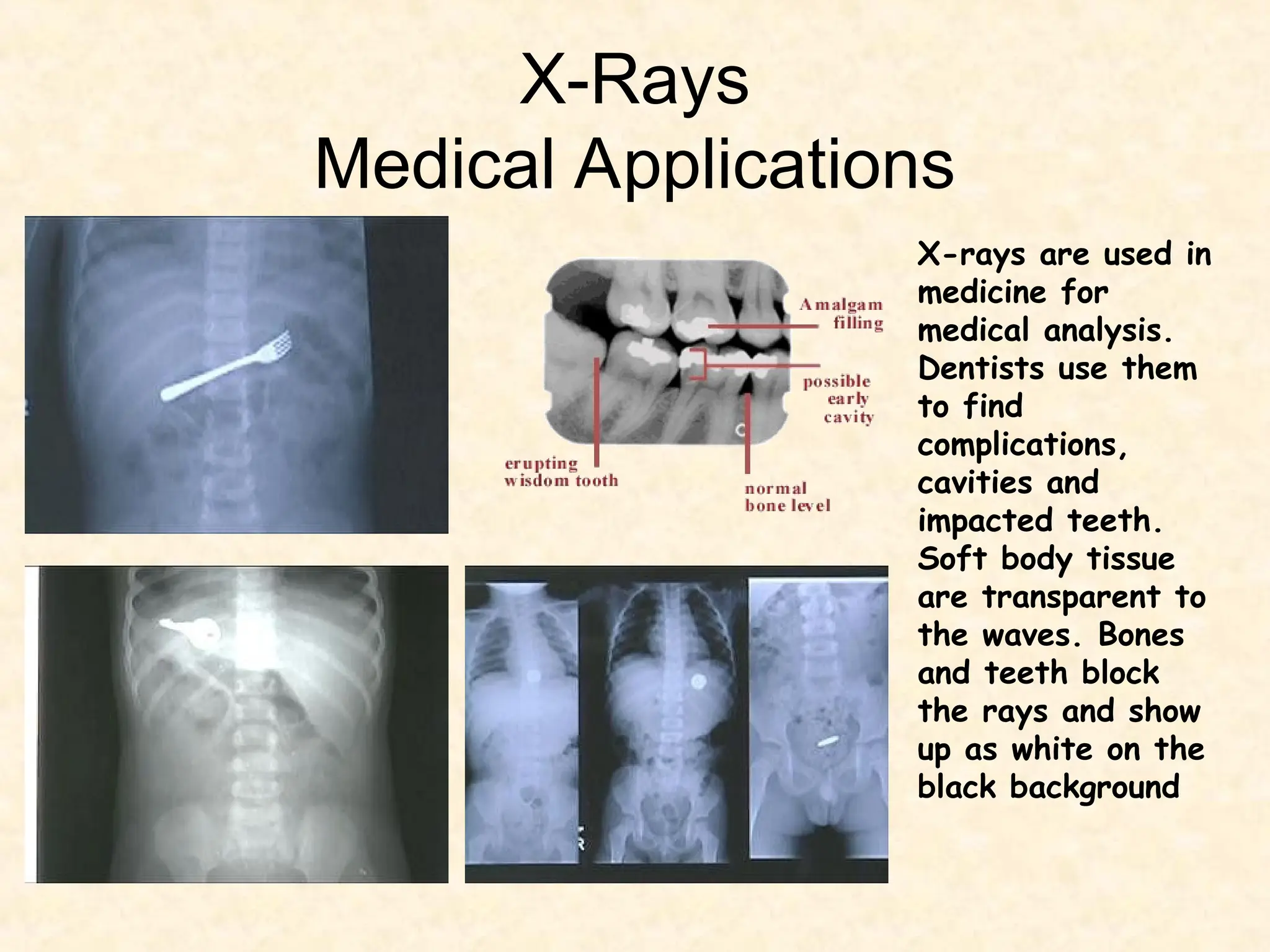
X-Rays
Medical Applications
X-rays are used in
medicine for
medical analysis.
Dentists use them
to find
complications,
cavities and
impacted teeth.
Soft body tissue
are transparent to
the waves. Bones
and teeth block
the rays and show
up as white on the
black background
Below are some x-rays showing objects which have been
swallowed by people. The examples show an open safety pin and a
child's stomach with all the pieces of a magnetic toy re-aligned
after he's swallowed them…one by one!
X-Rays and Mammograms
A mammogram (also called a
mammography exam) is a
safe, low-dose x-ray of the
breast. A high-quality
mammogram is the most
effective tool for detecting
breast cancer early. Early
detection of breast cancer
may allow more treatment
options. It could even mean
saving a woman’s breast or
her life.
The Physics of X - Rays
First of all, how do x-rays work? Well, they don't, of course, any more
than light 'works'. X-rays describe radiation which is part of the
electromagnetic spectrum (EMS). X – Rays have a very short
wavelength, generally the shorter the wavelength, the more penetrating
the wave.
The point about x-rays is that, unlike visible light, they pass straight
through some materials, including the materials that people are made
of. So, when a person is exposed to an x-ray, and a piece of film is
placed on the other side of them, a shadow of the inside of their body
is produced. This only works at all because the different tissues that
make up our bodies absorb x-rays to different extents.
The problem is, that we consist largely of water, so most of our soft
bits look much the same as far as x-rays are concerned. In fact, there
are really only three tissues that are sufficiently different from each
other in terms of x-ray absorption to show up on an x-ray film: bone,
air and soft tissue. These show up as grey/white areas on an x-ray.
X-Ray Diffraction
X-ray Crystallography
X-ray wavelengths are on the same order of
magnitude as atomic spacings.
Crystals thus make excellent diffraction gratings
Can use the geometry of the x-ray spots to
determine geometry of grating (i.e. the crystal)
nλ = 2d sin θ
Bragg Diffraction
• Diffraction from a three dimensional periodic structure such
as atoms in a crystal is called Bragg Diffraction.
• Similar to diffraction though grating.
• Consequence of interference between waves reflecting
from different crystal planes.
• Constructive interference is given by Bragg's law:
• Where λ is the wavelength, d is the distance between crystal
planes, θ is the angle of the diffracted wave. and n is an
integer known as the order of the diffracted beam.
nλ = 2d sin θ
Following Bragg's law, each dot (or reflection), in this
diffraction pattern forms from the constructive interference of
X-rays passing through a crystal. The data can be used to
determine the crystal's atomic structure.
How are X-Rays Produced?
X-Rays are
produced in
a special
type of tube
called… “An
X ray Tube!!”
Electrons are first emitted from a heated filament, by a process called thermionic emission.
They are then accelerated across the evacuated X-ray tube, under the action of a large voltage
across the tube, the filament forming the negative cathode and the target being positive anode.
On striking the target, the electrons lose most (about 99%) of their energy in low-energy
collisions with target atoms, resulting in a substantial heating of the target.
The rest of the electron energy (usually less than 1%) reappears as X-ray radiation.
Production
Of
X-Rays..
(continued)
•A rapidly-rotating anode is generally used. It forms the (tungsten) target surface on to which
the electron beam is focused. The target area under bombardment is constantly changing, thus
reducing local heat concentration. (You can often hear the whirring of the anode motor during the
taking of an X-ray.
Copper, being an excellent heat conductor, is used to hold the anode in place.
Oil, which circulates in the outer housing,, helps with convective cooling (as well as providing
electrical insulation).
Production
Of
X-Rays..
(continued)
X-Ray Imaging
• Imaging with x-rays involves exposing a part of the body to a
small dose of ionizing radiation to produce pictures of the
inside of the body.
• Making an X-ray image
It is relatively easy to make an X-ray image, or
roentgenogram-all that is needed is an X-ray source and a
film wrapped in black paper on which to record the image.
Unfortunately, X-ray cannot be focused to make
a picture as with a camera. X-ray images are basically
images of the shadows cast on film by the various structures
in the body; they were once called skiagraphs, which is
Greek for shadow graphs.
The production of a radiograph
• The x-ray tube generates the x-rays and the
x-rays are produced and spread out
through space.
• A collimator is placed in the beam’s path
and most x-rays get absorbed in the
collimator.
• A fraction get through and a narrow beam
strikes the patient.
• These x-rays are mostly absorbed by the
body but about 1% make it out and are used
to expose a film or to create electrical signals
in a detector for image formation.
Wolbarst, Physics of Radiology

Mr. ali mushtaq chest and other X-Rays uses.ppt

  • 1.
    X-Rays Medical Applications X-rays areused in medicine for medical analysis. Dentists use them to find complications, cavities and impacted teeth. Soft body tissue are transparent to the waves. Bones and teeth block the rays and show up as white on the black background
  • 2.
    Below are somex-rays showing objects which have been swallowed by people. The examples show an open safety pin and a child's stomach with all the pieces of a magnetic toy re-aligned after he's swallowed them…one by one!
  • 3.
    X-Rays and Mammograms Amammogram (also called a mammography exam) is a safe, low-dose x-ray of the breast. A high-quality mammogram is the most effective tool for detecting breast cancer early. Early detection of breast cancer may allow more treatment options. It could even mean saving a woman’s breast or her life.
  • 4.
    The Physics ofX - Rays First of all, how do x-rays work? Well, they don't, of course, any more than light 'works'. X-rays describe radiation which is part of the electromagnetic spectrum (EMS). X – Rays have a very short wavelength, generally the shorter the wavelength, the more penetrating the wave. The point about x-rays is that, unlike visible light, they pass straight through some materials, including the materials that people are made of. So, when a person is exposed to an x-ray, and a piece of film is placed on the other side of them, a shadow of the inside of their body is produced. This only works at all because the different tissues that make up our bodies absorb x-rays to different extents. The problem is, that we consist largely of water, so most of our soft bits look much the same as far as x-rays are concerned. In fact, there are really only three tissues that are sufficiently different from each other in terms of x-ray absorption to show up on an x-ray film: bone, air and soft tissue. These show up as grey/white areas on an x-ray.
  • 5.
  • 6.
    X-ray Crystallography X-ray wavelengthsare on the same order of magnitude as atomic spacings. Crystals thus make excellent diffraction gratings Can use the geometry of the x-ray spots to determine geometry of grating (i.e. the crystal) nλ = 2d sin θ
  • 7.
    Bragg Diffraction • Diffractionfrom a three dimensional periodic structure such as atoms in a crystal is called Bragg Diffraction. • Similar to diffraction though grating. • Consequence of interference between waves reflecting from different crystal planes. • Constructive interference is given by Bragg's law: • Where λ is the wavelength, d is the distance between crystal planes, θ is the angle of the diffracted wave. and n is an integer known as the order of the diffracted beam. nλ = 2d sin θ Following Bragg's law, each dot (or reflection), in this diffraction pattern forms from the constructive interference of X-rays passing through a crystal. The data can be used to determine the crystal's atomic structure.
  • 8.
    How are X-RaysProduced? X-Rays are produced in a special type of tube called… “An X ray Tube!!”
  • 9.
    Electrons are firstemitted from a heated filament, by a process called thermionic emission. They are then accelerated across the evacuated X-ray tube, under the action of a large voltage across the tube, the filament forming the negative cathode and the target being positive anode. On striking the target, the electrons lose most (about 99%) of their energy in low-energy collisions with target atoms, resulting in a substantial heating of the target. The rest of the electron energy (usually less than 1%) reappears as X-ray radiation. Production Of X-Rays.. (continued)
  • 10.
    •A rapidly-rotating anodeis generally used. It forms the (tungsten) target surface on to which the electron beam is focused. The target area under bombardment is constantly changing, thus reducing local heat concentration. (You can often hear the whirring of the anode motor during the taking of an X-ray. Copper, being an excellent heat conductor, is used to hold the anode in place. Oil, which circulates in the outer housing,, helps with convective cooling (as well as providing electrical insulation). Production Of X-Rays.. (continued)
  • 11.
    X-Ray Imaging • Imagingwith x-rays involves exposing a part of the body to a small dose of ionizing radiation to produce pictures of the inside of the body. • Making an X-ray image It is relatively easy to make an X-ray image, or roentgenogram-all that is needed is an X-ray source and a film wrapped in black paper on which to record the image. Unfortunately, X-ray cannot be focused to make a picture as with a camera. X-ray images are basically images of the shadows cast on film by the various structures in the body; they were once called skiagraphs, which is Greek for shadow graphs.
  • 12.
    The production ofa radiograph • The x-ray tube generates the x-rays and the x-rays are produced and spread out through space. • A collimator is placed in the beam’s path and most x-rays get absorbed in the collimator. • A fraction get through and a narrow beam strikes the patient. • These x-rays are mostly absorbed by the body but about 1% make it out and are used to expose a film or to create electrical signals in a detector for image formation. Wolbarst, Physics of Radiology

Editor's Notes

  • #7 Following Bragg's law, each dot (or reflection), in this diffraction pattern forms from the constructive interference of X-rays passing through a crystal. The data can be used to determine the crystal's atomic structure.