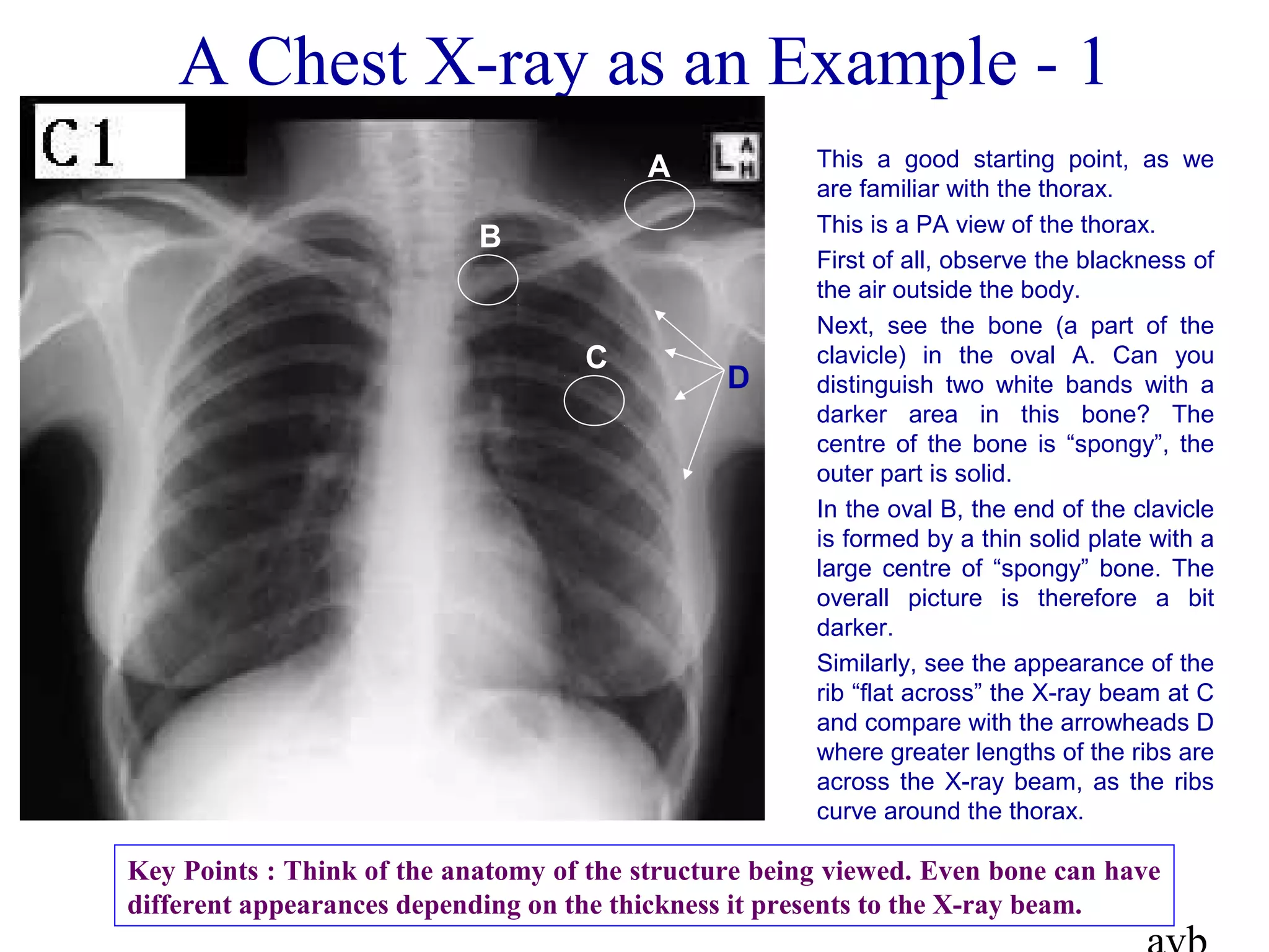
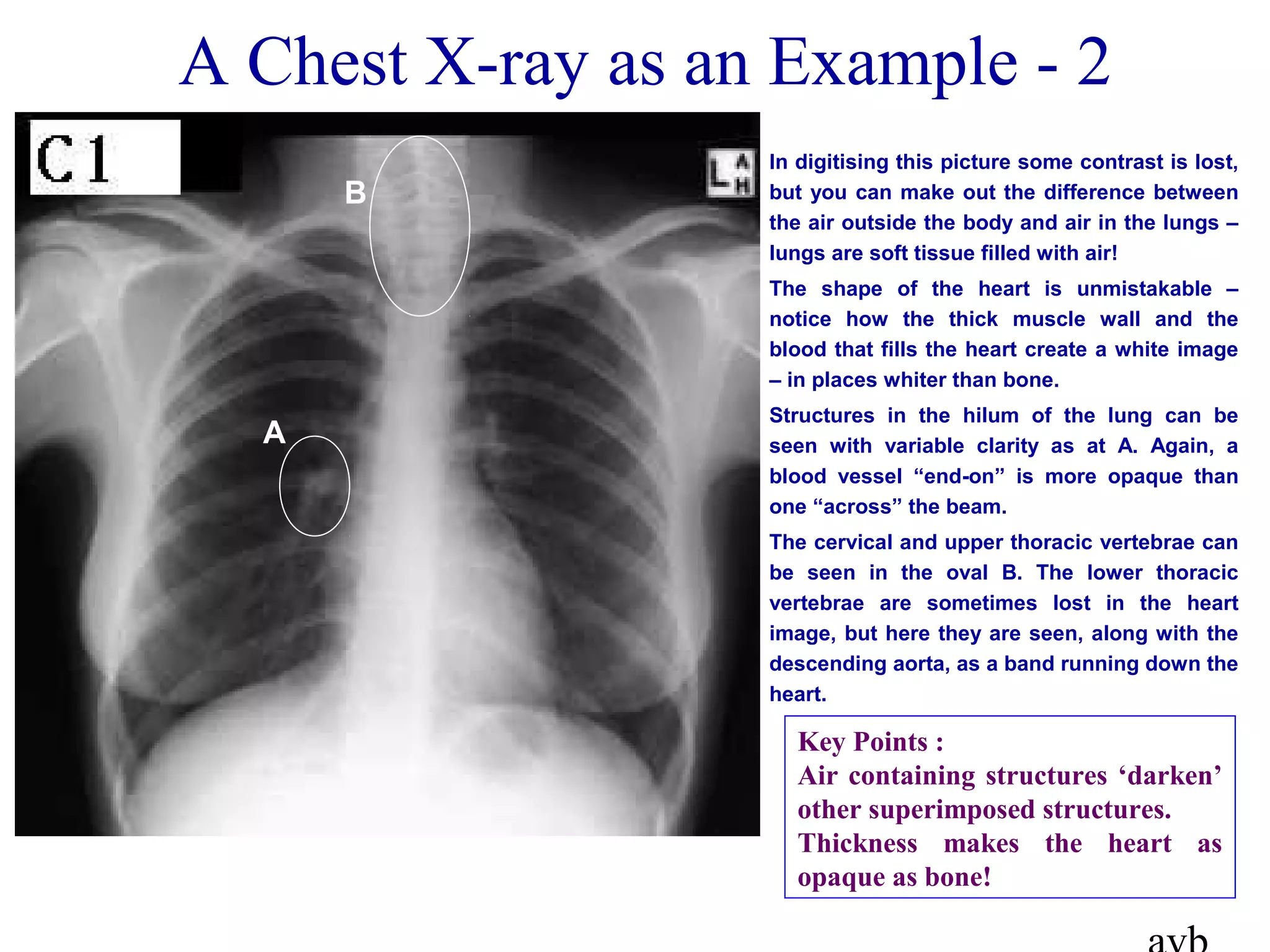
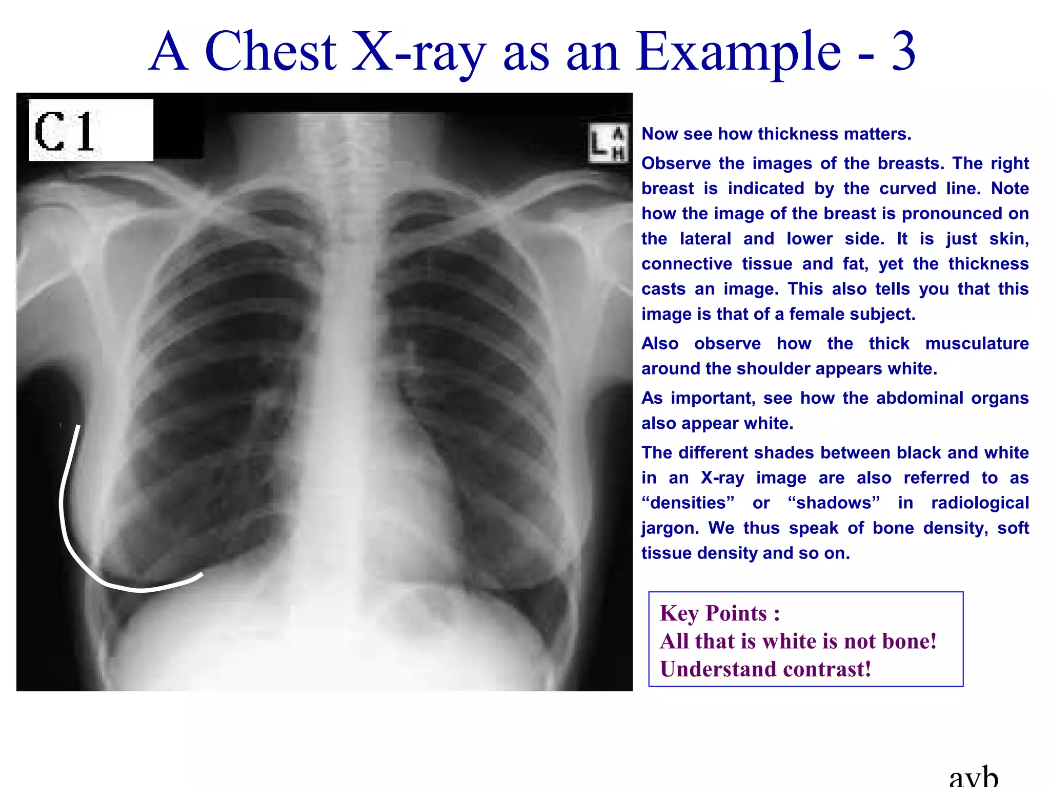
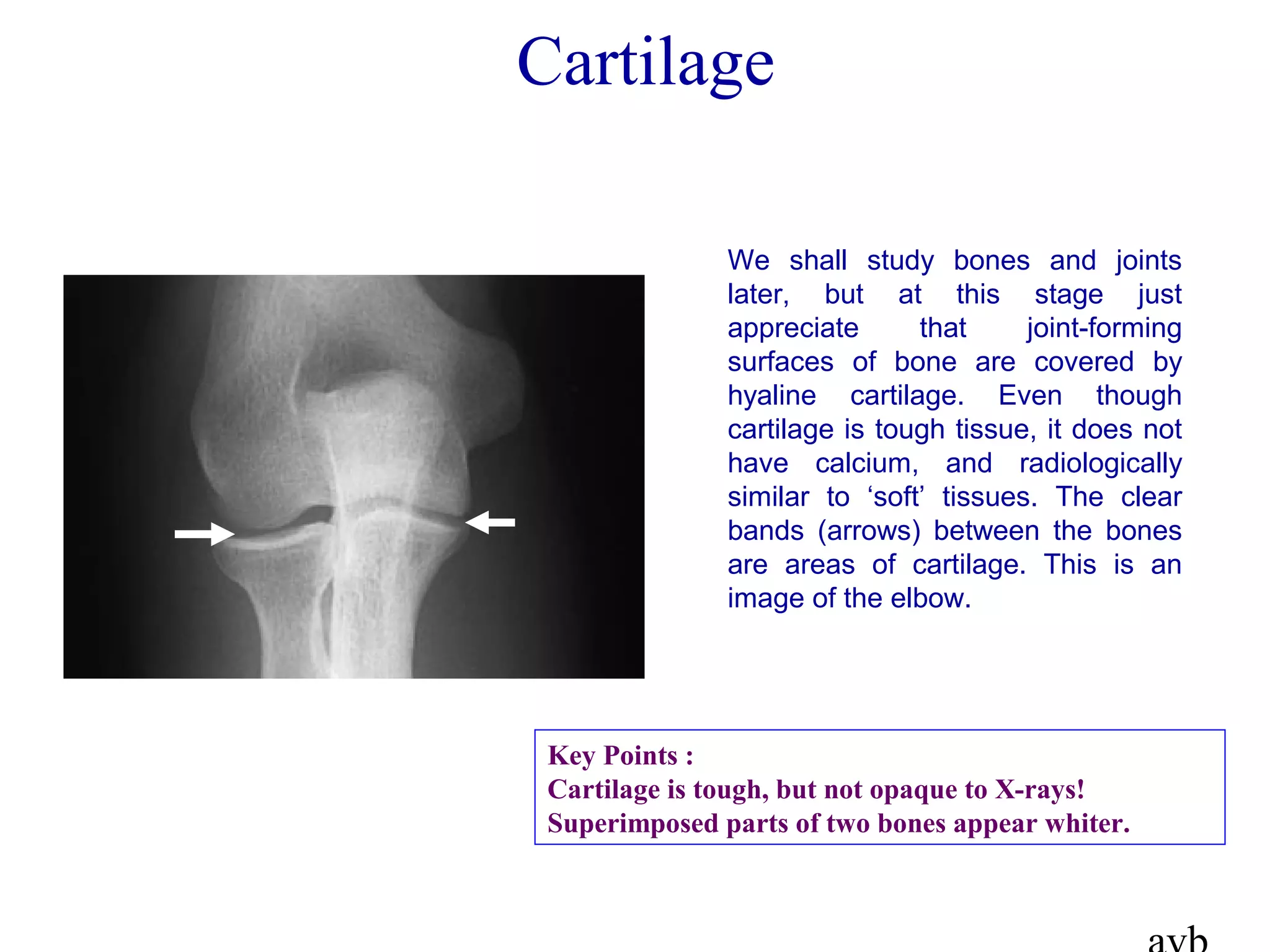
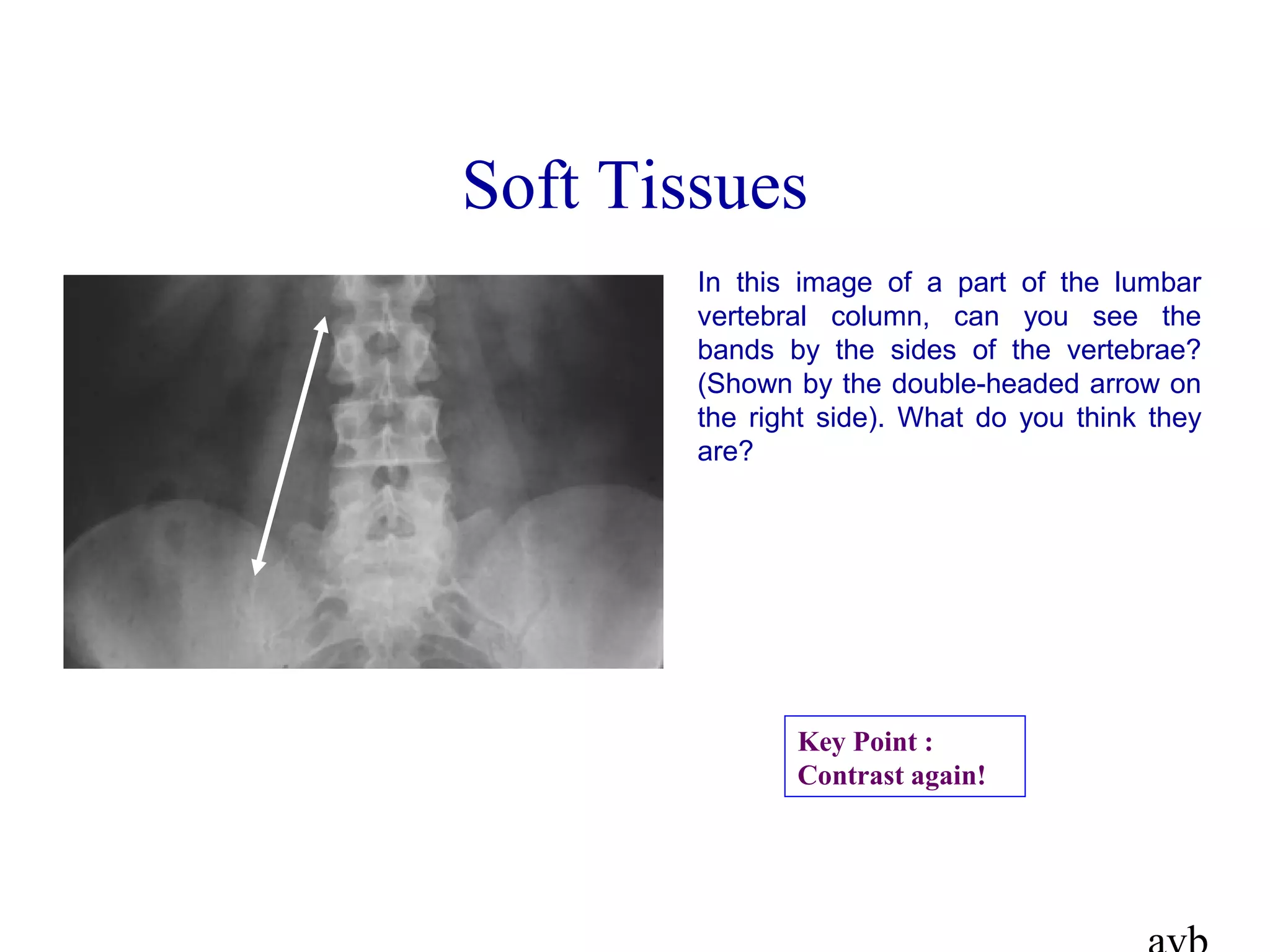
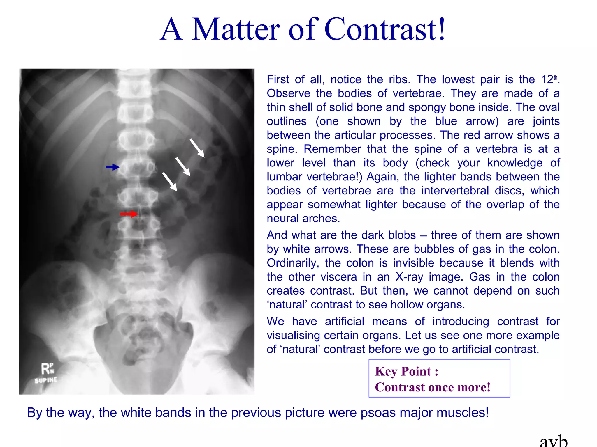
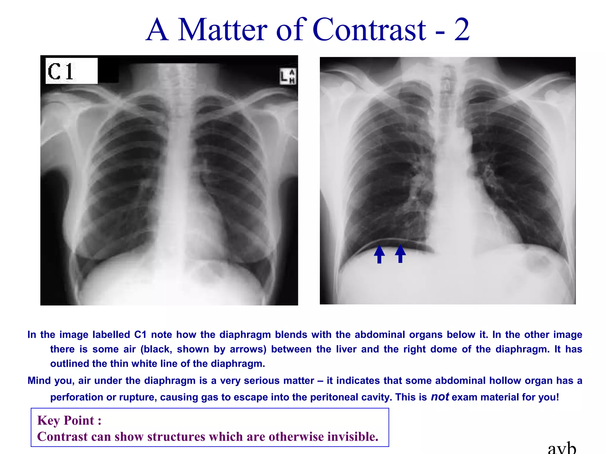
The document provides an introduction to imaging techniques for anatomy and human biology students. It aims to help students understand and interpret images of the normal human body, with an emphasis on conventional radiographic images. The summaries introduce x-rays and how they are produced in an x-ray tube. X-ray imaging involves passing x-rays through the body and exposing photographic film, with darker areas indicating more x-ray absorption. The level of absorption depends on tissue characteristics like atomic mass, with heavier elements like calcium and iron absorbing more. This creates contrast between tissues like bone, soft tissue and air in x-ray images.


![[1] MEDICAL IMAGING, X-RAY.pdf](https://cdn.slidesharecdn.com/ss_thumbnails/1medicalimagingx-ray-230209024903-74d059f1-thumbnail.jpg?width=640&height=640&fit=bounds)


