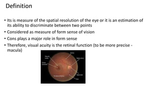
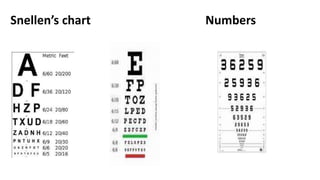
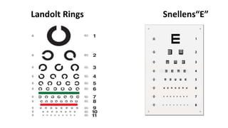
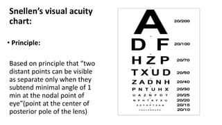
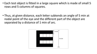
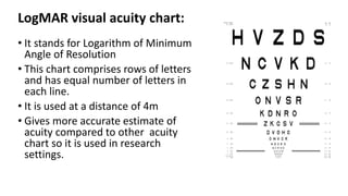
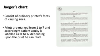
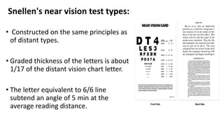
Visual Acuity In
Adults
Dr Bebika Phuyal
1st year resident
Department of ophthalmology
Definition
• Its is measure of the spatial resolution of the eye or it is an estimation of
its ability to discriminate between two points
• Considered as measure of form sense of vision
• Cons plays a major role in form sense
• Therefore, visual acuity is the retinal function (to be more precise -
macula)
Visual perception is the integration of
1. Light sense
2. Form sense
3. Sense of contrast
4. Color sense
1. Light sense
• Awareness of light
• Light is electromagnetic wave which does not need medium for
propagation
• Media of eye are permeable to visible rays between 390 & 600
nm
• Cornea absorbs rays shorter then 295 nm
• Therefore, rays between 295 & 600 nm only can reach lens. Lens
absorbs rays ‹350nm
• Therefore, rays between 350 & 600 nm can reach retina in phakic
eye; and those between 295nm and 600nm in aphakic eyes
2. Form sense:
• ability to discriminate between the shape of object
3. Sense of contrast:
• ability of eye to perceive slight changes in the luminance between
regions which are not separated by definite border
4. Color sense:
• ability of eye to discriminate between different color excited by light
of different wavelengths.
Axes of the eyeball:
• Optical axis :
line passing through center of cornea(P), center of lens(N) and meets the
retina(R) on the nasal side of the fovea
• Visual axis :
line joining fixation point(O), nodal point(N) & fovea(F).
• Fixation axis :
line joining fixation point(O) & center of rotation(C).
Visual angle:
• Angle subtended at the nodal point of the eye by the physical
dimension of a object in the visual field.
• Two adjacent points can be seen clearly & discretely only, when
these two points (say A and B in Fig above) produce a visual angle not
less than 1 min.
Visual angles include:
• Alpha angle: angle formed between optical axis & visual axis at nodal
point.
• Gamma angle: angle formed between optical axis & fixation axis at
center of rotation of eyeball.
• Kappa angle: angle formed between visual axis & pupillary line.
Practically, only kappa angle is measured and has clinical significance
Component of visual acuity
1. Minimum visible (Detection acuity)
2. Minimum separable (Resolution acuity)
3. Minimum legible (Recognition acuity)
4. Minimum Discriminable (Hyperacuity)
1. Minimum visible (Detection acuity):
• Ability to determine whether or not an object is present in an
otherwise empty visual field.
• The limit for this kind of acuity is ~ 30 arc second.
• Smallest dot size/ line width you can detect
Eg Catford Drum test
2. Minimum separable (Resolution acuity)
• Ability to locate and resolve a minimum difference between two
targets
• Normal angular threshold of discrimination for resolution is 30-60
seconds of an arc.
• Smallest separation between dots or between bars in a grating you
can resolve
Eg preferential looking test
3. Minimum legible(Recognition Acuity )
• Ability to recognize and identify a series of targets
• Not only discriminates spatial characteristics of test
pattern but also identifies the pattern with which
he has previous experience (identification of faces,
letters, symbols, pictures etc.)
Eg: smallest letter size you discriminate and
recognize in Snellen Optotypes or broken circles in
Landolt’s ring
4.Minimum Discriminable (Hyperacuity)
• Refer to spatial distinction by an observer when the threshold is
much lower than the ordinary acuity.
• Best example is
• Vernier Acuity: ability to determine whether or not 2 parallel &
straight lines are aligned in frontal plane.
• The threshold of Vernier acuity are in the range of few seconds
(2-10s) of arc
Factors affecting visual acuity
1. Stimulus related factors
2. Observer related factors
1. Stimulus related factors
• Luminance of test objects
• Geometrical configuration of the stimulus
• Contrast of the stimulus from the surround
• Influence of wavelength
• Exposure duration
• Interaction effects of the two targets
2. Observer related factors:
• Retinal locus of stimulation
• Pupil size
• Accommodation
• Effect of the eye movement
• Optical element of eye
• Developmental aspects
Optotypes
• Letters (snellen)
• Landolt Rings (Landolt ‘C’)
• Numbers
• Figures
• Tumbling E
Snellen’s chart Numbers
Landolt Rings Snellens“E”
Clinical measurement of visual acuity
A. Distant Visual Acuity
B. Near Visual Acuity
A. Distant visual acuity
Requirement:
1. Proper room illumination.
2. Normal testing distance (distance VA): 6m ( if no adequate space
use mirror)
3. Distance visual acuity chart ( can be self illuminating chart or we
have to illuminate the chart )
4. Occluder
5. Pinhole
Steps:
• Unaided VA( without correction)
• Aided VA( with patient wearing own glasses)
• Pinhole VA (with pinhole)
Distance Acuity chart:
• Snellen’s distance acuity chart
• Bailey-Lovie charts
• Landolt ring (or C) chart
• Illiterate E chart
Snellen’s visual acuity
chart:
• Principle:
Based on principle that “two
distant points can be visible
as separate only when they
subtend minimal angle of 1
min at the nodal point of
eye”(point at the center of
posterior pole of the lens)
• Each test object is fitted in a large square which is made of small 5
rows and 5 columns of squares.
• Thus, at given distance, each letter subtends an angle of 5 min at
nodal point of the eye and the different part of the object are
separated by a distance of 1 min of arc.
Unaided visual acuity
Aided visual acuity
Pinhole visual acuity
• Ideal size of the pinhole : 1.4mm
• Principle of the pinhole
pinhole allows only central rays of
light, and which do not undergo
refraction by, cornea or lens, to
pass through the eye
Examine both eyes
Incorrect method
•If the patient cannot see the top line from the
6m: slowly walk towards chart every 1m
Count fingers (CF)
Hand movements
Perception of light
Projection of rays
• Check: superior, inferior, nasal and temporal segment
Landolt’s C test types
• Each broken ring subtends an
angle of 5 min
• Detection of orientation of
breakpoint in a circle
• Used for those who cannot read
• The gap can be of various
positions ( left, right, button and
top)
• Types of Notation
Decimal Notation:
Snellen Fraction notated
as a decimal
• 20/20 = 1.00
• 20/40 = 0.50
• 20/100 = 0.20
Percentage Acuity:
(Represent in %)
20/20 = 100%
20/200 = 10%
20/2000 = 1%
Minimum angle of resolution
• It is derived from dividing the denominator of the Snellen fraction by
the numerator.
• MAR is expressed in minutes of arc
• Reciprocal of Snellen Fraction or decimal acuity
• 6/6: MAR = 1.0 min of arc
• 60/6: MAR = 10.0 min of arc
• 36/6: MAR = 6 min of arc
LogMAR visual acuity chart:
• It stands for Logarithm of Minimum
Angle of Resolution
• This chart comprises rows of letters
and has equal number of letters in
each line.
• It is used at a distance of 4m
• Gives more accurate estimate of
acuity compared to other acuity
chart so it is used in research
settings.
Log MAR snellen’s equivalent
• The
logMAR
score is
simply
the base -
10 log of
the MAR
Comparison
of Snellen
and logMAR
Visual Acuity
Use of visual acuity:
• Visual function
• Refractive status of the eye
• Measure outcome of treatment
• Medicolegal purpose
Low Vision : Best corrected visual acuity in better eye <6/18 & or visual
field < 2 degree from the point of fixation
Blindness : Best corrected visual acuity in better eye <3/60 or visual
field <10 degree from the point of fixation
• WHO classification of visual impairment
• NORMAL VISION : 6/6 -6/18
• LOW VISION
• <6/18 - 6/60 : Moderate visual impairment
• <6/60 - 3/60 : severe visual impairment
• BLINDNESS
• <3/60— PL : legally blind
• No perception of light : Totally blind
B. Near visual acuity
• Near vision is tested by asking the patient to read a near vision
chart kept at a distance of 40cm
• Each eye should be tested separately.
• Near vision is recorded as smallest paragraph which the patient
can read comfortably.
Jaeger’s chart:
• Consist of ordinary printer's fonts
of varying sizes.
• Prints are marked from 1 to 7 and
accordingly patient acuity is
labelled as J1 to J7 depending
upon the print he can read
Roman test types:
• Consists of Times Roman
fonts with standard
spacing near vision is
recorded as N5, N6, N8,
N10, N12, N18, N36 and
N48.
Snellen's near vision test types:
• Constructed on the same principles as
of distant types.
• Graded thickness of the letters is about
1/17 of the distant vision chart letter.
• The letter equivalent to 6/6 line
subtend an angle of 5 min at the
average reading distance.
References
• American Academy of Ophthalmology. (2001). Optics,
Refraction and Contact Lenses (Basic & Clinical Science
Course) (Rev Ed). American Academy of Ophthalmology.
• Kanski, J., n.d. Clinical ophthalmology. 9th ed. Edinburgh:
Butterworth Heinemann/Elsevier
• S., & Tandon, R. (2019). Parsons’ Diseases of the Eye (23rd
ed.). Elsevier India.
Thankyou

Visual Acuity.pptx

  • 1.
    Visual Acuity In Adults DrBebika Phuyal 1st year resident Department of ophthalmology
  • 2.
    Definition • Its ismeasure of the spatial resolution of the eye or it is an estimation of its ability to discriminate between two points • Considered as measure of form sense of vision • Cons plays a major role in form sense • Therefore, visual acuity is the retinal function (to be more precise - macula)
  • 3.
    Visual perception isthe integration of 1. Light sense 2. Form sense 3. Sense of contrast 4. Color sense
  • 4.
    1. Light sense •Awareness of light • Light is electromagnetic wave which does not need medium for propagation • Media of eye are permeable to visible rays between 390 & 600 nm • Cornea absorbs rays shorter then 295 nm • Therefore, rays between 295 & 600 nm only can reach lens. Lens absorbs rays ‹350nm • Therefore, rays between 350 & 600 nm can reach retina in phakic eye; and those between 295nm and 600nm in aphakic eyes
  • 5.
    2. Form sense: •ability to discriminate between the shape of object 3. Sense of contrast: • ability of eye to perceive slight changes in the luminance between regions which are not separated by definite border 4. Color sense: • ability of eye to discriminate between different color excited by light of different wavelengths.
  • 6.
    Axes of theeyeball: • Optical axis : line passing through center of cornea(P), center of lens(N) and meets the retina(R) on the nasal side of the fovea • Visual axis : line joining fixation point(O), nodal point(N) & fovea(F). • Fixation axis : line joining fixation point(O) & center of rotation(C).
  • 7.
    Visual angle: • Anglesubtended at the nodal point of the eye by the physical dimension of a object in the visual field. • Two adjacent points can be seen clearly & discretely only, when these two points (say A and B in Fig above) produce a visual angle not less than 1 min.
  • 8.
    Visual angles include: •Alpha angle: angle formed between optical axis & visual axis at nodal point. • Gamma angle: angle formed between optical axis & fixation axis at center of rotation of eyeball. • Kappa angle: angle formed between visual axis & pupillary line. Practically, only kappa angle is measured and has clinical significance
  • 9.
    Component of visualacuity 1. Minimum visible (Detection acuity) 2. Minimum separable (Resolution acuity) 3. Minimum legible (Recognition acuity) 4. Minimum Discriminable (Hyperacuity)
  • 10.
    1. Minimum visible(Detection acuity): • Ability to determine whether or not an object is present in an otherwise empty visual field. • The limit for this kind of acuity is ~ 30 arc second. • Smallest dot size/ line width you can detect Eg Catford Drum test
  • 11.
    2. Minimum separable(Resolution acuity) • Ability to locate and resolve a minimum difference between two targets • Normal angular threshold of discrimination for resolution is 30-60 seconds of an arc. • Smallest separation between dots or between bars in a grating you can resolve Eg preferential looking test
  • 12.
    3. Minimum legible(RecognitionAcuity ) • Ability to recognize and identify a series of targets • Not only discriminates spatial characteristics of test pattern but also identifies the pattern with which he has previous experience (identification of faces, letters, symbols, pictures etc.) Eg: smallest letter size you discriminate and recognize in Snellen Optotypes or broken circles in Landolt’s ring
  • 13.
    4.Minimum Discriminable (Hyperacuity) •Refer to spatial distinction by an observer when the threshold is much lower than the ordinary acuity. • Best example is • Vernier Acuity: ability to determine whether or not 2 parallel & straight lines are aligned in frontal plane. • The threshold of Vernier acuity are in the range of few seconds (2-10s) of arc
  • 14.
    Factors affecting visualacuity 1. Stimulus related factors 2. Observer related factors
  • 15.
    1. Stimulus relatedfactors • Luminance of test objects • Geometrical configuration of the stimulus • Contrast of the stimulus from the surround • Influence of wavelength • Exposure duration • Interaction effects of the two targets
  • 16.
    2. Observer relatedfactors: • Retinal locus of stimulation • Pupil size • Accommodation • Effect of the eye movement • Optical element of eye • Developmental aspects
  • 17.
    Optotypes • Letters (snellen) •Landolt Rings (Landolt ‘C’) • Numbers • Figures • Tumbling E
  • 18.
  • 19.
  • 20.
    Clinical measurement ofvisual acuity A. Distant Visual Acuity B. Near Visual Acuity
  • 21.
    A. Distant visualacuity Requirement: 1. Proper room illumination. 2. Normal testing distance (distance VA): 6m ( if no adequate space use mirror) 3. Distance visual acuity chart ( can be self illuminating chart or we have to illuminate the chart ) 4. Occluder 5. Pinhole
  • 22.
    Steps: • Unaided VA(without correction) • Aided VA( with patient wearing own glasses) • Pinhole VA (with pinhole)
  • 23.
    Distance Acuity chart: •Snellen’s distance acuity chart • Bailey-Lovie charts • Landolt ring (or C) chart • Illiterate E chart
  • 24.
    Snellen’s visual acuity chart: •Principle: Based on principle that “two distant points can be visible as separate only when they subtend minimal angle of 1 min at the nodal point of eye”(point at the center of posterior pole of the lens)
  • 25.
    • Each testobject is fitted in a large square which is made of small 5 rows and 5 columns of squares. • Thus, at given distance, each letter subtends an angle of 5 min at nodal point of the eye and the different part of the object are separated by a distance of 1 min of arc.
  • 26.
  • 27.
  • 28.
    Pinhole visual acuity •Ideal size of the pinhole : 1.4mm • Principle of the pinhole pinhole allows only central rays of light, and which do not undergo refraction by, cornea or lens, to pass through the eye
  • 29.
  • 30.
  • 31.
    •If the patientcannot see the top line from the 6m: slowly walk towards chart every 1m
  • 32.
  • 33.
  • 34.
  • 35.
    Projection of rays •Check: superior, inferior, nasal and temporal segment
  • 36.
    Landolt’s C testtypes • Each broken ring subtends an angle of 5 min • Detection of orientation of breakpoint in a circle • Used for those who cannot read • The gap can be of various positions ( left, right, button and top)
  • 37.
    • Types ofNotation Decimal Notation: Snellen Fraction notated as a decimal • 20/20 = 1.00 • 20/40 = 0.50 • 20/100 = 0.20 Percentage Acuity: (Represent in %) 20/20 = 100% 20/200 = 10% 20/2000 = 1%
  • 38.
    Minimum angle ofresolution • It is derived from dividing the denominator of the Snellen fraction by the numerator. • MAR is expressed in minutes of arc • Reciprocal of Snellen Fraction or decimal acuity • 6/6: MAR = 1.0 min of arc • 60/6: MAR = 10.0 min of arc • 36/6: MAR = 6 min of arc
  • 39.
    LogMAR visual acuitychart: • It stands for Logarithm of Minimum Angle of Resolution • This chart comprises rows of letters and has equal number of letters in each line. • It is used at a distance of 4m • Gives more accurate estimate of acuity compared to other acuity chart so it is used in research settings.
  • 40.
    Log MAR snellen’sequivalent • The logMAR score is simply the base - 10 log of the MAR
  • 41.
  • 42.
    Use of visualacuity: • Visual function • Refractive status of the eye • Measure outcome of treatment • Medicolegal purpose
  • 43.
    Low Vision :Best corrected visual acuity in better eye <6/18 & or visual field < 2 degree from the point of fixation Blindness : Best corrected visual acuity in better eye <3/60 or visual field <10 degree from the point of fixation
  • 44.
    • WHO classificationof visual impairment • NORMAL VISION : 6/6 -6/18 • LOW VISION • <6/18 - 6/60 : Moderate visual impairment • <6/60 - 3/60 : severe visual impairment • BLINDNESS • <3/60— PL : legally blind • No perception of light : Totally blind
  • 45.
    B. Near visualacuity • Near vision is tested by asking the patient to read a near vision chart kept at a distance of 40cm • Each eye should be tested separately. • Near vision is recorded as smallest paragraph which the patient can read comfortably.
  • 46.
    Jaeger’s chart: • Consistof ordinary printer's fonts of varying sizes. • Prints are marked from 1 to 7 and accordingly patient acuity is labelled as J1 to J7 depending upon the print he can read
  • 47.
    Roman test types: •Consists of Times Roman fonts with standard spacing near vision is recorded as N5, N6, N8, N10, N12, N18, N36 and N48.
  • 48.
    Snellen's near visiontest types: • Constructed on the same principles as of distant types. • Graded thickness of the letters is about 1/17 of the distant vision chart letter. • The letter equivalent to 6/6 line subtend an angle of 5 min at the average reading distance.
  • 49.
    References • American Academyof Ophthalmology. (2001). Optics, Refraction and Contact Lenses (Basic & Clinical Science Course) (Rev Ed). American Academy of Ophthalmology. • Kanski, J., n.d. Clinical ophthalmology. 9th ed. Edinburgh: Butterworth Heinemann/Elsevier • S., & Tandon, R. (2019). Parsons’ Diseases of the Eye (23rd ed.). Elsevier India.
  • 50.

Editor's Notes

  • #14 It is due to higher cortical centers