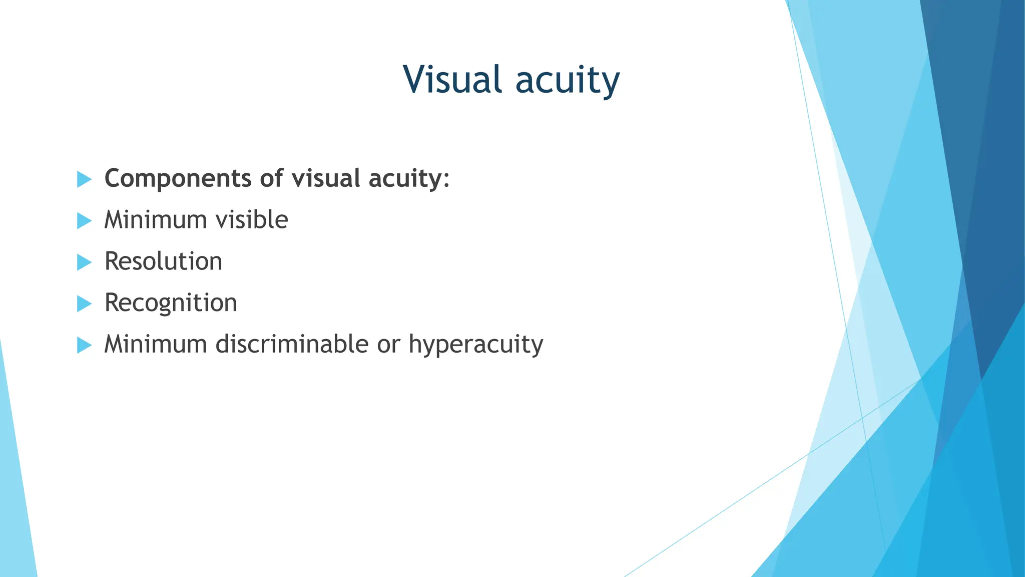
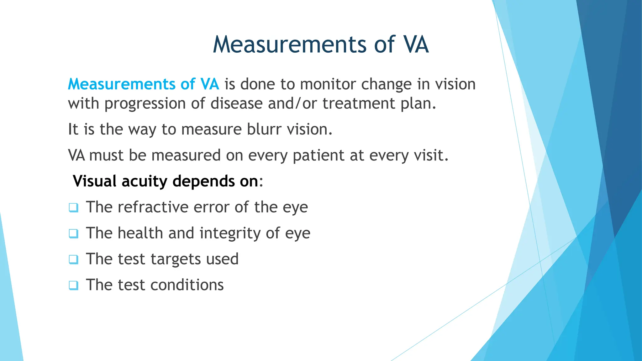
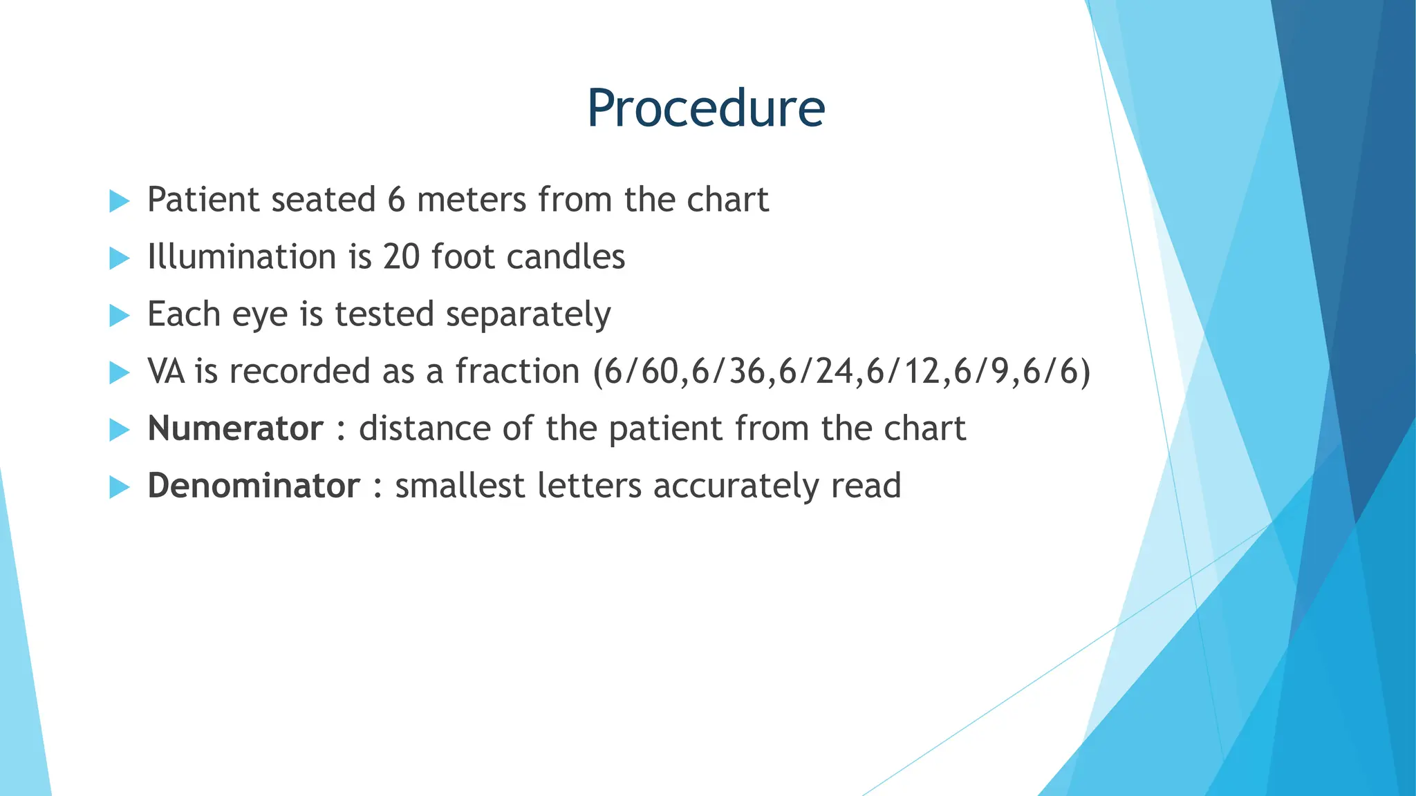
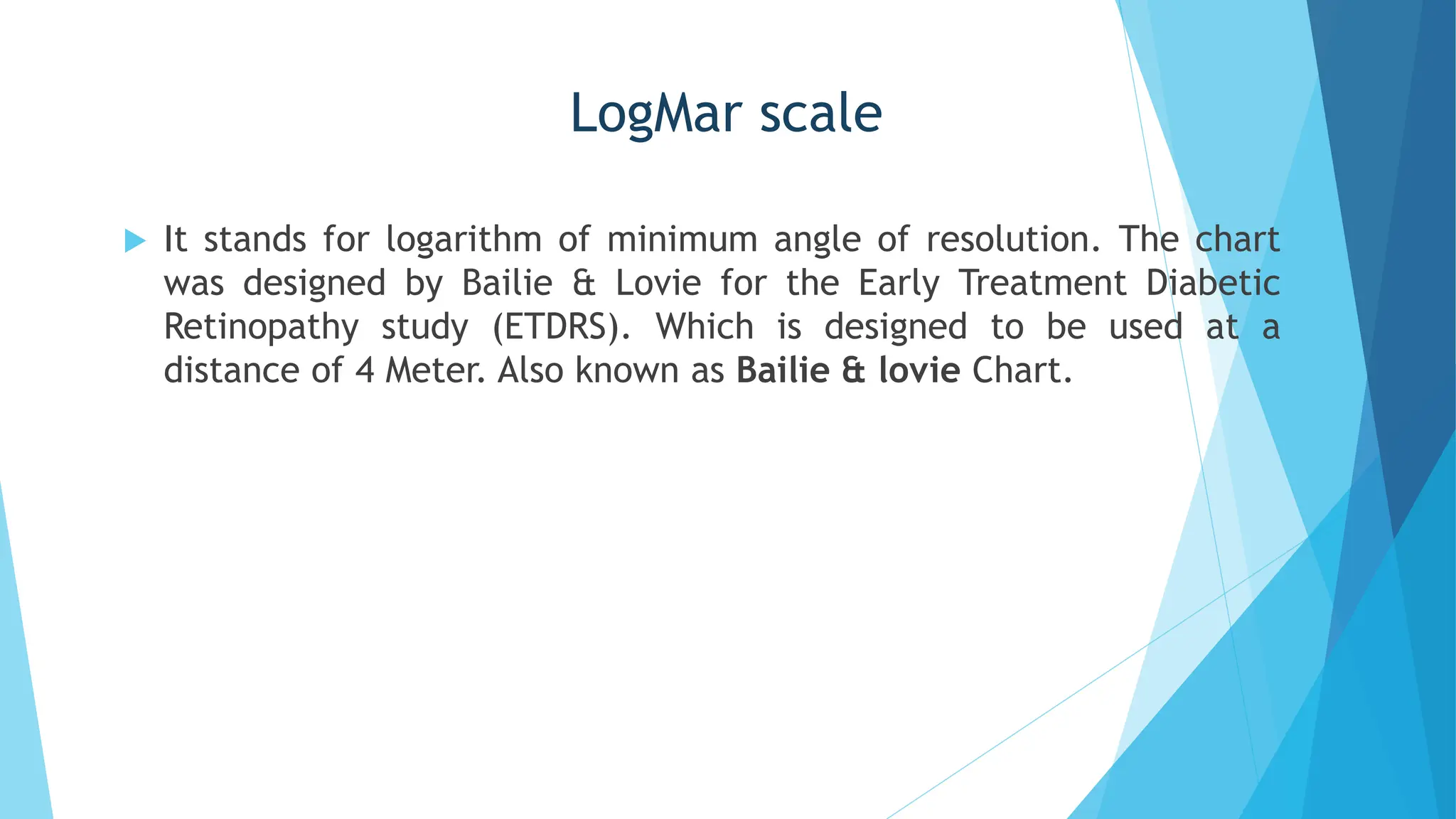
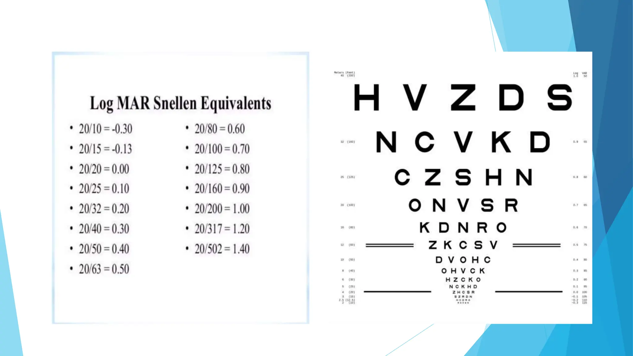
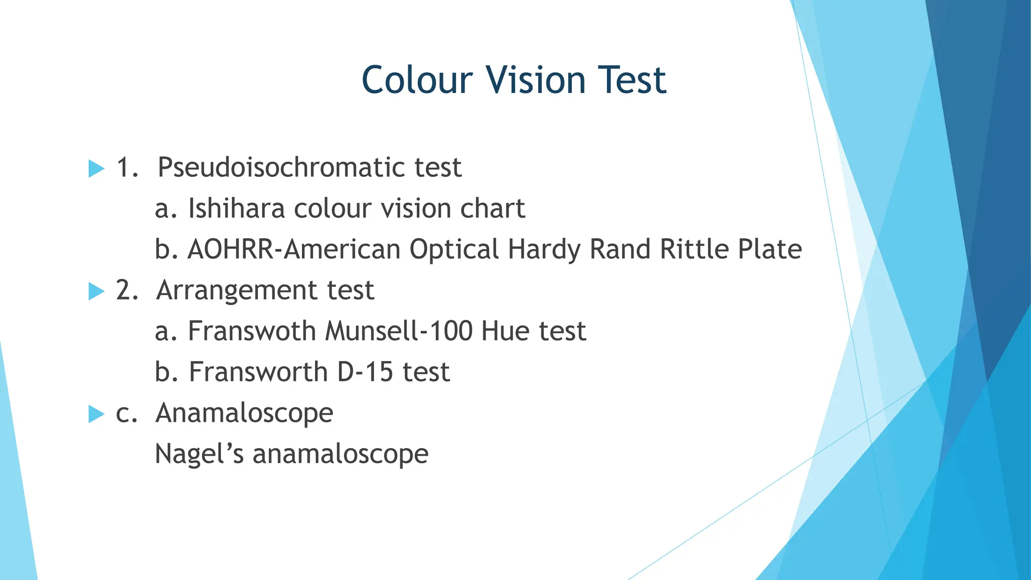
Visual acuity examination
for adult
CHAIRMAN : DR. MD. ASHIQUR RAHMAN AKANDA
Associate Professor
Department of Pediatric ophthalmology, NIO&H
MODERATOR : DR. CHANDAN KUMAR PAUL
Junior Consultant
Department of Pediatric ophthalmology, NIO&H
PRESENTER : DR. SONIA ISLAM
Phase A Resident NIO&H
Visual acuity
Definition: Visual acuity is the resolving power of eye by
which objects are distinguished clearly from other.
Visual acuity refers to the spatial limit of visual
discrimination.
Components of vision:
 Visual acuity
 Field of vision
 Colour vision
 Contrast sensitivity
 Light Brightness sensitivity
Visual acuity
 Components of visual acuity:
 Minimum visible
 Resolution
 Recognition
 Minimum discriminable or hyperacuity
Measurements of VA
Measurements of VA is done to monitor change in vision
with progression of disease and/or treatment plan.
It is the way to measure blurr vision.
VA must be measured on every patient at every visit.
Visual acuity depends on:
 The refractive error of the eye
 The health and integrity of eye
 The test targets used
 The test conditions
Clinical measurement of VA
 Criteria:
 Normal testing distance (distance visual acuity ) -6m
 For near -33cm/40cm.
 Illuminated visual acuity chart
 Proper room illumination.
 Steps :
 Unaided VA ( OD,OS,OU)
 Pinhole VA (OD,OS)
 Aided VA(OD,OS,OU)
DISTANCE VISUAL ACUITY CHARTS
 Snellen visual acuity chart -6m
 bailey-lovie (logMar) -4m/2m/1m
 Landolt ring chart (C chart)
 Tumbling E chart
Procedure
 Patient seated 6 meters from the chart
 Illumination is 20 foot candles
 Each eye is tested separately
 VA is recorded as a fraction (6/60,6/36,6/24,6/12,6/9,6/6)
 Numerator : distance of the patient from the chart
 Denominator : smallest letters accurately read
Procedure
 If the patient can not see the top line from 6m, he is
asked to slowly move towards the chart till he can read
the top line (5/60,4/60,3/60,2/60,1/60).
 If the patient is unable to see even from 1 m, he is asked
to count fingers of the examiner.
 When a patient fails to count fingers ,the examiner moves
his hands close to the patient's face & ask whether he
could appreciate the movement or not (HM +/-).
 When the patient cannot appreciate hand movement,
perception to light is noted.(PL+/-)with projection of rays
in four quadrants.
Formula
Near Acuity Chart
 Reduced snellel chart
 N chart
 M chart
 Jaeger chart
Procedure (Near Vision)
 Near vision tested by asking the patient to read a near
vision chart kept at distance of 16 inches (40cm).
 Each eye should be tested separately and respectively .
 Record the binocular acuity achieved.
 The near vision is recorded as the smallest type which the
patient can read comfortably
Jaeger’s Chart
 Consists of ordinary printer’s front of
varying sizes
 Prints are marked from 1 to 7 and
accordingly patient’s acuity is labeled
as J1 to J7 depending upon the print he
can read
Testing Pinhole Visual Acuity
 Position the patient and occlude the eye not being tested.
 Ask the patient to hold the pin whole occlude in front of eye that is being
tested. That patient’s habitual correction may be worn.
 Instruct the patient to look at the distance chart through the single
pinhole.
 Instruct the patient to use small hand or eye movement to align pinhole
to resolve the sharpest image on the chart .
 Ask the patient to read the line with smallest letters that are legible to
determined on the previous vision test without use of pinhole
 Record the Snellen acuity obtained and precede or follow it with the
abbreviation PH
Picture
Mechanism of Pinhole
LogMar scale
 It stands for logarithm of minimum angle of resolution. The chart
was designed by Bailie & Lovie for the Early Treatment Diabetic
Retinopathy study (ETDRS). Which is designed to be used at a
distance of 4 Meter. Also known as Bailie & lovie Chart.
Steps
 Set the patient 4 meters from the chart.
 Cover one eye, if cannot read, then move 1 meter & add 0.6.
 Repeat second eye.
 If still unable to read, then use other option.
 Example: It one read 0.4 line plus 3 letters of 0.3 line, he or she
will have a score of 0.4-(0.2x0.3)=0.34.
19
Colour vision
 This is the inability to distinguish certain colour .
 Unexplained reduction in visual acuity e.g. 20/25
 One or more cone type is missing or defective to any
extend.
 Abnormal colour matching and colour confusion .
Colour Vision Chart
Colour Vision Test
 1. Pseudoisochromatic test
a. Ishihara colour vision chart
b. AOHRR-American Optical Hardy Rand Rittle Plate
 2. Arrangement test
a. Franswoth Munsell-100 Hue test
b. Fransworth D-15 test
 c. Anamaloscope
Nagel’s anamaloscope
Some Diseases Associated with Reduced VA:
 Refractive errors
 Cataract
 Retinal detachment
 Retinopathy of prematurity
 Glaucoma
 Optic nerve atrophy
 Albinism
 Nystagmus
VA is the most fundamental and basic clinical examination in
eye. Meticulous visual acuity assessment is essential. We all
should practice it. VA is impotent for clinically and medico-
legally. VA acuity is the opening door for examination of
eye.
Take Home Message
Visual acuity examination(adult) Update (1).pptx

Visual acuity examination(adult) Update (1).pptx

  • 1.
    Visual acuity examination foradult CHAIRMAN : DR. MD. ASHIQUR RAHMAN AKANDA Associate Professor Department of Pediatric ophthalmology, NIO&H MODERATOR : DR. CHANDAN KUMAR PAUL Junior Consultant Department of Pediatric ophthalmology, NIO&H PRESENTER : DR. SONIA ISLAM Phase A Resident NIO&H
  • 2.
    Visual acuity Definition: Visualacuity is the resolving power of eye by which objects are distinguished clearly from other. Visual acuity refers to the spatial limit of visual discrimination. Components of vision:  Visual acuity  Field of vision  Colour vision  Contrast sensitivity  Light Brightness sensitivity
  • 3.
    Visual acuity  Componentsof visual acuity:  Minimum visible  Resolution  Recognition  Minimum discriminable or hyperacuity
  • 4.
    Measurements of VA Measurementsof VA is done to monitor change in vision with progression of disease and/or treatment plan. It is the way to measure blurr vision. VA must be measured on every patient at every visit. Visual acuity depends on:  The refractive error of the eye  The health and integrity of eye  The test targets used  The test conditions
  • 5.
    Clinical measurement ofVA  Criteria:  Normal testing distance (distance visual acuity ) -6m  For near -33cm/40cm.  Illuminated visual acuity chart  Proper room illumination.  Steps :  Unaided VA ( OD,OS,OU)  Pinhole VA (OD,OS)  Aided VA(OD,OS,OU)
  • 6.
    DISTANCE VISUAL ACUITYCHARTS  Snellen visual acuity chart -6m  bailey-lovie (logMar) -4m/2m/1m  Landolt ring chart (C chart)  Tumbling E chart
  • 7.
    Procedure  Patient seated6 meters from the chart  Illumination is 20 foot candles  Each eye is tested separately  VA is recorded as a fraction (6/60,6/36,6/24,6/12,6/9,6/6)  Numerator : distance of the patient from the chart  Denominator : smallest letters accurately read
  • 8.
    Procedure  If thepatient can not see the top line from 6m, he is asked to slowly move towards the chart till he can read the top line (5/60,4/60,3/60,2/60,1/60).  If the patient is unable to see even from 1 m, he is asked to count fingers of the examiner.  When a patient fails to count fingers ,the examiner moves his hands close to the patient's face & ask whether he could appreciate the movement or not (HM +/-).  When the patient cannot appreciate hand movement, perception to light is noted.(PL+/-)with projection of rays in four quadrants.
  • 10.
  • 11.
    Near Acuity Chart Reduced snellel chart  N chart  M chart  Jaeger chart
  • 12.
    Procedure (Near Vision) Near vision tested by asking the patient to read a near vision chart kept at distance of 16 inches (40cm).  Each eye should be tested separately and respectively .  Record the binocular acuity achieved.  The near vision is recorded as the smallest type which the patient can read comfortably
  • 13.
    Jaeger’s Chart  Consistsof ordinary printer’s front of varying sizes  Prints are marked from 1 to 7 and accordingly patient’s acuity is labeled as J1 to J7 depending upon the print he can read
  • 15.
    Testing Pinhole VisualAcuity  Position the patient and occlude the eye not being tested.  Ask the patient to hold the pin whole occlude in front of eye that is being tested. That patient’s habitual correction may be worn.  Instruct the patient to look at the distance chart through the single pinhole.  Instruct the patient to use small hand or eye movement to align pinhole to resolve the sharpest image on the chart .  Ask the patient to read the line with smallest letters that are legible to determined on the previous vision test without use of pinhole  Record the Snellen acuity obtained and precede or follow it with the abbreviation PH
  • 16.
  • 17.
  • 18.
    LogMar scale  Itstands for logarithm of minimum angle of resolution. The chart was designed by Bailie & Lovie for the Early Treatment Diabetic Retinopathy study (ETDRS). Which is designed to be used at a distance of 4 Meter. Also known as Bailie & lovie Chart.
  • 19.
    Steps  Set thepatient 4 meters from the chart.  Cover one eye, if cannot read, then move 1 meter & add 0.6.  Repeat second eye.  If still unable to read, then use other option.  Example: It one read 0.4 line plus 3 letters of 0.3 line, he or she will have a score of 0.4-(0.2x0.3)=0.34. 19
  • 21.
    Colour vision  Thisis the inability to distinguish certain colour .  Unexplained reduction in visual acuity e.g. 20/25  One or more cone type is missing or defective to any extend.  Abnormal colour matching and colour confusion .
  • 22.
  • 23.
    Colour Vision Test 1. Pseudoisochromatic test a. Ishihara colour vision chart b. AOHRR-American Optical Hardy Rand Rittle Plate  2. Arrangement test a. Franswoth Munsell-100 Hue test b. Fransworth D-15 test  c. Anamaloscope Nagel’s anamaloscope
  • 25.
    Some Diseases Associatedwith Reduced VA:  Refractive errors  Cataract  Retinal detachment  Retinopathy of prematurity  Glaucoma  Optic nerve atrophy  Albinism  Nystagmus
  • 26.
    VA is themost fundamental and basic clinical examination in eye. Meticulous visual acuity assessment is essential. We all should practice it. VA is impotent for clinically and medico- legally. VA acuity is the opening door for examination of eye. Take Home Message