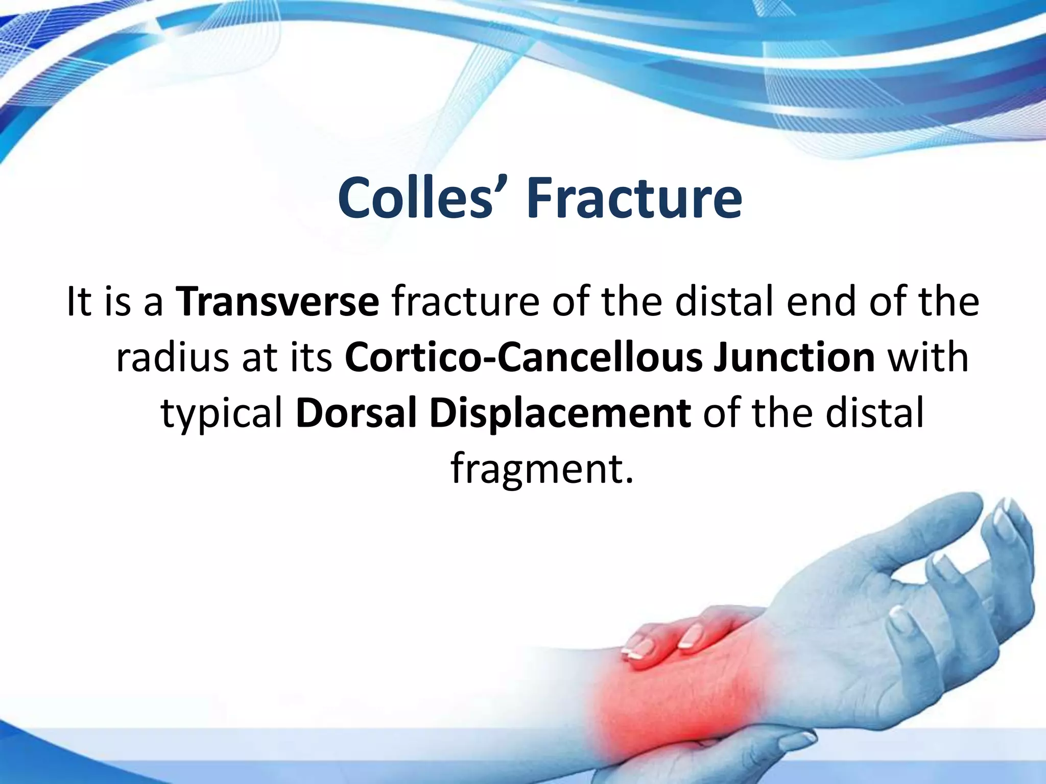
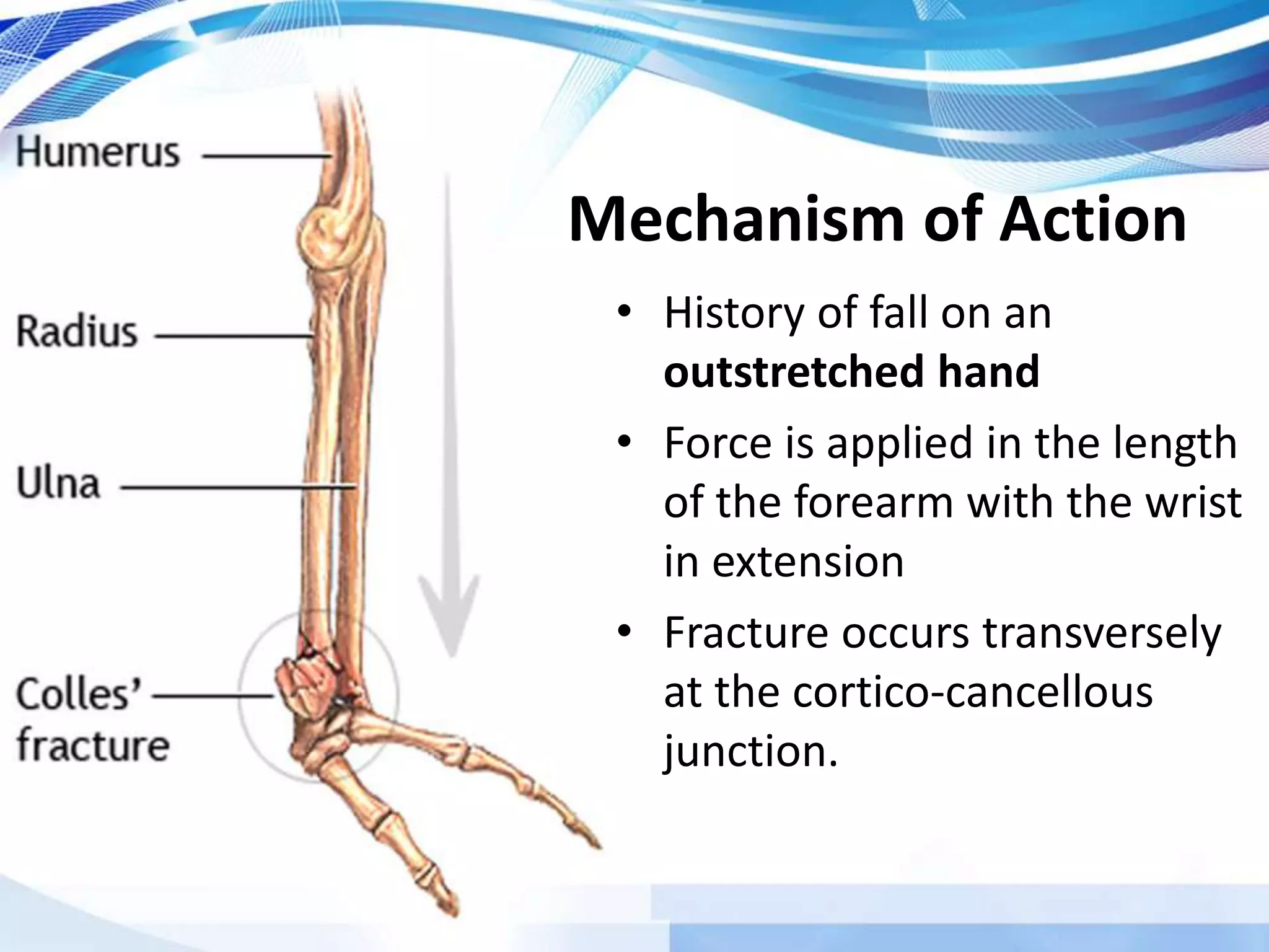
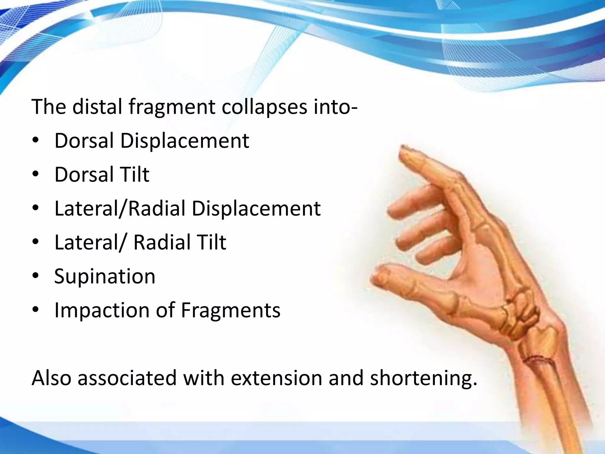
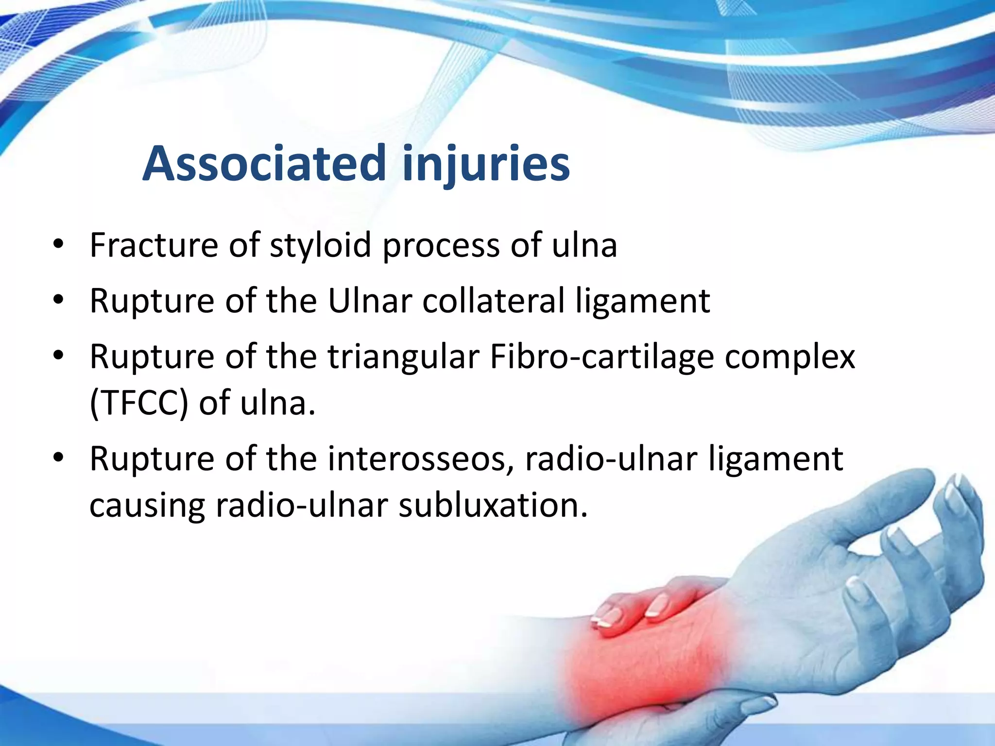
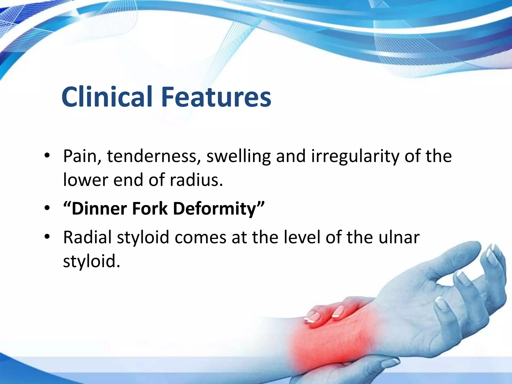
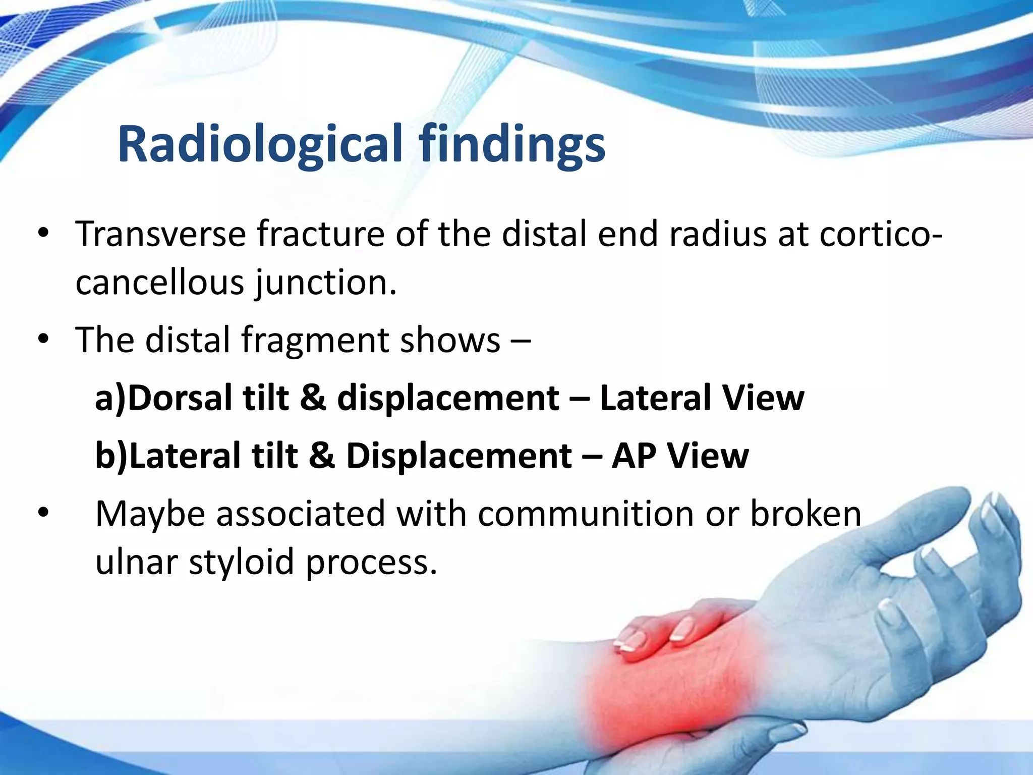
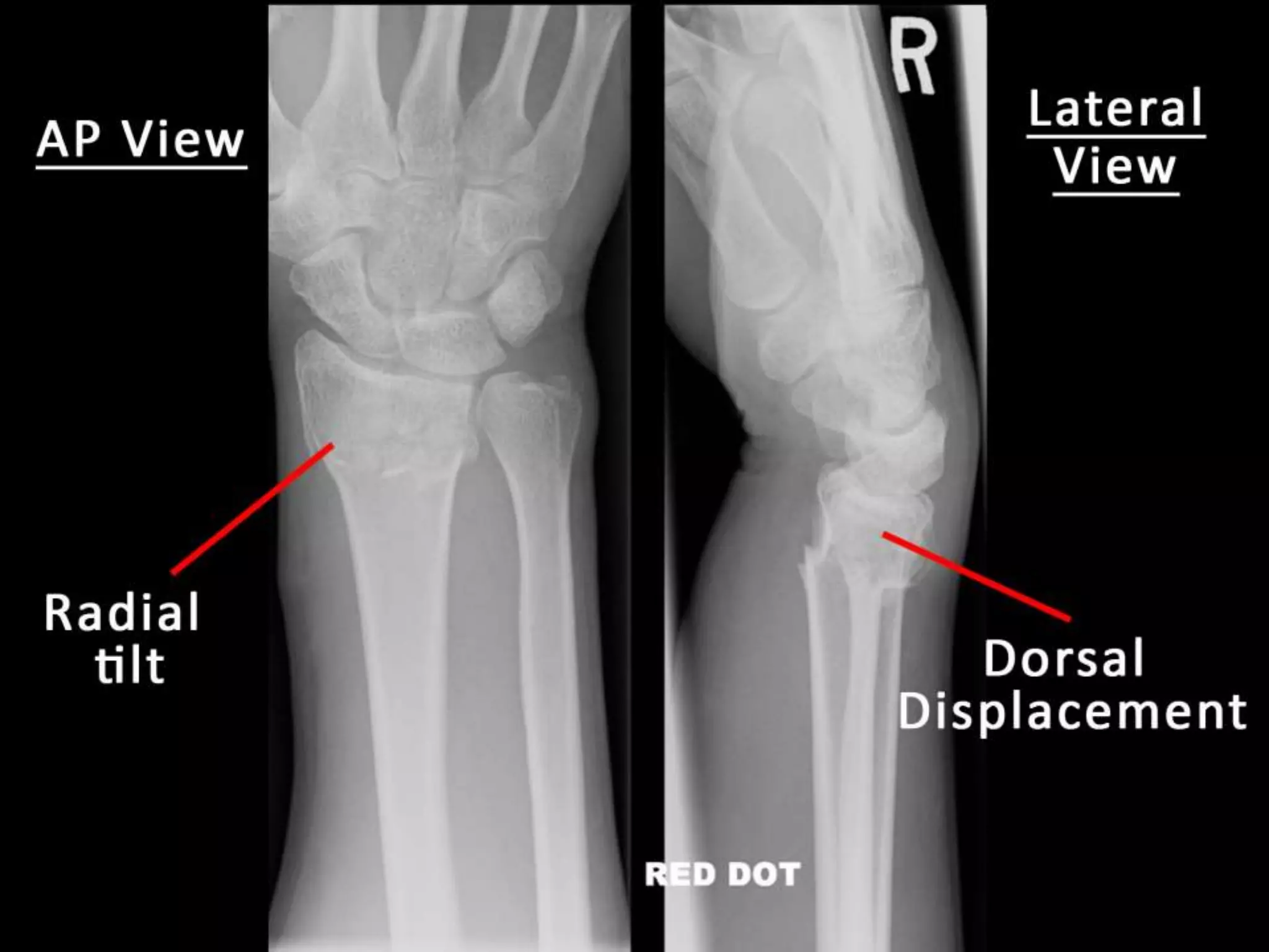
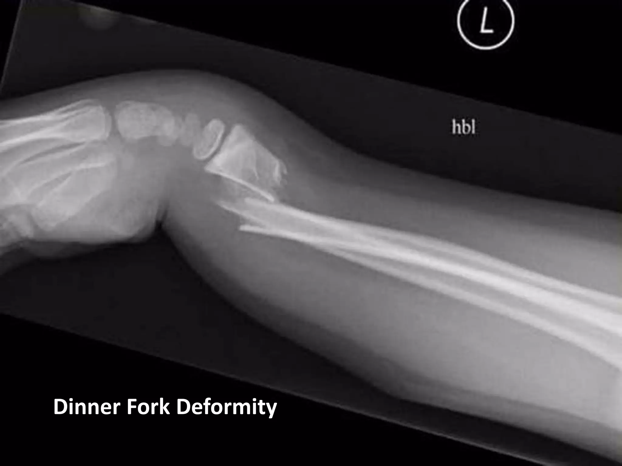
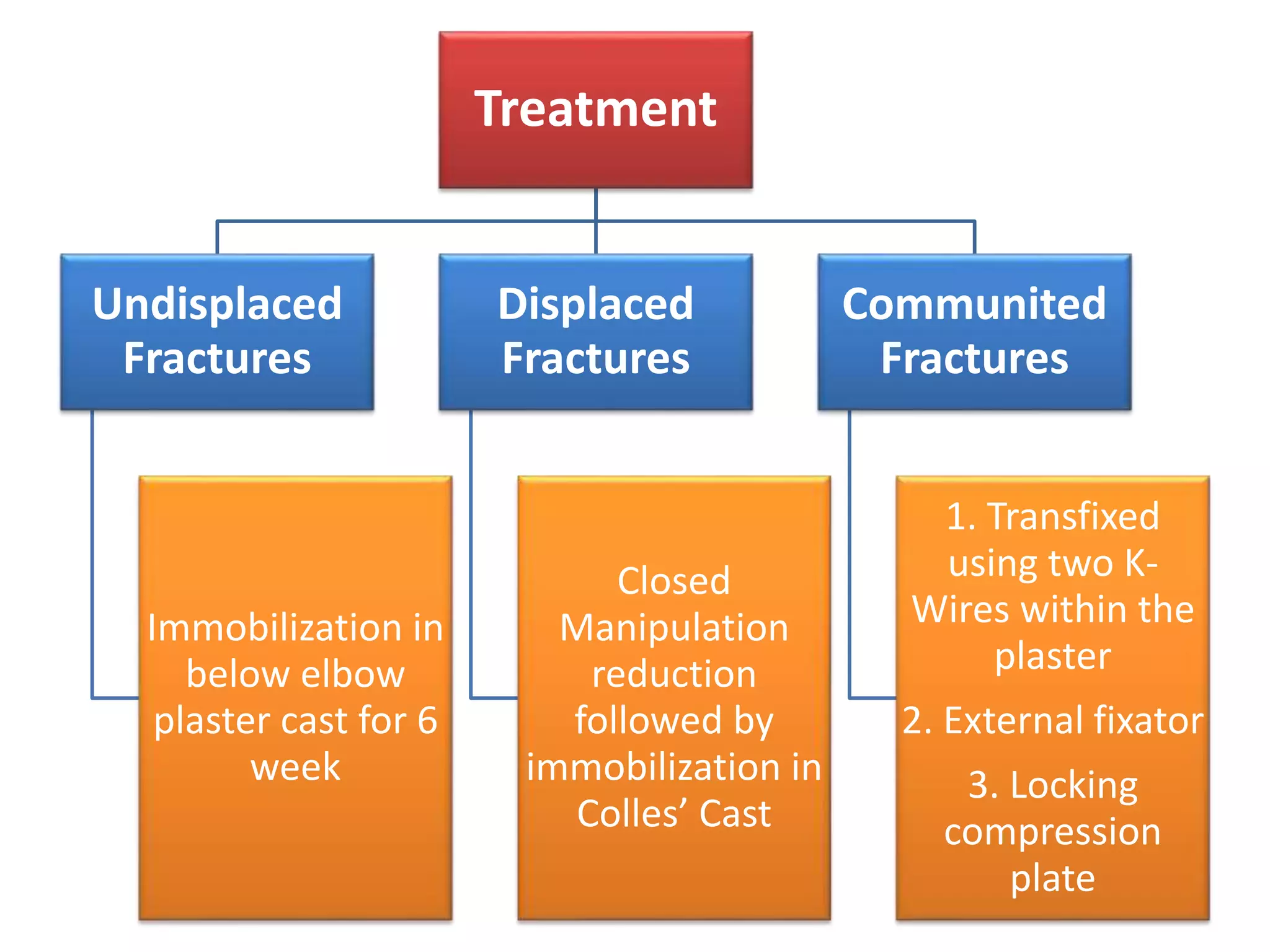
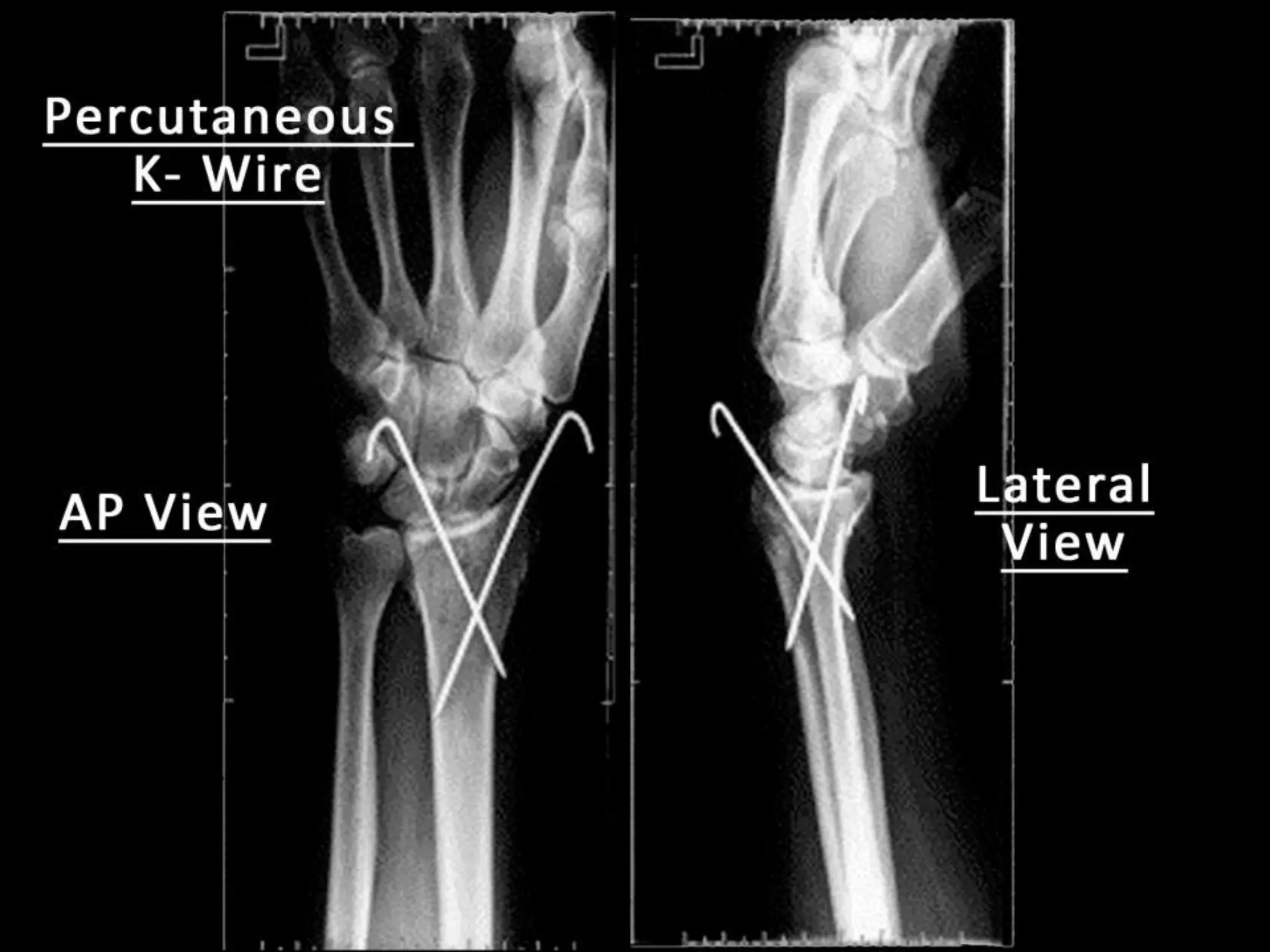
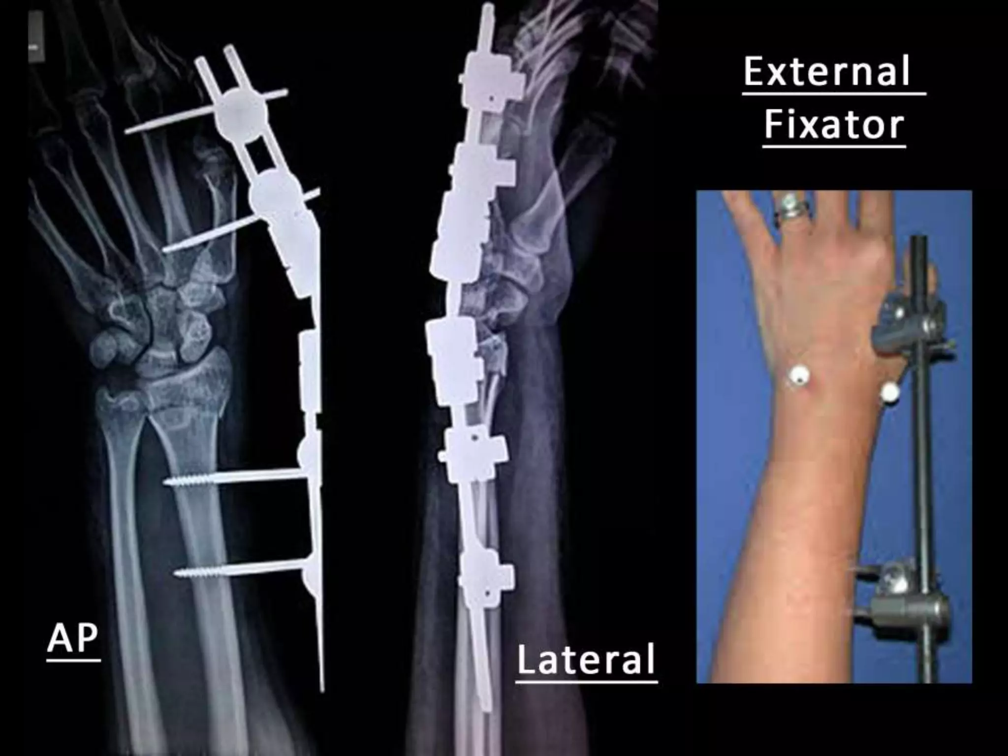
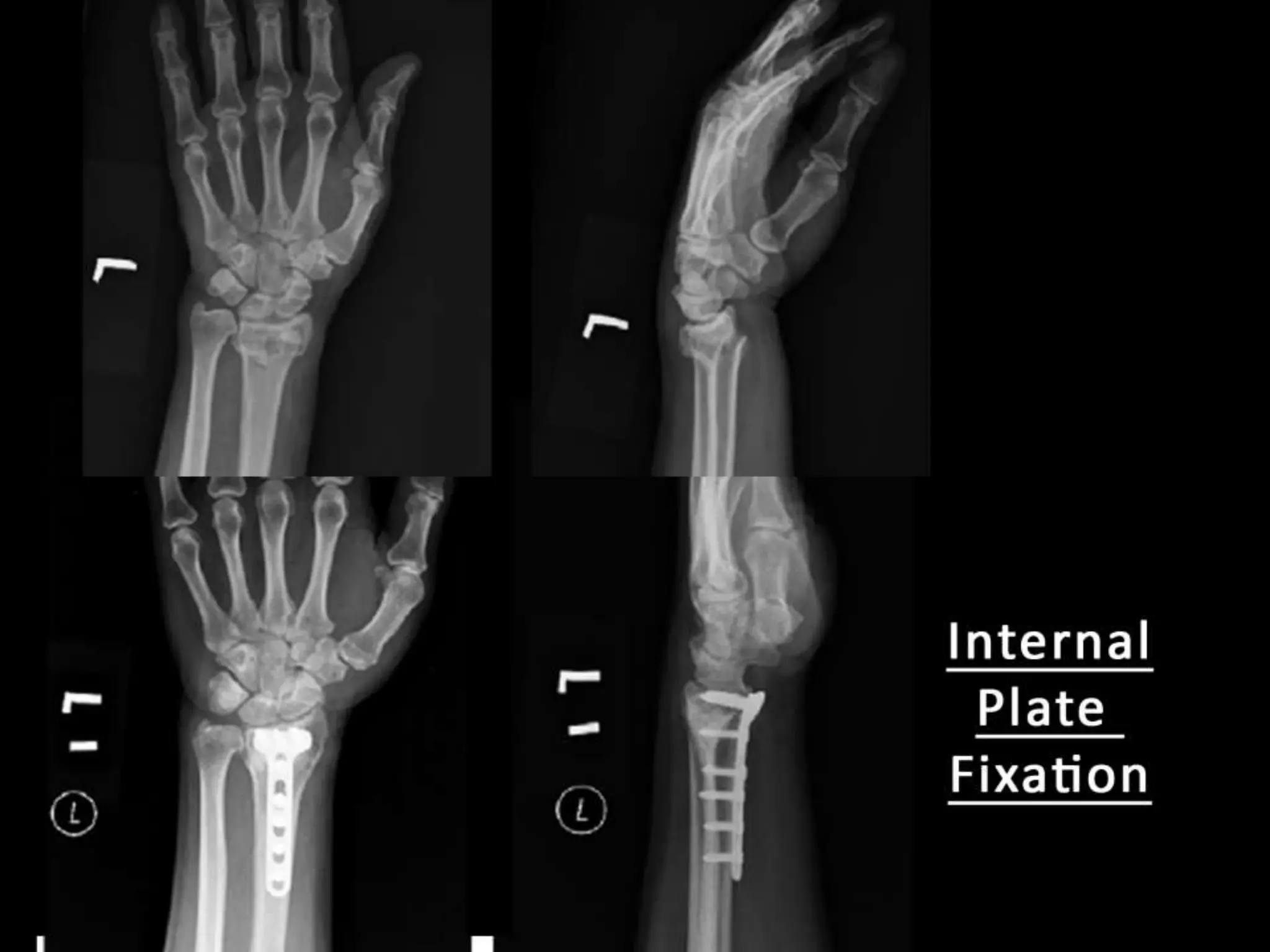
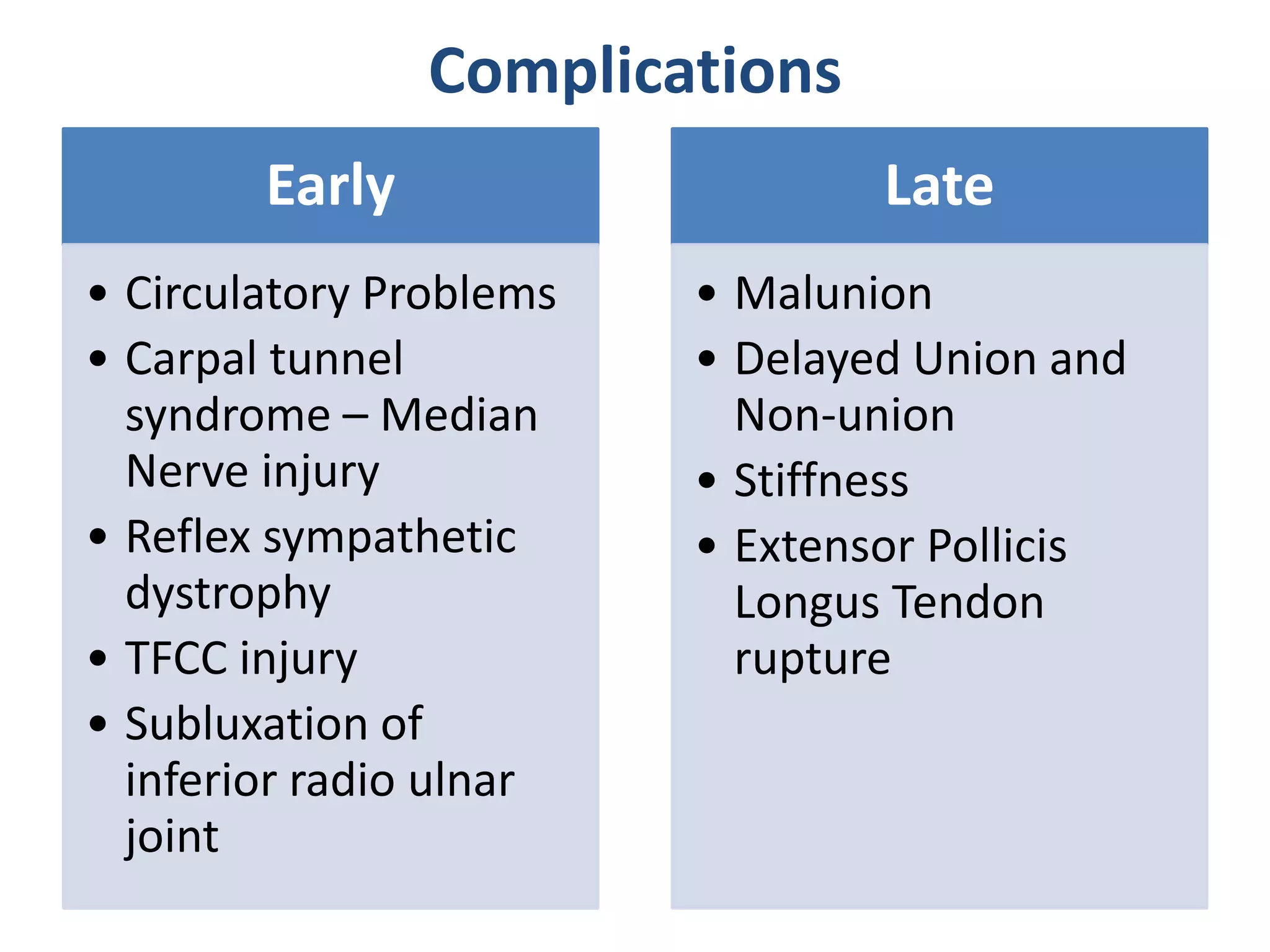
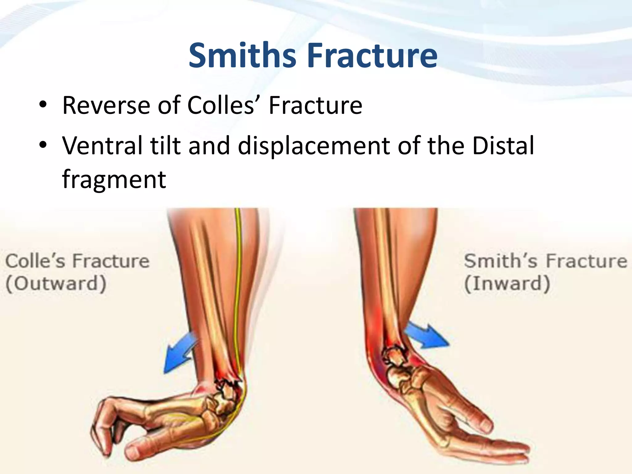
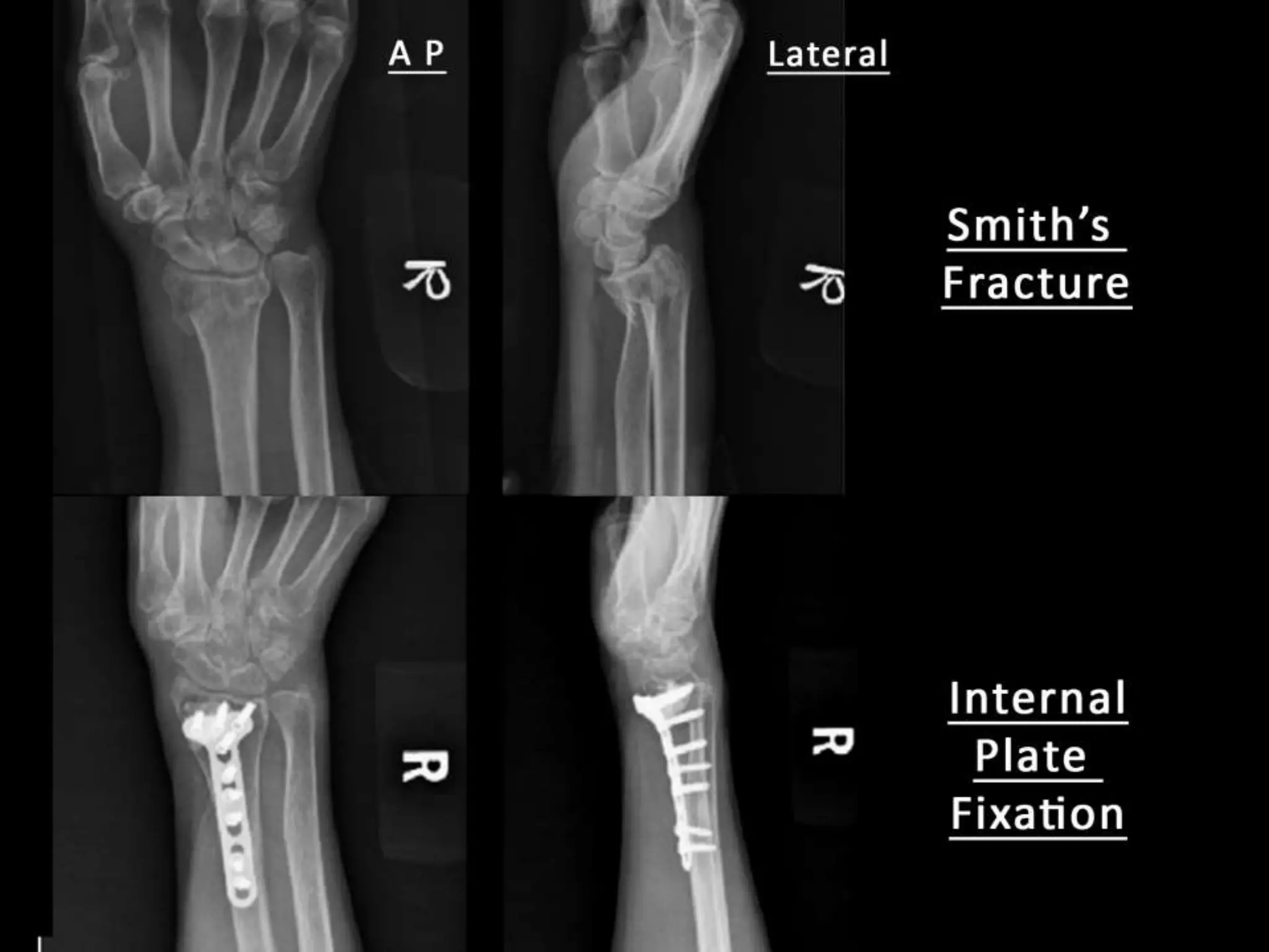
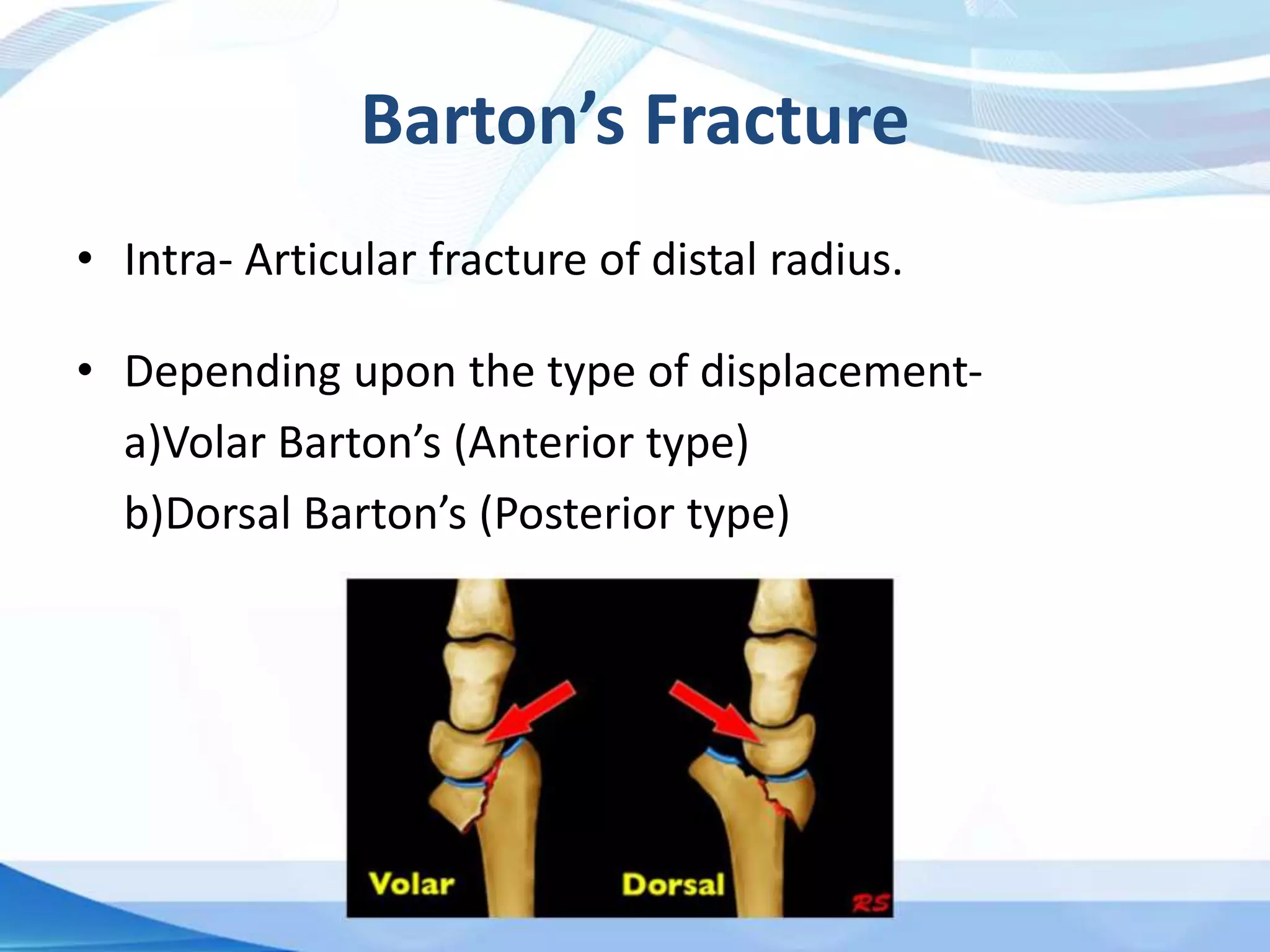
• Colles’ Fracture
• Smith’s Fracture
• Barton’s Fracture
• Described by Abraham
Colles in 1814.
• It is the most common of all
fractures in elderly.
• Usually in older women.
• Fall on a out stretched hand.
Colles’ Fracture
It is a Transverse fracture of the distal end of the
radius at its Cortico-Cancellous Junction with
typical Dorsal Displacement of the distal
fragment.
• Distal end of the radius
articulates with the carpal
bones and the ulna
• Normally the articular
surface of radius faces
ventrally and medially.
• Tip of radial styloid is 1cm
distal to the tip of the ulnar
styloid.
Relevant Anatomy
Mechanism of Action
• History of fall on an
outstretched hand
• Force is applied in the length
of the forearm with the wrist
in extension
• Fracture occurs transversely
at the cortico-cancellous
junction.
The distal fragment collapses into-
• Dorsal Displacement
• Dorsal Tilt
• Lateral/Radial Displacement
• Lateral/ Radial Tilt
• Supination
• Impaction of Fragments
Also associated with extension and shortening.
Associated injuries
• Fracture of styloid process of ulna
• Rupture of the Ulnar collateral ligament
• Rupture of the triangular Fibro-cartilage complex
(TFCC) of ulna.
• Rupture of the interosseos, radio-ulnar ligament
causing radio-ulnar subluxation.
Clinical Features
• Pain, tenderness, swelling and irregularity of the
lower end of radius.
• “Dinner Fork Deformity”
• Radial styloid comes at the level of the ulnar
styloid.
Dinner Fork Deformity
Radiological findings
• Transverse fracture of the distal end radius at cortico-
cancellous junction.
• The distal fragment shows –
a)Dorsal tilt & displacement – Lateral View
b)Lateral tilt & Displacement – AP View
• Maybe associated with communition or broken
ulnar styloid process.
Dinner Fork Deformity
Treatment
Undisplaced
Fractures
Immobilization in
below elbow
plaster cast for 6
week
Displaced
Fractures
Closed
Manipulation
reduction
followed by
immobilization in
Colles’ Cast
Communited
Fractures
1. Transfixed
using two K-
Wires within the
plaster
2. External fixator
3. Locking
compression
plate
Closed reduction and
manipulation
• Under general or
regional anesthesia.
• “Shaking Hand
Position”
a.Firm longitudinal
traction to disimpact.
b.Distal fragment into
palmar flexion and
Ulnar deviation.
Colles’ Cast
• Colles’ Cast is used to immobilize undisplaced or
reduced fracture.
• It is below elbow cast with
a.Palmar Flexion
b.Ulnar Deviation
a.
b.
Complications
Early
• Circulatory Problems
• Carpal tunnel
syndrome – Median
Nerve injury
• Reflex sympathetic
dystrophy
• TFCC injury
• Subluxation of
inferior radio ulnar
joint
Late
• Malunion
• Delayed Union and
Non-union
• Stiffness
• Extensor Pollicis
Longus Tendon
rupture
Smiths Fracture
• Reverse of Colles’ Fracture
• Ventral tilt and displacement of the Distal
fragment
• Clinical features - “Garden Spade Deformity”
• X-Ray – Ventral tilt and displacement
• Treatment-
Conservative - closed reduction and proper cast
immobilization for 6 weeks.
Surgical - K-wire or plate is used.
Barton’s Fracture
• Intra- Articular fracture of distal radius.
• Depending upon the type of displacement-
a)Volar Barton’s (Anterior type)
b)Dorsal Barton’s (Posterior type)
• Extends from articular surface of the radius to either
its anterior and posterior cortices.
• The small distal fragment is displaced along with the
carpals.
• Treatment
a) Closed Reduction and Plaster Cast
b) Internal Fixation by Plate or K-wire
Apley’s System of
Orthopaedics and Fractures
– 9th edition
Essential Orthopaedics –
Maheshwari & Mhaskar
– 5th edition
Distal End Radius Fractures - Colles, Smiths & Bartons

Distal End Radius Fractures - Colles, Smiths & Bartons

  • 2.
    • Colles’ Fracture •Smith’s Fracture • Barton’s Fracture
  • 3.
    • Described byAbraham Colles in 1814. • It is the most common of all fractures in elderly. • Usually in older women. • Fall on a out stretched hand.
  • 4.
    Colles’ Fracture It isa Transverse fracture of the distal end of the radius at its Cortico-Cancellous Junction with typical Dorsal Displacement of the distal fragment.
  • 5.
    • Distal endof the radius articulates with the carpal bones and the ulna • Normally the articular surface of radius faces ventrally and medially. • Tip of radial styloid is 1cm distal to the tip of the ulnar styloid. Relevant Anatomy
  • 6.
    Mechanism of Action •History of fall on an outstretched hand • Force is applied in the length of the forearm with the wrist in extension • Fracture occurs transversely at the cortico-cancellous junction.
  • 7.
    The distal fragmentcollapses into- • Dorsal Displacement • Dorsal Tilt • Lateral/Radial Displacement • Lateral/ Radial Tilt • Supination • Impaction of Fragments Also associated with extension and shortening.
  • 8.
    Associated injuries • Fractureof styloid process of ulna • Rupture of the Ulnar collateral ligament • Rupture of the triangular Fibro-cartilage complex (TFCC) of ulna. • Rupture of the interosseos, radio-ulnar ligament causing radio-ulnar subluxation.
  • 9.
    Clinical Features • Pain,tenderness, swelling and irregularity of the lower end of radius. • “Dinner Fork Deformity” • Radial styloid comes at the level of the ulnar styloid.
  • 10.
  • 11.
    Radiological findings • Transversefracture of the distal end radius at cortico- cancellous junction. • The distal fragment shows – a)Dorsal tilt & displacement – Lateral View b)Lateral tilt & Displacement – AP View • Maybe associated with communition or broken ulnar styloid process.
  • 13.
  • 14.
    Treatment Undisplaced Fractures Immobilization in below elbow plastercast for 6 week Displaced Fractures Closed Manipulation reduction followed by immobilization in Colles’ Cast Communited Fractures 1. Transfixed using two K- Wires within the plaster 2. External fixator 3. Locking compression plate
  • 15.
    Closed reduction and manipulation •Under general or regional anesthesia. • “Shaking Hand Position” a.Firm longitudinal traction to disimpact. b.Distal fragment into palmar flexion and Ulnar deviation.
  • 16.
    Colles’ Cast • Colles’Cast is used to immobilize undisplaced or reduced fracture. • It is below elbow cast with a.Palmar Flexion b.Ulnar Deviation a. b.
  • 20.
    Complications Early • Circulatory Problems •Carpal tunnel syndrome – Median Nerve injury • Reflex sympathetic dystrophy • TFCC injury • Subluxation of inferior radio ulnar joint Late • Malunion • Delayed Union and Non-union • Stiffness • Extensor Pollicis Longus Tendon rupture
  • 21.
    Smiths Fracture • Reverseof Colles’ Fracture • Ventral tilt and displacement of the Distal fragment
  • 22.
    • Clinical features- “Garden Spade Deformity” • X-Ray – Ventral tilt and displacement • Treatment- Conservative - closed reduction and proper cast immobilization for 6 weeks. Surgical - K-wire or plate is used.
  • 24.
    Barton’s Fracture • Intra-Articular fracture of distal radius. • Depending upon the type of displacement- a)Volar Barton’s (Anterior type) b)Dorsal Barton’s (Posterior type)
  • 25.
    • Extends fromarticular surface of the radius to either its anterior and posterior cortices. • The small distal fragment is displaced along with the carpals. • Treatment a) Closed Reduction and Plaster Cast b) Internal Fixation by Plate or K-wire
  • 26.
    Apley’s System of Orthopaedicsand Fractures – 9th edition Essential Orthopaedics – Maheshwari & Mhaskar – 5th edition