Microscope
•
2 likes•1,155 views

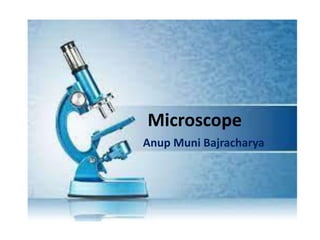
A microscope is an instrument used to observe very small organisms i.e. microorganisms. The microscope provides magnification and resolution which makes the image enlarged and fine. There are different types of microscopes ranging from simple to compound microscopes.

Recommended
More Related Content
What's hot
What's hot (20)
Similar to Microscope
Similar to Microscope (20)
More from Anup Bajracharya
More from Anup Bajracharya (20)
Recently uploaded
Recently uploaded (20)
Microscope
- 2. What is microscope? • "Micro" refers to tiny, • "scope" refers to view or look at. Microscopes are tools used to enlarge small objects so as they can be observed and studied. Microscopes range from a simple magnifying glass to the expensive electron microscope.
- 3. History of Microscopes • Timeline of the Microscope • 14th century: spectacles first made in Italy • 1590: Two Dutch spectacle-makers and father-and-son team, Hans and Zacharias Janssen, create the first microscope. • 1667: Robert Hooke's famous "Micrographia" is published, which outlines Hooke's various studies using the microscope. • 1675: Anton van Leeuwenhoek, who used a microscope with one lens to observe insects and other specimen. Leeuwenhoek was the first to observe bacteria. • 1830: Joseph Jackson Lister discovers that using weak lenses together at various distances provided clear magnification.
- 4. • 1878: A mathematical theory linking resolution to light wavelength is invented by Ernst Abbe. • 1903: Richard Zsigmondy invents the ultra microscope, which allows for observation of specimens below the wavelength of light. • 1932: Transparent biological materials are studied for the first time using Frits Zernike’s invention of the phase-contrast microscope. • 1938: Just six years after the invention of the phase contrast microscope comes the electron microscope, developed by Ernst Ruska, who realized that using electrons in microscopy enhanced resolution. • 1981: 3-D specimen images possible with the invention of the scanning tunneling microscope by Gerd Binnig and Heinrich Rohrer. History of Microscopes Ernst Ruska
- 5. Classification of Microscopes Simple microscope Compound microscope 1. It has a single lens system. 1. It has two or more lens system. 2. Eg:-Magnifying glass. 2. Eg:-Compound microscope basically used in the laboratory. Microscope can be classified on the basis of various properties. On the basis of lens system, microscopes are of two types.
- 6. Light Microscope Electron Microscope 1. It uses light as a source of illumination. 1. It uses electron or electron gun as a source of illumination. 2. Lens used in glass lens 2. Lens used is electromagnetic lens. 3. Here, vacuum is not needed. 3. Vacuum must be created since electrons can travel only in vacuum. 4. Resolution is low comparing to the electron microscope. 4. Resolution is very high capable of viewing particles of size 0.1 to 0.2 nanometer (nm). 5. Image can be directly seen by human eye. 5. Since, electrons can’t be seen by human eye. Electrons are converted into an amazing image by striking in fluorescent screen. Classification of Microscopes
- 9. Bright-Field Microscopy The standard instrument used in the laboratory to observe microorganisms is the bright field compound microscope. In bright field microscopy, the microscopic field is bright and the microorganisms appear dark because they absorb some of the light.
- 10. Principle of Microscopy • 1. Magnification The microscope produces an enlarged image of the object which is examined through it. This enlargement is known as its magnification and is measured in diameter • Eg: A magnifying lens which gives an image 36 time as large as that of the object is said to have a magnification of 36 diameter or 36X(x=times).
- 11. Which of these images would be viewed at a higher power of magnification? 1.Magnification We can see better details with higher the powers of magnification, but we cannot see as much of the image.
- 13. Fill the gaps….
- 14. 2. Resolution • The ability of microscope to distinguish two closely spaced objects as separate and distinct entities called resolution. • Eg. The human eyes has the resolving power of 0.25mm which means that two dots is placed 0.25mm apart can be distinguished as two dots. • If the distance between the two dots is less than 0.25,only one dot will be seen. • Resolving Power(R.P.) = 𝜆 2.𝑁.𝐴 whereλ wavelength of light ,N.A=Numerical Aperture
- 15. Resolution : Resolution is the quality of image that is measured by the smallest distance which could be seen clearly without the blur due to diffraction. Resolving power : The ability of an optical instrument to separate or distinguish small or closely adjacent objects through the image formation is said to be resolving power of the instrument.
- 18. Oil immersion • Immersion Oil is a special oil used in microscope work with the highest power objective lenses (ie 100x lens). • In light microscopy, oil immersion is a technique used to increase the resolving power of a microscope. • This is achieved by immersing both the objective lens and the specimen in a transparent oil of high refractive index, thereby increasing the numerical aperture of the objective lens.
- 19. Immersion Oil and Refractive Index
- 20. Microscope
- 21. Parts of Microscope • Body: the portion of the microscope which contains the lenses. • Revolving Nosepiece: the assembly which contains the objective lenses. • Stage: the platform upon which the object to be examined is placed • Sub-stage Assembly: the parts below the stage; these include the condenser, iris diaphragm, and illuminator. • Condenser Lens: a lens assembly beneath the stage which concentrates light on the object being examined. • Diaphragm: a series of holes which control the amount of light which reaches the object being examined.
- 22. • Coarse and Fine Adjustment Knobs: these knobs adjust the distance between the objective lens and the stage. This distance is called the working distance. • Illuminator: the light source. It may be built into the base or it may not be part of the scope at all. Parts of Microscope COARSE ADJUSTMENT KNOB — rapid control which allows for quick focusing by moving the objective lens or stage up and down. It is used for initial focusing. FINE ADJUSTMENT KNOB — Slow but precise control used to fine focus the image when viewing at the higher magnifications.
- 23. Handle and care of Microscope Always keep your microscope covered when not in use even if the microscope is stored in a cabinet. Always use two hands when moving the microscope. When carrying your microscope, hold it by the base and the metal support arm.
- 24. If using immersion oil, always ensure the objectives are cleaned immediately after use. Objective, eyepieces and condenser may be removed for cleaning. Use only lens paper and lens cleaner.
- 25. Turn the microscope off after use. Do not keep the light on all day as this will shorten the bulb’s life.