Guide to Radiology Equipment and Imaging Modalities
•Download as PPTX, PDF•
2 likes•503 views

Radiology uses various imaging technologies to diagnose and treat diseases. The document outlines different types of radiology equipment and procedures including X-ray, CT, MRI, ultrasound, mammography, PET, SPECT, nuclear medicine, radiotherapy, fluoroscopy, DEXA, and interventional radiology. Key equipment includes X-ray machines, CT and MRI scanners, ultrasound machines, gamma cameras, linear accelerators, C-arms, and cyberknives. Radiologists use these tools to examine organ structure and function, guide procedures, and deliver radiation therapy for cancer treatment.

Recommended
More Related Content
What's hot
What's hot (20)
Similar to Guide to Radiology Equipment and Imaging Modalities
Similar to Guide to Radiology Equipment and Imaging Modalities (20)
More from Ankit Mishra
More from Ankit Mishra (16)
Recently uploaded
Recently uploaded (20)
Guide to Radiology Equipment and Imaging Modalities
- 1. Introduction about radiology equipments
- 2. Radiology Diagnostic radiology/Radiography X-Ray Computed tomography Magnetic Resonance imaging Ultrasonography Fluoroscopy DEXA Mammography Radiotherapy Linear accelerator Brachytherapy Implant therapy Cyber knife Cobalt 60 Etc. Nuclear medicine Positron Emitted Tomography Single photon Emitted Tomography Gamma Camera, Etc. Interventional radiology C-arm
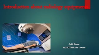
- 3. • Radiology:- Radiology is a branch of medicine that uses imaging technology to diagnose and treat disease. Radiology may be divided into two different areas, diagnostic radiology and interventional radiology. • Diagnostic radiology:- Diagnostic radiology refers to the field of medicine that uses non- invasive imaging scans to diagnose a patient. The tests and equipment used sometimes involves low doses of radiation to create highly detailed images of an area. • Nuclear medicine:- Nuclear medicine is a specialized area of radiology that uses very small amounts of radioactive materials, or radiopharmaceuticals, to examine organ function and structure. Nuclear medicine imaging is a combination of many different disciplines. These include chemistry, physics, mathematics, computer technology, and medicine. This branch of radiology is often used to help diagnose and treat abnormalities very early in the progression of a disease, such as thyroid cancer. • Radiotherapy:- Radiation therapy (also called radiotherapy) is a cancer treatment that uses high doses of radiation to kill cancer cells and shrink tumors. At low doses, radiation is used in x-rays to see inside the body, as with x-rays of teeth or broken bones.
- 4. Interventional Radiology o Interventional radiology is a medical specialization that involves performing a range of imaging procedures to obtain images of the inside of the body. The interventional radiologist carefully interprets these images to diagnose injury and disease, and to perform a range of interventional medical procedures. o Interventional radiologist use imaging techniques such as X-rays, MRIs (magnetic resonance imaging) scans, fluoroscopy (an X-ray procedure that makes it possible to see internal organs in motion), CT (computed tomography) scans and ultrasounds. o Interventional radiologists perform a broad range of procedures such as treating tumors, taking organ biopsies or placing stents by inserting tiny instruments and thin plastic tubes (catheters) into the body via an artery or vein.
- 5. X-ray • X-ray is a form of energy which travel in any medium. • Discovered by Sir Wilhelm Conrad Roentgen in 1895. • X-rays are types of electromagnetic radiation probably most well-known for their ability to visualize a image of internal body. • X-rays are roughly classified into soft X-rays and hard X- rays. • Wavelength:- 0.01 to 10 nanometers • Frequency:- 3×10¹⁹ to 3×10¹⁶ Hz.
- 6. Computed Tomography • Also known as "CAT scanning" (Computed Axial Tomography). • Invented in 1972 by British engineer Godfrey Hounsfield • The first clinical CT scanners were installed between 1974 and 1976. • Uses ionizing radiation • scanned images are in axial section • Guide procedures such as surgery, biopsy and radiation therapy
- 7. Mammography • It is specialized medical imaging that uses a low- dose ionizing radiation . • Uses for breast imaging for early detection and diagnosis of breast diseases in women. • Three recent advances in mammography : 1. digital mammography, 2. computer-aided detection 3.breast tomosynthesis.
- 8. Magnetic resonance imaging • Invented by Paul C. Lauterbur . • On July 3, 1977, the first magnetic resonance imaging (MRI) exam on a live human patient was performed • Uses very high magnetic field • Scanning images are in 3 plane(axial, coronal, sagittal). • It is a non-invasive and painless
- 9. DEXA :- Dual energy X-ray absorptiometry • uses a very small dose of ionizing radiation • It is commonly used to diagnose osteoporosis, to assess an individual's risk for developing osteoporotic fractures. • It is simple, quick and noninvasive. • Bone densitometry, is an enhanced form of x-ray technology that is used to measure bone loss.
- 10. ultrasonography • It is a diagnostic imaging technique, or therapeutic application of ultrasound. • Safe for pregnant women scanning • Helps in FNAC and biopsy • Uses sound waves for scanning • A Doppler ultrasound study may be part of an ultrasound examination. • It uses transducer for scanning pts.
- 11. Fluoroscopy • Uses ionizing radiation for scanning • Performed to evaluate specific areas of the body, including the bones, muscles, and joints, as well as solid organs, such as the heart, lung, or kidneys. • A continuous X-ray beam is passed through the body part being examined. The beam is transmitted to a TV-like monitor so that the body part and its motion can be seen in detail.
- 12. Linear accelerator • First LINAC was developed by wide Roe in 928. • Used to accelerate or push the electron in linear direction. • It uses high radio frequency electromagnetic waves to accelerate the charge particle in a linear path inside the tube like structure which is called as accelerator wave guide.
- 13. Brachytherapy • It is a procedure that involves placing radioactive material inside pts. body. • It is one type of radiation therapy that's used to treat cancer. Brachytherapy is sometimes called internal radiation. • It can be used alone or in conjunction with other cancer treatments • Fewer side effects. • Preservation of organ structure and function.
- 14. Cobalt-60 • It is a synthetic radioactive isotope of cobalt with a half- life of 5.2713 years. • It is produced artificially in nuclear reactors. • It is formed when metal structures, such as steel rods, are exposed to neutron radiation
- 15. Cyber knife • It is a non-invasive treatment for cancerous and non-cancerous tumors and other conditions where radiation therapy is indicated. • Treatments are typically performed in 1 to 5 sessions. • It is the only radiation delivery system which features a linear accelerator (LINAC) directly mounted on a robot to deliver the high-energy x-rays or photons used in radiation therapy.
- 16. PET • Positron emission tomography (PET) uses small amounts of radioactive materials called radiotracers or radiopharmaceutical. • Nuclear medicine imaging procedures are noninvasive. With the exception of intravenous injections, they are usually painless. • It comes with PET CT or PET MRI, also called fusion imaging.
- 17. SPECT Single photon emitted computed tomography
- 18. Gamma camera • Also called a scintillation camera or Anger camera • A device used to image gamma radiation emitting radioisotopes, a technique known as scintilography. • predominant nuclear medicine imaging machine currently in use.
- 19. C-arm • Uses ionizing radiation for scanning • Uses in Cathlab for interventional procedures • A C-arm provides high-resolution, real-time fluoroscopic x-ray imaging during surgical, orthopedic, and emergency care procedures.