
Recommended
More Related Content
Similar to первая лекция иностр.pptx
Similar to первая лекция иностр.pptx (20)
Recently uploaded
Recently uploaded (20)
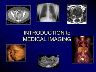
первая лекция иностр.pptx
- 2. Objectives • List the main diagnostic imaging modalities used in medical practice • Explain the basic principles of the main types of imaging modalities • Describe advantages of the different imaging techniques • List disadvantages of the different imaging techniques
- 3. Medical Imaging X-Rays: ionizing radiation Radiography CT scan (Computed Tomography) Gamma rays Nuclear Medicine Sound waves Ultrasound Magnetic fields/radiofrequency waves MRI (Magnetic Resonance Imaging)
- 4. Who Does Imaging? • Radiologist – Consultant: Diagnostic, Subspecialties – Interventionalist • Radiation Oncologist: Treatment planning • Cardiologist: Invasive, Non-Invasive • Vascular Surgeon: Endovascular procedures • Other specialists – Usually for procedure guidance (Ob-Gyn, Internal medicine, Orthopedics, etc.)
- 5. Training • Radiologist – Mandatory clinical internship – 4 years general radiology residency – Physics, radiation protection, radiobiology, technology, diagnosis, anatomy, pathology, physiology, etc. – 1 year fellowship in subspecialty • Cardiologist, vascular surgeon, others – intergrated into training program
- 7. X-rays • Dr. Wilhelm C. Roentgen at University of Wurzburg, 1895 – Discovered and named X-rays • Awarded first Nobel Prize for Physics in 1901
- 8. X-Rays • Radiography – Plain film radiography - without added contrast material – Contrast radiography – with contrast material – Computed Radiography (CR) – Fluoroscopy: done in real time • Barium studies: Upper GI, BE • Angiography • CT (Computed Tomography), aka CAT scans (Computer Assisted Tomography)
- 9. Radiographs • Electromagnetic waves (X-rays) are produced in an X-ray tube by converting electrical energy into an electromagnetic wave • Electrons are accelerated from an electrically negative cathode to a positive target anode • Energy is released and converted into heat and X-rays
- 10. Radiographs • Images produced by electromagnetic waves (X-rays) • produced by an X-ray tube • pass thru the body • are absorbed by the different tissues • reach the film and • expose the film Radiographic cassette
- 11. Computed Radiography (CR) • Produces digital radiographic images • Instead of film, a phosphor plate is exposed to X-rays • Laser beam scans the plate • Light is released, intensified and converted to electron stream • Converted by computer into digital image • Viewed on a monitor • Transferred over networks
- 12. Radiographic Densities • Air Black • Fat Dark gray • Water* Light gray • Bone White • Calcium White • Metal Very white *Water=soft tissue: organs, muscles, blood vessels, masses
- 13. Radiodensity Radiodensity is a function of: 1. Composition (atomic number) 2. Thickness of object 3. Strength of X-ray Radiodensity as function of thickness of object Radiolucency is the opposite
- 14. Radiodensity • If an object is thick and dense, less radiation passes thru to reach the film – Radiodense – Film is underexposed and stays light • Air gives no obstruction to X-rays – Radiolucent – Film gets overexposed and turns black • Bone absorbs radiation, less radiation reaches the film – Film is underexposed and stays white
- 15. Abdominal Radiograph: KUB • Air Black • Fat Dark gray • Water Light gray • Bone White • Calcium White • Metal Very white *Water=soft tissue: organs, muscles, blood vessels, masses
- 16. Radiographs Composed of overlapping radiodensities, overlapping shadows Tissues stacked in front of each other Need 90 degree projection for anatomic placement
- 17. Radiographs Perpendicular projections are necessary to localize structures in the body
- 19. Patient Position for Chest X-Ray Posteroanterior (PA) Chest: X-ray beam passes from posterior to anterior RT LT
- 20. Erect Position Free intraabdominal air: Pneumoperitoneum
- 21. Portable Films • Patient is too ill to go to Radiology Department • Less optimal • Portable X-ray unit • X-ray film is behind patient • X-rays pass through patient from anterior to posterior
- 22. Abdominal Radiographs • Erect/upright • Supine • Prone • Decubitus • Oblique/rotated
- 23. Contrast Radiography • Injection, ingestion or placement of radiopaque material into the body for contrast enhancement • Oral, rectal contrast: Barium, gastrograffin
- 25. Caution: Radiation Exposure • Radiation workers follow safety guidelines • Women of child-bearing age should be questioned about possibility of pregnancy before abdominal X-ray. • Ask about LMP and check pregnancy test, if in doubt.
- 28. Computed Tomography • Ionizing radiation used to obtain cross-sectional images of the body • Table moves through large donut-shaped scanner – Fast moving X-ray tube (thin X-ray beam rotates) – Numerous electronic detectors • Rapid acquisition of images • Contrast agents necessary for most scans – Oral – Intravenous iodinated
- 29. Computed Tomography • Multidetector CT scans – Advancement from tomographic imaging (slices) to volume imaging – Produce a volume of data that can be manipulated – Reconstruct at 1-10 mm increments – Axial, sagittal, coronal, 3D reconstructions
- 30. Computed Tomography Coronal and sagittal reconstructions
- 31. Computed Tomography • Large field of view – Entire cross-section of body • Improved differentiation of soft tissue densities • Excellent spatial resolution – Small 3-4 mm lymph nodes, vessels • Automated scanners – Less operator-dependent • Ultra-fast scanners – Suspended respiration – Less bowel motion artifact – Trauma ADVANTAGES
- 33. CT of the Female Pelvis • Exposure to ionizing radiation • Allergic reactions to intravenous contrast – Mild to severe (anaphylaxis) • Contrast nephropathy – May cause renal failure – Caution in diabetics with nephropathy • Problems of dehydration – Cautious use in multiple myeloma, sickle cell disease • Soft tissue differentiation not as good as MRI DISADVANTAGES
- 34. CT Windows Windowing displays the image in differing shades of gray Correspond to brightness and contrast Soft tissue window Lung (air) window
- 35. CT Windows Soft tissue window Bone window Windowing displays the image in differing shades of gray Correspond to brightness and contrast
- 36. CT Diagnosis • Trauma Shattered spleen Fractured sacrum
- 37. CT Diagnosis • Pulmonary embolism • Intravenous contrast material is necessary
- 38. CT Diagnosis • Angiography • Bowel disorders Aortic aneurysm Perforated sigmoid diverticulitis
- 39. CT 3D Volume Rendering Aorta, plaque Aortic arch CT Angiogram
- 41. Diagnostic Ultrasound • Audible sound 20Hz-20KHz • Ultrasound >20Hz • Medical ultrasound 1-20 MHz • Transducer sends high frequency sound waves into the body and gathers their reflections (echoes) • Converts echoes into electronic signals • Displays images on a monitor
- 42. Diagnostic Ultrasound • Sound travels very well through fluid • Ultrasound is good for anything containing fluid, as long as there is no interference for the sound beam to reach the fluid
- 43. Diagnostic Ultrasound • Interferences to sound waves: air, bone, metal, thick tissues, deep structures
- 44. Diagnostic Ultrasound • Multiplanar imaging in real-time • Non-invasive, safe, no radiation – Pregnancy, Pediatrics • Relatively inexpensive • Widely available • Portable, bedside • Good contrast of tissue layers in many organs ADVANTAGES
- 45. Diagnostic Ultrasound • Good contrast of tissues layers ADVANTAGES
- 46. Pelvic Ultrasound versus CT Ultrasound CT Ovarian Neoplasm
- 47. Diagnostic Ultrasound • Relatively small field of view • Operator dependent: inconsistent reproducibility • Depends on sound penetration – Air, bone, obesity DISADVANTAGES
- 48. Diagnostic Ultrasound • Trade-off between depth (beam penetration) and resolution – For deeper penetration need lower frequency transducers, resulting in lower resolution DISADVANTAGES 3.5 MHz 12 MHz
- 49. Doppler Ultrasound • Evaluation of blood flow – Patency, direction, character of flow in vessels – Vascularity in a mass Carotid artery Testicular flow
- 50. FD = (FR - FT ) = 2 • FT • v • cos c v FT FR The Doppler Equation The relationship of Doppler frequency shift to velocity of a moving object C =1540m/sec speed of sound in tissues
- 51. 3D Ultrasound 1st Trimester fetus 3rd Trimester fetus
- 52. Magnetic Resonance Imaging Magnetic fields Radiofrequency waves
- 53. Magnetic Resonance Imaging • Patient placed inside a large cylinder-shaped magnet • Radio waves 10,000 - 30,000 stronger the earth’s magnetic field are sent thru body • Nuclei of body’s (hydrogen) atoms shift position • As they move back they send out radio waves • Scanner detects the signals • Creates image based on location & strength of signals
- 54. Magnetic Resonance Imaging • Large field of view – Cross-section of entire body – Volume imaging (from tomographic imaging, slices) • No ionizing radiation as with CT • Not as operator dependent as ultrasound • Much fewer contrast allergies and less risk of contrast nephropathy than with iodinated agent used in CT • Excellent contrast resolution among tissue layers, esp. fat, hemorrhage ADVANTAGES
- 55. Magnetic Resonance Imaging Large field of view Excellent Contrast Resolution
- 56. Ultrasound vs CT vs MRI Field of View, Excellent Contrast Resolution Ultrasound MRI CT
- 57. Magnetic Resonance Imaging • Exquisite neuroanatomical detail • Musculoskeletal disorders • Cardiovascular
- 58. Magnetic Resonance Imaging • Motion related artifacts – Bowel peristalsis – Respiratory motion • Cost $$$ • Image production is time-consuming with complicated protocols & scan time • Intravenous contrast is often required to improve tissue differentiation • Claustrophobia • Obesity DISADVANTAGES
- 59. Magnetic Resonance Imaging • Contraindication in patients with ferromagnetic metallic objects, implants, foreign bodies: – Metallic fragments in eye – Cochlear implants – Cardiac pacemakers – Brain aneurysm clips – Certain heart valves – Neurological stimulators • Orthopedic devices are not harmful to patient, but create artifacts • Intrauterine devices are safe METALLIC OBJECTS
- 61. Nuclear Medicine • Uses small amounts of radioactive material for diagnosis and treatment • Molecular Imaging: images reflect biological processes that take place at the cellular and subcellular levels • Evaluates physiological function rather than anatomic structure • Uses radiopharmaceuticals: agents that have trace amounts of radioactive atoms attached • Radiation emitted from the patient is imaged by a gamma camera, SPECT or PET scanner
- 62. Nuclear Medicine Applications • Oncology: tumor localization, staging, metastases • Cardiology: myocardial perfusion scans • Gastrointestinal: acute cholecystitis, biliary tract, GI bleeding • Pulmonary: ventilation, perfusion • Infectious disease: localize infections (subtle) • Therapy, e.g. I-131 for thyrotoxicosis, thyroid cancer
- 63. Common Radionuclides • Intravenous administration – Technetium-99m – Iodine-123 and 131 – Thallium-210 – Gallium-67 – Fluorine-18 Fluorodeoxyglucose – Indium-111 labeled leukocytes • Inhaled gaseous/aerosol radionuclides – Xenon-133 – Krypton-81m – Technicium-99m gas
- 64. Gamma Camera
- 66. PET Scan: Cancer Detection • Positron Emission Tomography • 18F-fluorodeoxyglucose (FDG) • Primarily used for diagnosis, staging & monitoring of cancers: lung, breast, cervical, colorectal, esophagus, head & neck, lymphoma, melanoma Lymphoma pre and post chemotherapy
- 67. PET-CT Scanner Combined PET scanner and CT scanner
- 68. PET-CT • Imaging by “fusion” of anatomy and physiology • Superimposition of the anatomic images of a CT scan and the co-registration of physiologic uptake of a radionuclide agent (molecular imaging)
- 69. PET-CT Scans Fusion imaging: Lingular mass CT PET PET-CT
- 70. Summary • Medical imaging is essential for medical practice • Interpretation of various imaging modalities requires training and experience • Radiologist specializes in various imaging modalities – Consultant – Interventionalist
- 71. Sample Question Of the following imaging modalities, which test has the least harmful effects for a fetus? a. CT b. Ultrasound c. MRI d. Nuclear medicine
- 72. Thank you for your attention!