Implementation of Lower Leg Bone Fracture Detection from X Ray Images
kent_komine_assip_poster
1. Measurement of Muscle Volume using freehand 3D Ultrasound Imaging
Kent Komine, Dr. Nelson Cortes, and Dr. Siddhartha Sikdar
Abstract ResultsMethods
Acknowledgements
Introduction
Conclusions
• One possible consequence of ACL reconstruction
surgery is the loss of muscle volume and strength.
Unfortunately, this leads to the impairment of the
individual to safely perform activities of daily living.
Muscle volume is typically measured with MRI, but
new novel technology may allow volume measure at
the clinician’s office using ultrasound imaging.
• Additionally, muscle volume is directly related to the
ability for a person to torque their joints, in this case
the knee. Muscle volume of the rectus femoris is
significant because it is correlated with the ability to
perform activities of daily living.
• After a patient undergoes ACL reconstruction surgery,
unwanted side-effects may emerge, often times
affecting the muscles surrounding the knee. By
measuring the volume of the rectus femoris after the
surgery, physicians can better detect possible
complications and develop targeted intervention
plans for each individual.
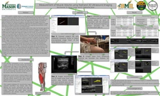
Step 1. Calibrated the Ascension
3D Guidance trakSTAR™ motion
tracking sensor to work in
conjunction with the Ultrasonix
Sonix RP ultrasound machine to
ensure accuracy for 3D
reconstructions later on.
Step 4. Images reconstructed
into 3D model using “slices” and
position data. Muscle volume
and cross-sectional area can be
quantified directly in Stradwin.
Step 3. Images taken from ultrasound
machine are recorded into Stradwin for
analysis.
We would like to extend our gratitude to Khalid Almuhanna, Oladipo Eddo, and all of the
test subjects for their invaluable assistance and participation in the study!
• The data demonstrates that freehand 3D ultrasound imaging is a feasible technique to
quantify and analyze muscle volume.
• Using ultrasound imaging to acquire 3D reconstructions of the muscle may be a practical
alternative to using MRI.
Step 2. Scanned subject’s leg with
ultrasound transducer probe to collect
cross-sectional image “slices” of the
rectus femoris muscle.
When patients undergo anterior cruciate ligament (ACL) reconstruction surgery, it is
crucial to analyze any effects on the body’s functional ability that may lead to early
development of osteoarthritis. One way to do this is by examining the volume and cross
sectional area of the rectus femoris muscle. The objective of our study is to assess the
ability of freehand 3D ultrasound imaging in reconstructing 3D images of the rectus
femoris. In doing so, we hope to determine if freehand 3D ultrasound imaging is a viable
alternative to magnetic resonance imaging (MRI) when acquiring reconstructions of the
rectus femoris for its volume and cross sectional area.
For the study, we recruited nine healthy subjects (five males, four females) ages 21 to
46. We utilized an Ultrasonix Sonix RP® ultrasound machine to capture cross sectional
images of the rectus femoris and an Ascension 3D Guidance trakSTAR™ motion tracker to
locate where the images were captured in relation to the muscle. To synchronize the
images to the location data, we calibrated the motion tracker with the ultrasound probe to
ensure accurate tempo-spatial 3D reconstructions. The images and location data were then
recorded into Stradwin, a freehand 3D ultrasound acquisition program that pieces together
image “slices” according to respective spatial location into a single 3D reconstruction. Once
the 3D image is constructed, users can analyze the images and specifically segment the
surfaces of the rectus femoris, which is quantifiable in volume and cross sectional area.
The results of the tests conducted demonstrate the reliability and feasibility of
freehand 3D ultrasound imaging. In the ultrasound phantom tests, 3D reconstructions
showed an average error of 2.4% in volume when compared with actual volumes. In the
anatomical distances test, the measured distance deviated an average of 3.5% from the
actual distance. Lastly, in the reproducibility test, the difference in volume of the 3D
reconstructions between the first scan and the second scan was 2.2mL, or 1.49% from the
average of the two scans. If implemented in healthcare, freehand 3D ultrasound imaging
will allow for muscle reconstructions to be conducted more conveniently and affordably
than MRI.
Reproducibility Test
Volume
Reconstruction 1 147.097mL
Reconstruction 2 149.319mL
Average 148.208mL
Deviation from Average 1.49%
Anatomical Distances Test
Actual Ultrasound Error
Image 1 200.0mm 185.3mm 7.4%
Image 2 200.0mm 196.8mm 1.6%
Image 3 200.0mm 203.0mm 1.5%
Average 200.0mm 195.0 3.5%
Phantom Test (Model 525)
Actual Ultrasound Error
Diameter 8.0mm 7.9mm 1.25%
Length 160.0mm 161.4mm 0.88%
CSA 50.3mm2 49.0mm2 2.58%
Volume 8.042mL 7.891mL 1.88%
Phantom Test (Model 539)
Actual Ultrasound Error
Diameter 8.0mm 7.9mm 1.25%
Length 57.5mm 57.3mm 0.35%
CSA 50.3mm2 49.0mm2 2.58%
Volume 2.892mL 2.808mL 2.90%
Figure 1. Human leg muscles with the
rectus femoris highlighted in red.
Figure 2. Calibration mode in Stradwin
Figure 3. Scanning of upper leg with ultrasound probe.
Figure 4. Ultrasound image taken from ultrasound machine.
Figure 5. 3D reconstruction of the rectus femoris drawn out in Stradwin.
Figure 9.
Reconstruction
of phantom
model 525.
Figure 10.
Reconstruction
of phantom
model 539.
Figure 7.
Reconstruction 1
of the subject’s
rectus femoris
from the
reproducibility
test.
Figure 8.
Reconstruction 2
of the subject’s
rectus femoris
from the
reproducibility
test.
Figure 6. Comparison of anatomical distances with distances measured in ultrasound images.