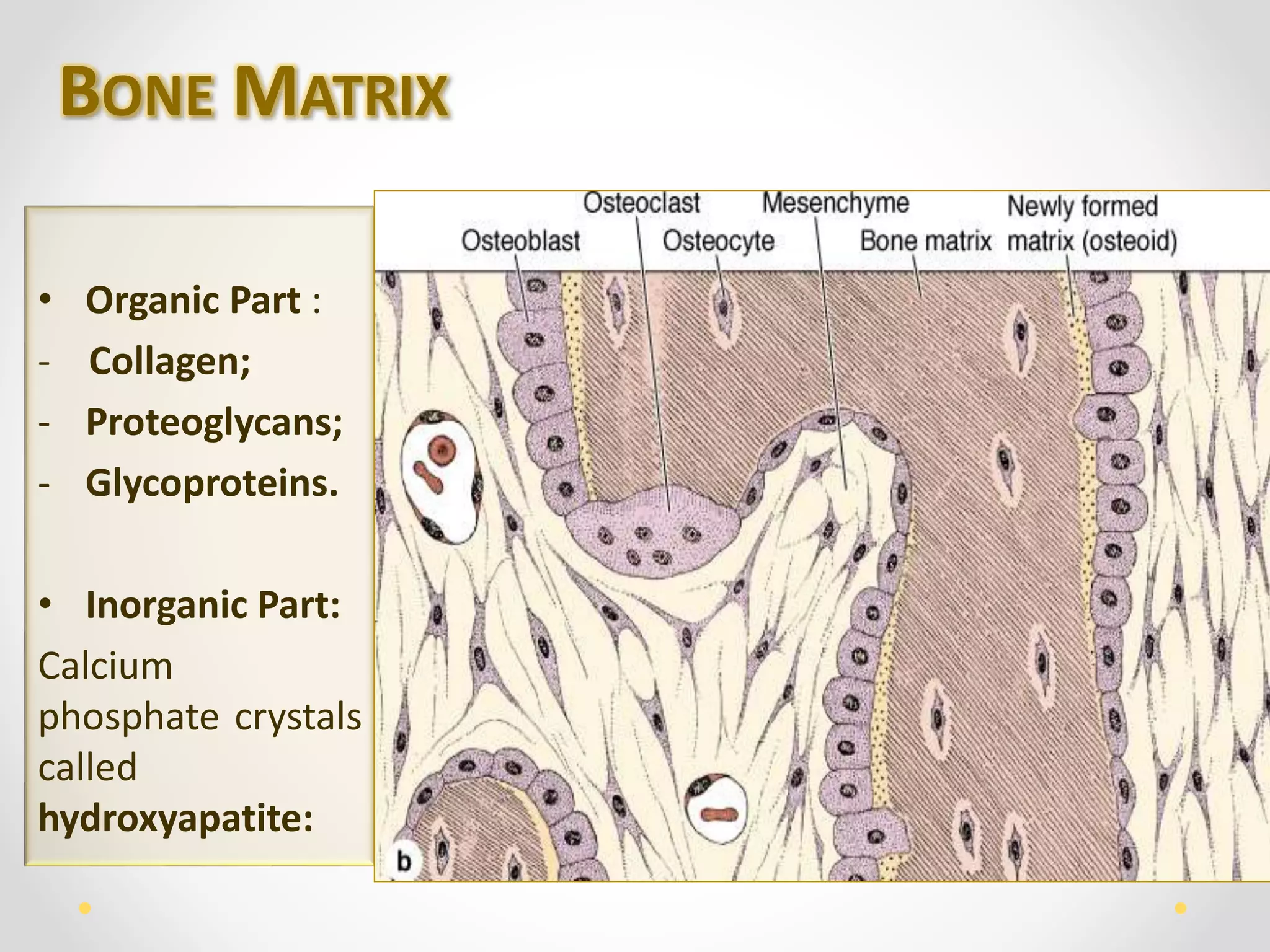
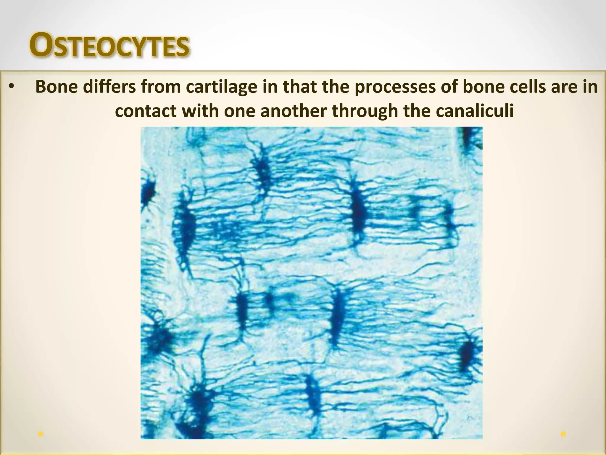
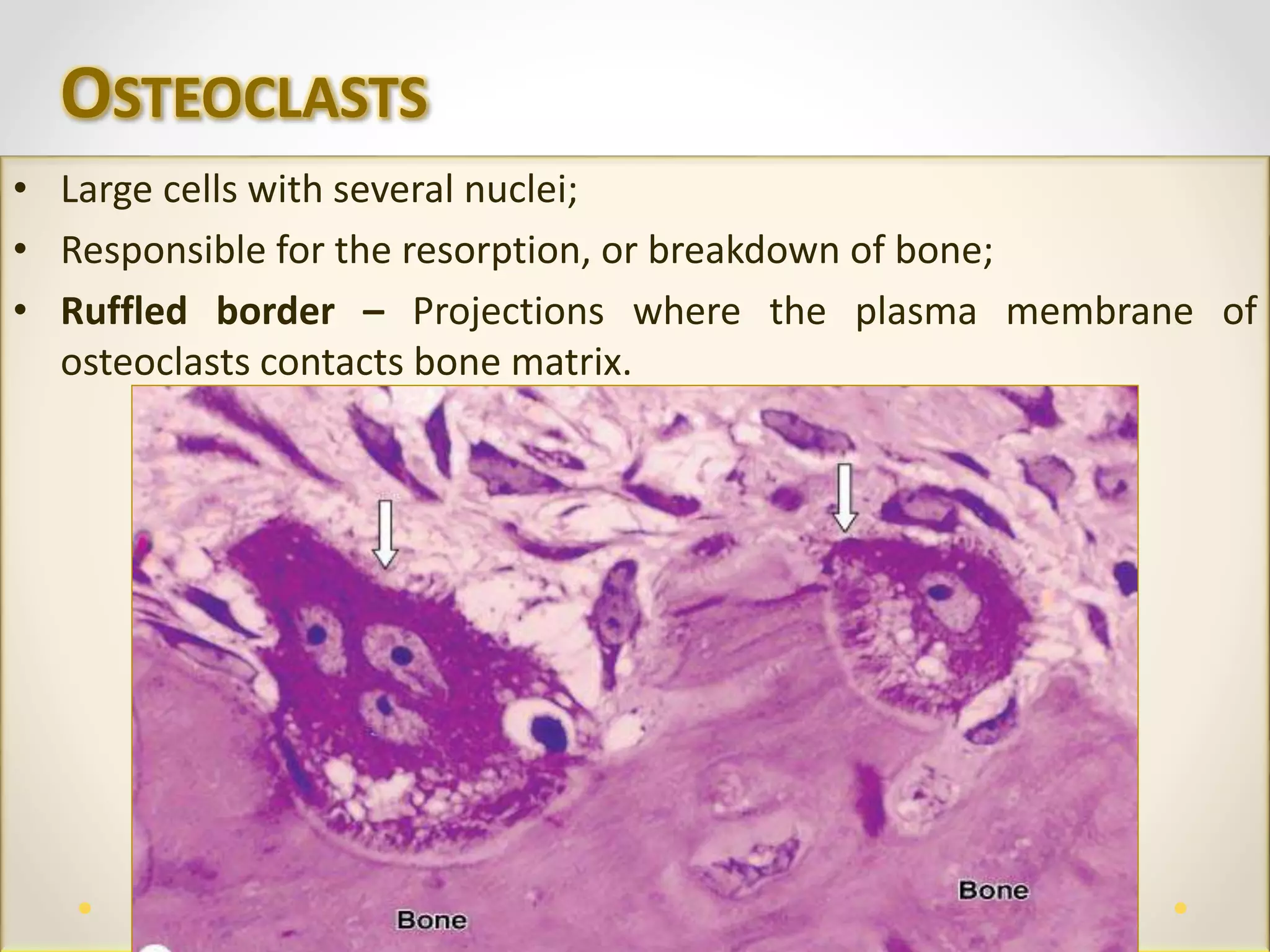
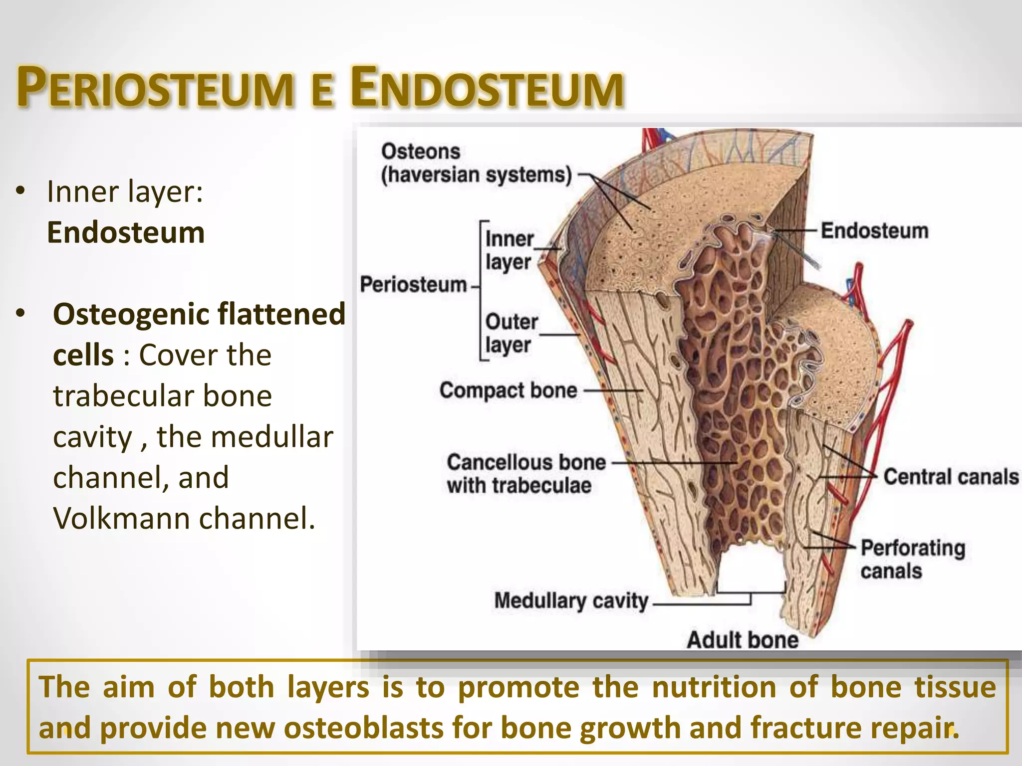
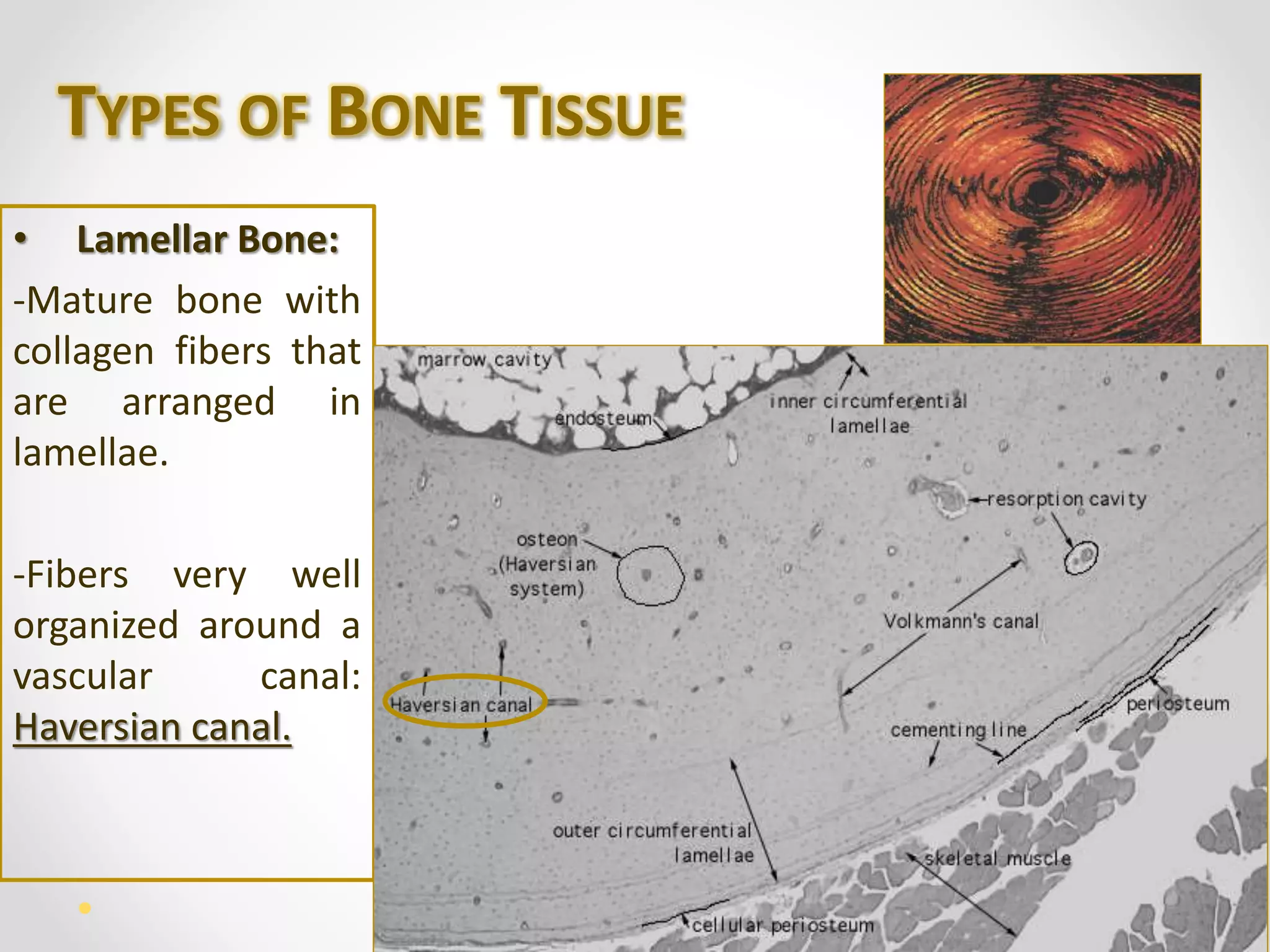
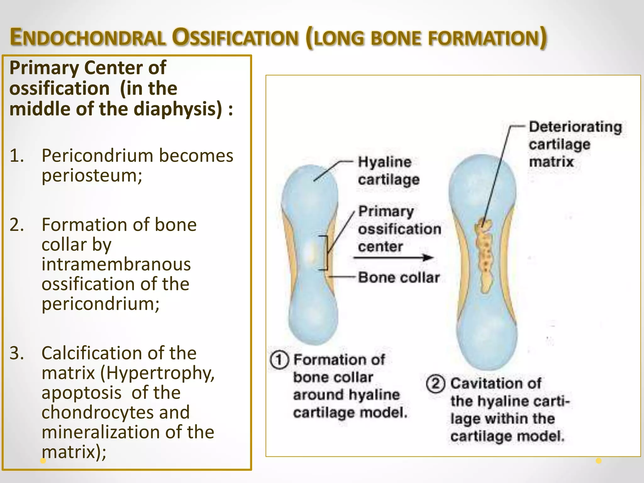
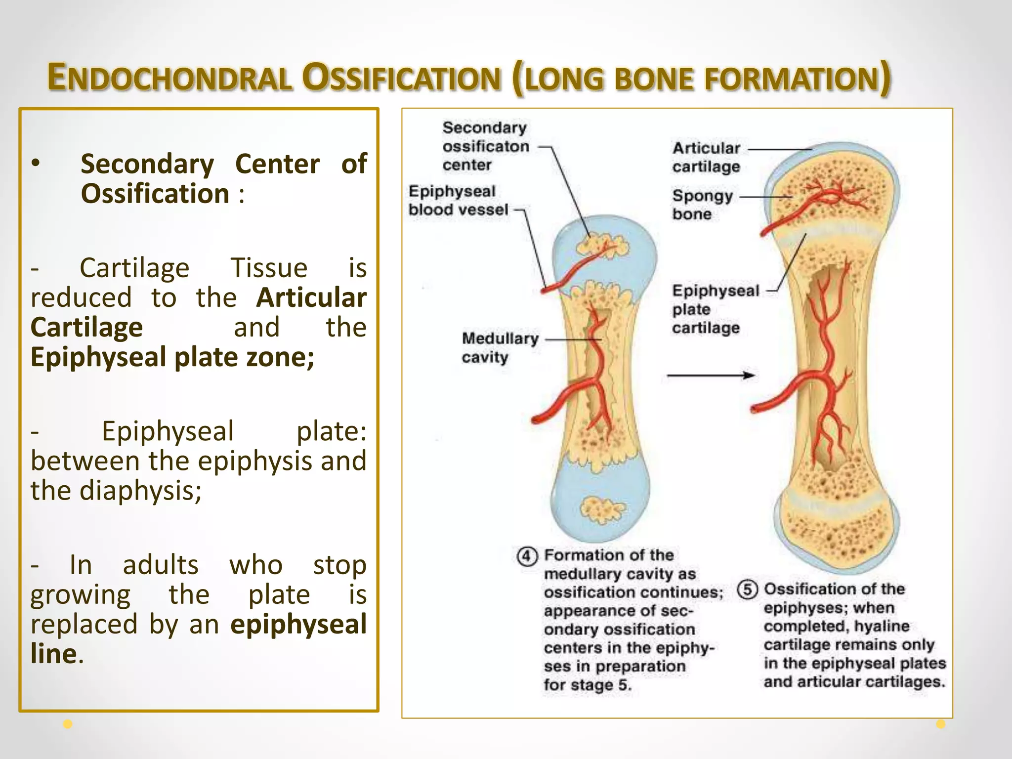
HUMAN BONE 
TISSUE
CONSTITUTION OF BONE TISSUE 
- Extracellular Bone Matrix; 
- Bone Cells. 
Functions: 
1. Support; 
2. Protection; 
3. Movement; 
4. Storage.
BONE MATRIX 
• Organic Part : 
- Collagen; 
- Proteoglycans; 
- Glycoproteins. 
• Inorganic Part: 
Calcium 
phosphate crystals 
called 
hydroxyapatite:
BONE MATRIX 
• The collagen and mineral components: Responsible for the 
major functional characteristics of bone. 
Without 
mineral 
Without 
collagen 
Flexibility Fragile
BONE CELLS 
• Produce the bone matrix, become entrapped within it, and break it down 
so that new matrix can replace the old matrix. 
Bone cells are categorized as: 
 Osteoblasts; Osteocytes; Osteoclasts.
OSTEOBLASTS 
• Extensive endoplasmic reticulum and numerous ribosomes;
OSTEOBLASTS 
• Produce collagen and proteoglycans, which are packaged into vesicles 
by the Golgi apparatus and released from the cell by exocytosis; 
• Form vesicles that accumulate calcium ions (Ca2+), phosphate ions 
(PO2 
4), and various enzymes used to form hydroxyapatite crystals.
OSTEOCYTES 
• They produce components needed to maintain the bone matrix; 
• Lacunae: Spaces occupied 
by the osteocyte cell 
bodies; 
• Canaliculi: Spaces occupied 
by the osteocyte cell 
processes;
OSTEOCYTES 
• Bone differs from cartilage in that the processes of bone cells are in 
contact with one another through the canaliculi
OSTEOCLASTS 
• Large cells with several nuclei; 
• Responsible for the resorption, or breakdown of bone; 
• Ruffled border – Projections where the plasma membrane of 
osteoclasts contacts bone matrix.
OSTEOCLASTS 
• Hydrogen ions are pumped across the ruffled border and produce an 
acid environment : Decalcification of the mineralized bone matrix; 
By endocytosis 
some of the 
breakdown 
products are 
taken into the 
osteoclast. 
BONE REABSORPTION
PERIOSTEUM E ENDOSTEUM 
• Layers of osteogenic cells and conjuntive tissue that covers the 
internal and external surfaces of the bones. 
• Outer layer: periosteum; 
-Collagen fibers – Sharpey's 
fibers penetrate the bone 
and the periosteum hold 
firmly to the bone; 
- Fibroblasts.
PERIOSTEUM E ENDOSTEUM 
• Inner layer: 
Endosteum 
• Osteogenic flattened 
cells : Cover the 
trabecular bone 
cavity , the medullar 
channel, and 
Volkmann channel. 
The aim of both layers is to promote the nutrition of bone tissue 
and provide new osteoblasts for bone growth and fracture repair.
TYPES OF BONE TISSUE 
• Compact bone 
- No visible cavities 
• Cancellous Bone 
- Full of interconnecting channels
TYPES OF BONE TISSUE 
•Short bones (tarsals in the foot, 
e.g.): 
-Cancellous bone in the center; 
-Compact bone recoating all its 
peripheral surface. 
•Flat bones (cranium,e.g.): 
-Two thin layers of compact bone; 
-Enclosing between them: variable 
quantity of cancellous bone. 
•Interstices of the cancellous bone 
and the medullary cavity in the 
diaphysis of the long bones are 
occupied by bone marrow.
TYPES OF BONE TISSUE 
• Primary Bone 
(Temporary): 
- First bone tissue that 
appears in 
embryonic 
development and in 
fraction repair. 
- Collagen fibers are 
arranged in irregular 
arrays.
TYPES OF BONE TISSUE 
• Lamellar Bone: 
-Mature bone with 
collagen fibers that 
are arranged in 
lamellae. 
-Fibers very well 
organized around a 
vascular canal: 
Haversian canal.
OSTEOGENESIS 
• Intramembranous ossification: 
-In which osteoblasts differentiate directly from mesenchyme and begin 
secreting osteoid; 
-Most flat bones are produced and is so called because it takes place 
within condensations of embryonic mesenchymal tissue. 
Groups of 
mesenchymal cells in 
a "membrane" or 
sheet of this 
embryonic tissue, 
round up and 
differentiate as 
osteoblasts producing 
osteoid.
INTRAMEMBRANOUS OSSIFICATION: 
Cells trapped in the 
calcifying matrix 
differentiate as 
osteocytes. 
Woven bone is produced in 
this manner, with 
vascularized internal 
spaces that will form the 
marrow cavity and 
surrounded on both sides 
by developing periosteum.
INTRAMEMBRANOUS OSSIFICATION: 
Remodeling of the 
woven bone produces 
the 
two layers of compact 
lamellar bone with 
cancellous bone in 
between, which is 
characteristic of flat 
bones. 
• The starting point for bone formation is 
called a Primary ossification center. 
• The ossification centers of a bone grow 
radially and finally fuse together, replacing 
the original connective tissue.
OSTEOGENESIS 
• Endochondral ossification: 
- Cartilage is present during the process; 
- It is the process responsible for the formation of short and long bones.
ENDOCHONDRAL OSSIFICATION (LONG BONE FORMATION) 
Primary Center of 
ossification (in the 
middle of the diaphysis) : 
1. Pericondrium becomes 
periosteum; 
2. Formation of bone 
collar by 
intramembranous 
ossification of the 
pericondrium; 
3. Calcification of the 
matrix (Hypertrophy, 
apoptosis of the 
chondrocytes and 
mineralization of the 
matrix);
ENDOCHONDRAL OSSIFICATION (LONG BONE FORMATION) 
4. 
The blood vessels 
from the 
periosteum which 
carry 
osteoprogenitor 
cells: 
Invade the cavity 
left by the 
chondrocytes and 
the cells proliferate 
and differentiate 
into osteoblasts;
ENDOCHONDRAL OSSIFICATION (LONG BONE FORMATION) 
5. Formation of the 
trabeculae (Primary 
bone): 
The osteoblasts secrete 
osteoid which will 
mineralize and form 
trabecula. 
Osteoclasts break down 
spongy bone to form 
the medullary cavity .
ENDOCHONDRAL OSSIFICATION (LONG BONE FORMATION) 
• Secondary Center of 
Ossification : 
- Cartilage Tissue is 
reduced to the Articular 
Cartilage and the 
Epiphyseal plate zone; 
- Epiphyseal plate: 
between the epiphysis and 
the diaphysis; 
- In adults who stop 
growing the plate is 
replaced by an epiphyseal 
line.
ENDOCHONDRAL OSSIFICATION (LONG BONE FORMATION) 
In the epiphyseal plate, five layers are distinguished: 
Normal resting hyaline 
cartilage 
Chondrocytes undergo rapid 
mitosis 
Chondrocytes stop mitosis 
and begin to hypertrophy 
Chondrocytes are either 
dying or dead 
Osteoprogenitor cells invade the 
area and differentiate into 
osteoblasts
BONE REMODELING 
When bone becomes old and it’s replaced by new bone 
Osteoclasts: remove old bone 
Osteoblasts: deposit new bone
BONE REMODELING 
It is also 
responsible 
for the 
formation of 
new 
osteons in 
compact 
bone
BONE REMODELING – NEW OSTEONS 
Within already 
existing osteons: 
- Osteoclasts enter 
a central canal 
through the blood 
vessels, remove 
bone from the 
center of the 
osteon; 
- New concentric 
lamellae are formed 
around the vessels.
BONE REMODELING – NEW OSTEONS 
Osteoclasts in the periosteum: 
- Remove bone: groove formation along the surface; 
- Periosteal capillaries lie within these grooves and become surrounded to 
form a tunnel as the osteoblasts of the periosteum form new bone; 
- Additional lamellae then are added to the inside of the tunnel until an osteon 
results.
BONE REMODELING 
Bone is constantly being removed by osteoclasts, and new bone 
is being formed by osteoblasts: 
• The relative 
thickness of 
compact bone is 
maintained; 
• It leaves behind 
portions of older 
bone - interstitial 
lamellae.
BONE REMODELING 
It can modify the strength of the bone in response to the amount of stress 
applied to it. 
Mechanical stress 
applied to bone 
increases osteoblast 
activity 
• Applying weight to a 
broken bone speeds 
the healing process. 
Removal of mechanical 
stress decreases 
osteoblast activity
BONE REPAIR 
Bone can undergo repair following damage to it in four major steps 
1. Hematoma formation: 
- The blood vessels and surrounding periosteum are damaged: a 
hematoma forms; 
- The blood in a hematoma forms a clot .
BONE REPAIR 
2. Callus formation: 
- Callus : mass of tissue that forms at a fracture site and connects 
the broken ends of the bone.
BONE REPAIR 
2. Callus formation: 
- Internal callus: fibers and cartilage; 
- External callus: Bone–cartilage collar that stabilizes the ends of the 
broken bone .
BONE REPAIR 
3. Callus ossification: 
- Cartilage in the external callus and fibers and cartilage of the 
internal callus: replaced by woven, cancellous bone
BONE REPAIR 
4. Remodeling of bone 
- The woven bone of the internal callus and the dead bone adjacent to 
the fracture site are replaced by compact bone; 
- The internal callus is remodeled and the external callus is reduced in 
size by osteoclast activity.
CALCIUM HOMEOSTASIS 
Calcium moves into bone as osteoblasts build new bone 
and out of bone as osteoclasts break down bone 
Blood calcium 
levels too low 
Osteoclast 
activity 
increases 
More calcium is 
released by 
osteoclasts from 
bone into the 
blood 
Blood calcium 
levels increase 
Blood calcium 
levels too high 
Osteoclast 
activity 
decreases 
Less calcium is 
released by 
osteoclasts from 
bone into the 
blood 
Blood calcium 
levels decrease
CALCIUM HOMEOSTASIS 
Parathyroid hormone (PTH) from the parathyroid glands 
is the major regulator of blood calcium levels.
CALCIUM HOMEOSTASIS 
• PTH binds to its receptors on osteoblasts/stromal cells and these cells 
produce a receptor for activation of nuclear factor kappa B ligand (RANKL)- 
stimulating osteoclasts molecule
CALCIUM HOMEOSTASIS 
• Increased PTH inhibits the release from osteoblasts/stromal cells of a 
protein called osteoprotegerin (OPG) that inhibits the formation of 
osteoclasts because it binds to RANKL 
↑ PTH 
↑ RANKL 
↓ OPG 
↑ Osteoclasts 
↑ Calcium 
in the blood
CALCIUM HOMEOSTASIS 
• Calcitonin plays a minor role in calcium maintenance by inhibiting 
osteoclast activity
2. In the kidneys, 
PTH increases 
calcium 
reabsorption from 
the urine. 
. And promotes the 
formation of active 
vitamin D, which 
increases calcium 
absorption from the 
small intestine.
BONE DISORDERS 
Osteopetrosis Defect in the formation of osteoclasts. The bones harden, becoming denser. 
Rickets 
Softening of bones in children due to deficiency or impaired metabolism of 
Vitamin D. 
Osteomalacia 
Softening of the bones caused by defective bone mineralization secondary to 
inadequate amounts of available phosphorus and calcium. 
Osteoporosis 
Bones become fragile and more likely to fracture. The bone loses density, which 
measures the amount of calcium and minerals in the bone. 
Cretinism 
Stunted physical and mental growth due to untreated congenital deficiency of 
thyroid hormones . 
Giantism 
Abnormally increased height because of excessive cartilage and bone formation 
at the epiphyseal plates of long bones. 
Acromegaly 
Excess pituitary growth hormone secretion; it involves growth of connective 
tissue after the epiphyseal plates have ossified. 
Dwarfism A person is abnormally short, is the opposite of giantism
THANK YOU! 
Follow Us: 
http://biomedeeks.blogspot.pt/

Human Bone Tissue

  • 1.
  • 2.
    CONSTITUTION OF BONETISSUE - Extracellular Bone Matrix; - Bone Cells. Functions: 1. Support; 2. Protection; 3. Movement; 4. Storage.
  • 3.
    BONE MATRIX •Organic Part : - Collagen; - Proteoglycans; - Glycoproteins. • Inorganic Part: Calcium phosphate crystals called hydroxyapatite:
  • 4.
    BONE MATRIX •The collagen and mineral components: Responsible for the major functional characteristics of bone. Without mineral Without collagen Flexibility Fragile
  • 5.
    BONE CELLS •Produce the bone matrix, become entrapped within it, and break it down so that new matrix can replace the old matrix. Bone cells are categorized as:  Osteoblasts; Osteocytes; Osteoclasts.
  • 6.
    OSTEOBLASTS • Extensiveendoplasmic reticulum and numerous ribosomes;
  • 7.
    OSTEOBLASTS • Producecollagen and proteoglycans, which are packaged into vesicles by the Golgi apparatus and released from the cell by exocytosis; • Form vesicles that accumulate calcium ions (Ca2+), phosphate ions (PO2 4), and various enzymes used to form hydroxyapatite crystals.
  • 8.
    OSTEOCYTES • Theyproduce components needed to maintain the bone matrix; • Lacunae: Spaces occupied by the osteocyte cell bodies; • Canaliculi: Spaces occupied by the osteocyte cell processes;
  • 9.
    OSTEOCYTES • Bonediffers from cartilage in that the processes of bone cells are in contact with one another through the canaliculi
  • 10.
    OSTEOCLASTS • Largecells with several nuclei; • Responsible for the resorption, or breakdown of bone; • Ruffled border – Projections where the plasma membrane of osteoclasts contacts bone matrix.
  • 11.
    OSTEOCLASTS • Hydrogenions are pumped across the ruffled border and produce an acid environment : Decalcification of the mineralized bone matrix; By endocytosis some of the breakdown products are taken into the osteoclast. BONE REABSORPTION
  • 12.
    PERIOSTEUM E ENDOSTEUM • Layers of osteogenic cells and conjuntive tissue that covers the internal and external surfaces of the bones. • Outer layer: periosteum; -Collagen fibers – Sharpey's fibers penetrate the bone and the periosteum hold firmly to the bone; - Fibroblasts.
  • 13.
    PERIOSTEUM E ENDOSTEUM • Inner layer: Endosteum • Osteogenic flattened cells : Cover the trabecular bone cavity , the medullar channel, and Volkmann channel. The aim of both layers is to promote the nutrition of bone tissue and provide new osteoblasts for bone growth and fracture repair.
  • 14.
    TYPES OF BONETISSUE • Compact bone - No visible cavities • Cancellous Bone - Full of interconnecting channels
  • 15.
    TYPES OF BONETISSUE •Short bones (tarsals in the foot, e.g.): -Cancellous bone in the center; -Compact bone recoating all its peripheral surface. •Flat bones (cranium,e.g.): -Two thin layers of compact bone; -Enclosing between them: variable quantity of cancellous bone. •Interstices of the cancellous bone and the medullary cavity in the diaphysis of the long bones are occupied by bone marrow.
  • 16.
    TYPES OF BONETISSUE • Primary Bone (Temporary): - First bone tissue that appears in embryonic development and in fraction repair. - Collagen fibers are arranged in irregular arrays.
  • 17.
    TYPES OF BONETISSUE • Lamellar Bone: -Mature bone with collagen fibers that are arranged in lamellae. -Fibers very well organized around a vascular canal: Haversian canal.
  • 18.
    OSTEOGENESIS • Intramembranousossification: -In which osteoblasts differentiate directly from mesenchyme and begin secreting osteoid; -Most flat bones are produced and is so called because it takes place within condensations of embryonic mesenchymal tissue. Groups of mesenchymal cells in a "membrane" or sheet of this embryonic tissue, round up and differentiate as osteoblasts producing osteoid.
  • 19.
    INTRAMEMBRANOUS OSSIFICATION: Cellstrapped in the calcifying matrix differentiate as osteocytes. Woven bone is produced in this manner, with vascularized internal spaces that will form the marrow cavity and surrounded on both sides by developing periosteum.
  • 20.
    INTRAMEMBRANOUS OSSIFICATION: Remodelingof the woven bone produces the two layers of compact lamellar bone with cancellous bone in between, which is characteristic of flat bones. • The starting point for bone formation is called a Primary ossification center. • The ossification centers of a bone grow radially and finally fuse together, replacing the original connective tissue.
  • 21.
    OSTEOGENESIS • Endochondralossification: - Cartilage is present during the process; - It is the process responsible for the formation of short and long bones.
  • 22.
    ENDOCHONDRAL OSSIFICATION (LONGBONE FORMATION) Primary Center of ossification (in the middle of the diaphysis) : 1. Pericondrium becomes periosteum; 2. Formation of bone collar by intramembranous ossification of the pericondrium; 3. Calcification of the matrix (Hypertrophy, apoptosis of the chondrocytes and mineralization of the matrix);
  • 23.
    ENDOCHONDRAL OSSIFICATION (LONGBONE FORMATION) 4. The blood vessels from the periosteum which carry osteoprogenitor cells: Invade the cavity left by the chondrocytes and the cells proliferate and differentiate into osteoblasts;
  • 24.
    ENDOCHONDRAL OSSIFICATION (LONGBONE FORMATION) 5. Formation of the trabeculae (Primary bone): The osteoblasts secrete osteoid which will mineralize and form trabecula. Osteoclasts break down spongy bone to form the medullary cavity .
  • 25.
    ENDOCHONDRAL OSSIFICATION (LONGBONE FORMATION) • Secondary Center of Ossification : - Cartilage Tissue is reduced to the Articular Cartilage and the Epiphyseal plate zone; - Epiphyseal plate: between the epiphysis and the diaphysis; - In adults who stop growing the plate is replaced by an epiphyseal line.
  • 26.
    ENDOCHONDRAL OSSIFICATION (LONGBONE FORMATION) In the epiphyseal plate, five layers are distinguished: Normal resting hyaline cartilage Chondrocytes undergo rapid mitosis Chondrocytes stop mitosis and begin to hypertrophy Chondrocytes are either dying or dead Osteoprogenitor cells invade the area and differentiate into osteoblasts
  • 27.
    BONE REMODELING Whenbone becomes old and it’s replaced by new bone Osteoclasts: remove old bone Osteoblasts: deposit new bone
  • 28.
    BONE REMODELING Itis also responsible for the formation of new osteons in compact bone
  • 29.
    BONE REMODELING –NEW OSTEONS Within already existing osteons: - Osteoclasts enter a central canal through the blood vessels, remove bone from the center of the osteon; - New concentric lamellae are formed around the vessels.
  • 30.
    BONE REMODELING –NEW OSTEONS Osteoclasts in the periosteum: - Remove bone: groove formation along the surface; - Periosteal capillaries lie within these grooves and become surrounded to form a tunnel as the osteoblasts of the periosteum form new bone; - Additional lamellae then are added to the inside of the tunnel until an osteon results.
  • 31.
    BONE REMODELING Boneis constantly being removed by osteoclasts, and new bone is being formed by osteoblasts: • The relative thickness of compact bone is maintained; • It leaves behind portions of older bone - interstitial lamellae.
  • 32.
    BONE REMODELING Itcan modify the strength of the bone in response to the amount of stress applied to it. Mechanical stress applied to bone increases osteoblast activity • Applying weight to a broken bone speeds the healing process. Removal of mechanical stress decreases osteoblast activity
  • 33.
    BONE REPAIR Bonecan undergo repair following damage to it in four major steps 1. Hematoma formation: - The blood vessels and surrounding periosteum are damaged: a hematoma forms; - The blood in a hematoma forms a clot .
  • 34.
    BONE REPAIR 2.Callus formation: - Callus : mass of tissue that forms at a fracture site and connects the broken ends of the bone.
  • 35.
    BONE REPAIR 2.Callus formation: - Internal callus: fibers and cartilage; - External callus: Bone–cartilage collar that stabilizes the ends of the broken bone .
  • 36.
    BONE REPAIR 3.Callus ossification: - Cartilage in the external callus and fibers and cartilage of the internal callus: replaced by woven, cancellous bone
  • 37.
    BONE REPAIR 4.Remodeling of bone - The woven bone of the internal callus and the dead bone adjacent to the fracture site are replaced by compact bone; - The internal callus is remodeled and the external callus is reduced in size by osteoclast activity.
  • 38.
    CALCIUM HOMEOSTASIS Calciummoves into bone as osteoblasts build new bone and out of bone as osteoclasts break down bone Blood calcium levels too low Osteoclast activity increases More calcium is released by osteoclasts from bone into the blood Blood calcium levels increase Blood calcium levels too high Osteoclast activity decreases Less calcium is released by osteoclasts from bone into the blood Blood calcium levels decrease
  • 39.
    CALCIUM HOMEOSTASIS Parathyroidhormone (PTH) from the parathyroid glands is the major regulator of blood calcium levels.
  • 40.
    CALCIUM HOMEOSTASIS •PTH binds to its receptors on osteoblasts/stromal cells and these cells produce a receptor for activation of nuclear factor kappa B ligand (RANKL)- stimulating osteoclasts molecule
  • 41.
    CALCIUM HOMEOSTASIS •Increased PTH inhibits the release from osteoblasts/stromal cells of a protein called osteoprotegerin (OPG) that inhibits the formation of osteoclasts because it binds to RANKL ↑ PTH ↑ RANKL ↓ OPG ↑ Osteoclasts ↑ Calcium in the blood
  • 42.
    CALCIUM HOMEOSTASIS •Calcitonin plays a minor role in calcium maintenance by inhibiting osteoclast activity
  • 43.
    2. In thekidneys, PTH increases calcium reabsorption from the urine. . And promotes the formation of active vitamin D, which increases calcium absorption from the small intestine.
  • 44.
    BONE DISORDERS OsteopetrosisDefect in the formation of osteoclasts. The bones harden, becoming denser. Rickets Softening of bones in children due to deficiency or impaired metabolism of Vitamin D. Osteomalacia Softening of the bones caused by defective bone mineralization secondary to inadequate amounts of available phosphorus and calcium. Osteoporosis Bones become fragile and more likely to fracture. The bone loses density, which measures the amount of calcium and minerals in the bone. Cretinism Stunted physical and mental growth due to untreated congenital deficiency of thyroid hormones . Giantism Abnormally increased height because of excessive cartilage and bone formation at the epiphyseal plates of long bones. Acromegaly Excess pituitary growth hormone secretion; it involves growth of connective tissue after the epiphyseal plates have ossified. Dwarfism A person is abnormally short, is the opposite of giantism
  • 45.
    THANK YOU! FollowUs: http://biomedeeks.blogspot.pt/