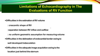
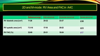
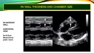
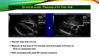
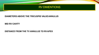
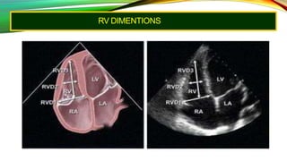
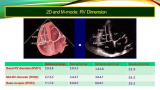
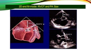
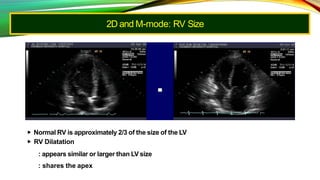
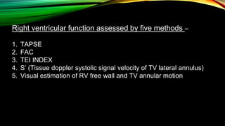
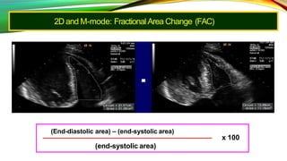
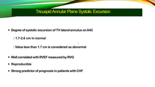
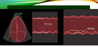
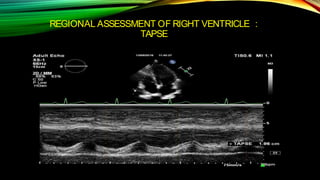
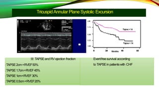
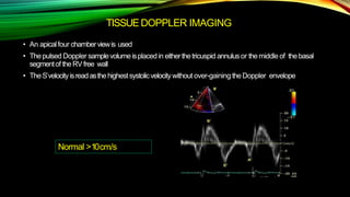
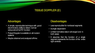
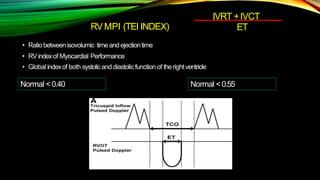
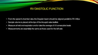
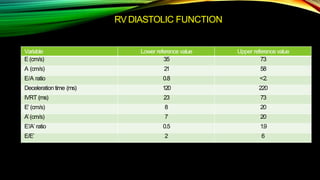
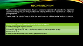
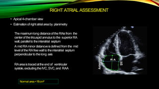
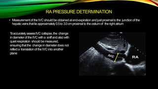
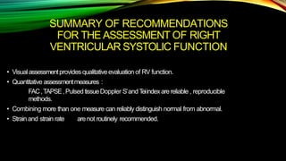
ECHO ASSESMENT OF RV FUNCTION
DR.AZIZULHAQUE
REGISTRARCARDIOLOGY
DHAKAMEDICALCOLLEGE
HOSPITAL
RIGHT VENTRICULAR ANATOMY
Limitations of Echocardiography in The
Evaluations of RV Function
▶Difficulties in the estimation of RV volume
: crescentic shape of RV
:separation between RV inflow and outflow
: no uniform geometric assumption for measuringvolume
▶Difficulties in the delineation of endocardial border owing to
well developed trabeculation
▶Difficulties in the adequate image acquisition owing to the
location just behind the sternum
Limitations of Echocardiography in The
Evaluations of RV Function
▶ Difficult to standardize the evaluation method of RV function
: Variations in the direction or location of the RV are common
: Easily affected by preload, afterload, or LVfunction
▶Different complex contraction-relaxation mechanism among
the segments of the RV
▶Cannot image the entire RV in a single view
Function of the Right Ventricle
Why should we measure RV function?
▶ RV is not just a conduit of blood flow
: has its unique function
▶Prognostic significance in various clinicalsettings
▶Risk stratification or guide to optimal therapy
Function of theRight Ventricle
▶ Maintain adequate pulmonary artery perfusion pressure to
improve gas exchange
▶ Maintain low systemic venous pressure to prevent
congestion of tissues or organs
▶ Affect LV function
: limit LV preload in RV dysfunction
: Ventricular interdependence
▶ Prognostic significance in various clinical settings
2D and M-mode: RV Area and FACin A4C
▶ Wellcorrelated withRV function measured by radionuclide
ventriculography or MRI
▶ Good predictor of prognosis
▶ Limitations: fail to measure FAC due to inadequate RV tracing
Referencerange Mild abnormal Moderate abnormal Severe abnormal
RV diastolic area(cm2) 11-28 29-32 33-37 ≥38
RV systolic area(cm2) 7.5-16 17-19 20-22 ≥23
RV FAC(%) 32-60 25-31 18-24 ≤17
RV WALL THICKNESS AND CHAMBER SIZE
RV INFERIOR
WALL
SUBCOSTAL
VIEW
N=<0.5cm
Measured at
peak r wave
2D and M-mode: Thicknessof RV Free Wall
▶ Normal: less than 0.5 cm
▶ Measure at the level of TV chordae and at the peak of R wave of
ECG on subcostal view
▶ Well correlated with peak RV systolic pressure
RV DIMENTIONS
DIAMETERS ABOVE THE TRICUSPID VALVEANNULUS
MID RV CAVITY
DISTANCE FROM THE TV ANNULUS TO RVAPEX
RV DIMENTIONS
2D and M-mode: RV Dimension
Referencerange Mild abnormal Moderate abnormal Severe abnormal
Basal RV diameter (RVD1) 2.0-2.8 2.9-3.3 3.4-3.8 ≥3.9
Mid-RV diameter (RVD2) 2.7-3.3 3.4-3.7 3.8-4.1 ≥4.2
Base–to-apex (RVD3) 7.1-7.9 8.0-8.5 8.6-9.1 ≥9.2
2D and M-mode: RVOT and PA Size
Referencerange Mild abnormal Moderate abnormal Severeabnormal
RVOT diameters, cm
Above aortic
valve(RVOT1)
2.5-2.9 3.0-3.2 3.3-3.5 ≥3.6
Above pulmonic
valve(RVOT1)
1.7-2.3 2.4-2.7 2.8-3.1 ≥3.2
PA Diameters, cm
Belowpulmonic valve
(PA1)
1.5-2.1 2.2-2.5 2.6-2.9 ≥3.0
2D and M-mode: RV Size
▶ Normal RV is approximately 2/3 of the size of the LV
▶ RV Dilatation
: appears similar or larger than LV size
: shares the apex
Right ventricular function assessed by five methods –
1. TAPSE
2. FAC
3. TEI INDEX
4. S’ (Tissue doppler systolic signal velocity of TV lateral annulus)
5. Visual estimation of RV free wall and TV annular motion
2D and M-mode: Fractional Area Change (FAC)
(End-diastolic area) – (end-systolic area)
x 100
(end-systolic area)
TricuspidAnnular Plane Systolic Excursion
▶ Degree of systolic excursion of TV lateralannulus onA4C
: 1.7-2.6 cm in normal
: Value less than 1.7 cm is considered as abnormal
▶ WellcorrelatedwithRVEF measuredby RVG
▶ Reproducible
▶ Strong predictor of prognosis in patients with CHF
REGIONAL ASSESSMENT OF RIGHT VENTRICLE :
TAPSE
TricuspidAnnular Plane Systolic Excursion
※ TAPSEand RVejection fraction
:TAPSE2cm=RVEF50%
:TAPSE1.7cm=RVEF45%
:TAPSE1cm=RVEF30%
:TAPSE0.5cm=RVEF20%
Eventfreesurvival according
to TAPSEin patientswith CHF
TISSUEDOPPLER IMAGING
• An apicalfour chamberviewis used
• Thepulsed Doppler samplevolumeisplaced in eitherthetricuspid annulusor themiddle of thebasal
segmentof theRVfree wall
• TheS´velocityisreadasthehighestsystolicvelocitywithout over-gaining theDoppler envelope
Normal >10cm/s
TISSUE DOPPLER (S´)
Advantages
• A simple,reproducible techniquewith good
discriminatory ability to detect normal
versusabnormal RV function
• Pulsed Doppler isavailableon allmodern
systems
• Maybe obtained and analyzed off-line
Disadvantages
• Lessreproducible for nonbasal segments
• Isangle dependent
• Limitednormativedatain allranges'and in
both sexes
• It assumes that the function of a single
segment represents the function of the entire
right ventricle
RVMPI (TEI INDEX)
• Ratiobetweenisovolumic timeandejectiontime
• RVindexof Myocardial Performance
• Global indexof both systolicand diastolic function of theright ventricle
IVRT +IVCT
ET
Normal <0.40 Normal <0.55
If Heart rate> 100
Advantages
• Thisapproach isfeasiblein alarge
majorityof subjects
• TheMPI is reproducible
• Itavoidsgeometric assumptionsand
limitations of thecomplexRV geometry
• Thepulsed TDI method allows for
measurementof MPI aswellasS´,E´,and A´all
fromasingle image
Disadvantages
• TheMPI isunreliablewhenRVETandTR time
aremeasuredwithdiffering R-R intervals,asin
atrial fibrillation
• Itisload dependent and unreliablewhen RA
pressuresare elevated
RV DIASTOLIC FUNCTION
• From the apical 4-chamber view,the Doppler beam should be aligned parallel to RV inflow
• Sample volume is placed at the tips of the tricuspid valve leaflets
• Measureatheld end-expiration and/or taketheaverageof ≥5consecutivebeats
• Measurementsare essentiallythe sameasthose used for the left side
RV DIASTOLIC FUNCTION
Variable Lower reference value Upper reference value
E (cm/s) 35 73
A (cm/s) 21 58
E/A ratio 0.8 <2.
Deceleration time (ms) 120 220
IVRT (ms) 23 73
E’ (cm/s) 8 20
A’(cm/s) 7 20
E’/A’ ratio 0.5 1.9
E/E’ 2 6
RECOMMENDATION
• Measurementof RVdiastolic function should be consideredin patientswithsuspectedRV impairment
asamarkerof earlyor subtleRVdysfunction,or in patientswithknownRV impairmentasamarkerfor
poor prognosis
• TranstricupsidE/A ratio,E/E’ratio, and RAsizehavebeen mostvalidatedarethepreferred measures
Grading of RVDiastolic Dysfunctionshould be done as follows:
E/A ratio <0.8suggestsimpaired relaxation
E/A ratio 0.8-<2.0with anE/E’ ratio >6or diastolic prominence in thehepaticveins suggest
pseudonormal filling
E/A ratio >2.with deceleration time<120mssuggestsrestrictivefilling
RIGHT ATRIAL ASSESSMENT
• Apical 4-chamber view
• Estimationof right atrialareaby planimetry
Themaximumlong distanceof theRAis from the
centerof thetricuspid annulusto the superiorRA
wall,parallelto theinteratrial septum
A mid RAminor distancveisdefined from the mid
levelof theRAfreewallto theinteratrial septum
perpendicularto thelong axis
RAareaistracedattheend of ventricular
systole,excluding theIVC, SVC,and RAA
Normal area<18cm²
RA PRESSURE DETERMINATION
• Measurementof theIVC should be obtained atend-expiration and justproximalto the junction of the
hepaticveinsthatlieapproximately0.5to 3.0cmproximalto theostiumof theright atrium
ToaccuratelyassessIVC collapse,the change
in diameterof theIVC witha sniffand also with
quiet respiration should be measured,
ensuringthatthe changein diameterdoes not
reflecta translationof theIVC into another
plane
Estimation of RA pressure from IVC
diameter
IVC SIZE BSA
NORMAL 17 mm <1.55 m2
20 mm 1.55-<1.71 m2
21 mm >1.71 m2
IVC COLLAPSE RAP
size Normal IVC >50% 05 mm hg
Normal IVC <50% 10 mmhg
Dilated IVC >50% 15 mmhg
Dilated IVC <50% 20 mmhg
R V PATHOLOGY
1. RV VOLUME OVERLOAD
2. RV PRESSURE OVERLOAD
3. RV INFARCTION
4. ARVD
5. PULMONARY EMBOLISM
6. CARDIAC TEMPONADE
HEMODYNAMIC ASSESSMENT
Systolic pulmonary artery pressure
• Estimated withTR jet velocity using simplified Bernoulli equation ( provided there is no RVOT
obstruction )
RVSP=4(V)2+RApressure
• Normal peak RVSP is 35 to 36 mmHg assuming RApressure of 3 to 5 mmHg
Note :MeasureTRjetvelocityfromvariousviewsto getthehighest velocity
SYSTOLIC PULMONARY ARTERY
PRESSURE
HEMODYNAMIC ASSESSMENT
Pulmonary artery diastolic pressure(PADP )
•Estimatedfrom velocityof end diastolic pulmonary regurgitant jetusing PADP =
4(V)2+RA pressure
HEMODYNAMIC ASSESSMENT
Mean Pulmonary Pressure
Can be measured :
• MAP =1/3(SPAP)+2/3(PADP)
• = 0.6.PASP+2.1
• =79-0.45.AT
• = 4(EARLYPR
VELOCITY)2+RAPRESSURE
EXCEPTION
If the transducer is not parallel to the flow to the TR jet ,peak velocity of the jet will be reduced
and underestimation of PASP
Incorrectly estimating mean RA pressure from the IVC can lead to under or overestimation of
pulmonary pressure
SUMMARY OF RECOMMENDATIONS
FOR THE ASSESSMENT OF RIGHT
VENTRICULAR SYSTOLIC FUNCTION
• Visualassessmentprovides qualitativeevaluationof RV function.
• Quantitative assessmentmeasures :
FAC,TAPSE,PulsedtissueDoppler S’andTeiindexarereliable , reproducible
methods.
• Combining more than one measure can reliably distinguish normal from abnormal.
• Strainand strain rate arenot routinely recommended.
THANK YOU

Echo assesment of rv function

  • 1.
    ECHO ASSESMENT OFRV FUNCTION DR.AZIZULHAQUE REGISTRARCARDIOLOGY DHAKAMEDICALCOLLEGE HOSPITAL
  • 2.
  • 4.
    Limitations of Echocardiographyin The Evaluations of RV Function ▶Difficulties in the estimation of RV volume : crescentic shape of RV :separation between RV inflow and outflow : no uniform geometric assumption for measuringvolume ▶Difficulties in the delineation of endocardial border owing to well developed trabeculation ▶Difficulties in the adequate image acquisition owing to the location just behind the sternum
  • 5.
    Limitations of Echocardiographyin The Evaluations of RV Function ▶ Difficult to standardize the evaluation method of RV function : Variations in the direction or location of the RV are common : Easily affected by preload, afterload, or LVfunction ▶Different complex contraction-relaxation mechanism among the segments of the RV ▶Cannot image the entire RV in a single view
  • 6.
    Function of theRight Ventricle Why should we measure RV function? ▶ RV is not just a conduit of blood flow : has its unique function ▶Prognostic significance in various clinicalsettings ▶Risk stratification or guide to optimal therapy
  • 7.
    Function of theRightVentricle ▶ Maintain adequate pulmonary artery perfusion pressure to improve gas exchange ▶ Maintain low systemic venous pressure to prevent congestion of tissues or organs ▶ Affect LV function : limit LV preload in RV dysfunction : Ventricular interdependence ▶ Prognostic significance in various clinical settings
  • 8.
    2D and M-mode:RV Area and FACin A4C ▶ Wellcorrelated withRV function measured by radionuclide ventriculography or MRI ▶ Good predictor of prognosis ▶ Limitations: fail to measure FAC due to inadequate RV tracing Referencerange Mild abnormal Moderate abnormal Severe abnormal RV diastolic area(cm2) 11-28 29-32 33-37 ≥38 RV systolic area(cm2) 7.5-16 17-19 20-22 ≥23 RV FAC(%) 32-60 25-31 18-24 ≤17
  • 9.
    RV WALL THICKNESSAND CHAMBER SIZE RV INFERIOR WALL SUBCOSTAL VIEW N=<0.5cm Measured at peak r wave
  • 10.
    2D and M-mode:Thicknessof RV Free Wall ▶ Normal: less than 0.5 cm ▶ Measure at the level of TV chordae and at the peak of R wave of ECG on subcostal view ▶ Well correlated with peak RV systolic pressure
  • 11.
    RV DIMENTIONS DIAMETERS ABOVETHE TRICUSPID VALVEANNULUS MID RV CAVITY DISTANCE FROM THE TV ANNULUS TO RVAPEX
  • 12.
  • 13.
    2D and M-mode:RV Dimension Referencerange Mild abnormal Moderate abnormal Severe abnormal Basal RV diameter (RVD1) 2.0-2.8 2.9-3.3 3.4-3.8 ≥3.9 Mid-RV diameter (RVD2) 2.7-3.3 3.4-3.7 3.8-4.1 ≥4.2 Base–to-apex (RVD3) 7.1-7.9 8.0-8.5 8.6-9.1 ≥9.2
  • 14.
    2D and M-mode:RVOT and PA Size
  • 15.
    2D and M-mode:RVOT and PA Size Referencerange Mild abnormal Moderate abnormal Severeabnormal RVOT diameters, cm Above aortic valve(RVOT1) 2.5-2.9 3.0-3.2 3.3-3.5 ≥3.6 Above pulmonic valve(RVOT1) 1.7-2.3 2.4-2.7 2.8-3.1 ≥3.2 PA Diameters, cm Belowpulmonic valve (PA1) 1.5-2.1 2.2-2.5 2.6-2.9 ≥3.0
  • 16.
    2D and M-mode:RV Size ▶ Normal RV is approximately 2/3 of the size of the LV ▶ RV Dilatation : appears similar or larger than LV size : shares the apex
  • 17.
    Right ventricular functionassessed by five methods – 1. TAPSE 2. FAC 3. TEI INDEX 4. S’ (Tissue doppler systolic signal velocity of TV lateral annulus) 5. Visual estimation of RV free wall and TV annular motion
  • 18.
    2D and M-mode:Fractional Area Change (FAC) (End-diastolic area) – (end-systolic area) x 100 (end-systolic area)
  • 19.
    TricuspidAnnular Plane SystolicExcursion ▶ Degree of systolic excursion of TV lateralannulus onA4C : 1.7-2.6 cm in normal : Value less than 1.7 cm is considered as abnormal ▶ WellcorrelatedwithRVEF measuredby RVG ▶ Reproducible ▶ Strong predictor of prognosis in patients with CHF
  • 21.
    REGIONAL ASSESSMENT OFRIGHT VENTRICLE : TAPSE
  • 22.
    TricuspidAnnular Plane SystolicExcursion ※ TAPSEand RVejection fraction :TAPSE2cm=RVEF50% :TAPSE1.7cm=RVEF45% :TAPSE1cm=RVEF30% :TAPSE0.5cm=RVEF20% Eventfreesurvival according to TAPSEin patientswith CHF
  • 23.
    TISSUEDOPPLER IMAGING • Anapicalfour chamberviewis used • Thepulsed Doppler samplevolumeisplaced in eitherthetricuspid annulusor themiddle of thebasal segmentof theRVfree wall • TheS´velocityisreadasthehighestsystolicvelocitywithout over-gaining theDoppler envelope Normal >10cm/s
  • 24.
    TISSUE DOPPLER (S´) Advantages •A simple,reproducible techniquewith good discriminatory ability to detect normal versusabnormal RV function • Pulsed Doppler isavailableon allmodern systems • Maybe obtained and analyzed off-line Disadvantages • Lessreproducible for nonbasal segments • Isangle dependent • Limitednormativedatain allranges'and in both sexes • It assumes that the function of a single segment represents the function of the entire right ventricle
  • 25.
    RVMPI (TEI INDEX) •Ratiobetweenisovolumic timeandejectiontime • RVindexof Myocardial Performance • Global indexof both systolicand diastolic function of theright ventricle IVRT +IVCT ET Normal <0.40 Normal <0.55
  • 27.
  • 28.
    Advantages • Thisapproach isfeasibleinalarge majorityof subjects • TheMPI is reproducible • Itavoidsgeometric assumptionsand limitations of thecomplexRV geometry • Thepulsed TDI method allows for measurementof MPI aswellasS´,E´,and A´all fromasingle image Disadvantages • TheMPI isunreliablewhenRVETandTR time aremeasuredwithdiffering R-R intervals,asin atrial fibrillation • Itisload dependent and unreliablewhen RA pressuresare elevated
  • 29.
    RV DIASTOLIC FUNCTION •From the apical 4-chamber view,the Doppler beam should be aligned parallel to RV inflow • Sample volume is placed at the tips of the tricuspid valve leaflets • Measureatheld end-expiration and/or taketheaverageof ≥5consecutivebeats • Measurementsare essentiallythe sameasthose used for the left side
  • 30.
    RV DIASTOLIC FUNCTION VariableLower reference value Upper reference value E (cm/s) 35 73 A (cm/s) 21 58 E/A ratio 0.8 <2. Deceleration time (ms) 120 220 IVRT (ms) 23 73 E’ (cm/s) 8 20 A’(cm/s) 7 20 E’/A’ ratio 0.5 1.9 E/E’ 2 6
  • 31.
    RECOMMENDATION • Measurementof RVdiastolicfunction should be consideredin patientswithsuspectedRV impairment asamarkerof earlyor subtleRVdysfunction,or in patientswithknownRV impairmentasamarkerfor poor prognosis • TranstricupsidE/A ratio,E/E’ratio, and RAsizehavebeen mostvalidatedarethepreferred measures Grading of RVDiastolic Dysfunctionshould be done as follows: E/A ratio <0.8suggestsimpaired relaxation E/A ratio 0.8-<2.0with anE/E’ ratio >6or diastolic prominence in thehepaticveins suggest pseudonormal filling E/A ratio >2.with deceleration time<120mssuggestsrestrictivefilling
  • 32.
    RIGHT ATRIAL ASSESSMENT •Apical 4-chamber view • Estimationof right atrialareaby planimetry Themaximumlong distanceof theRAis from the centerof thetricuspid annulusto the superiorRA wall,parallelto theinteratrial septum A mid RAminor distancveisdefined from the mid levelof theRAfreewallto theinteratrial septum perpendicularto thelong axis RAareaistracedattheend of ventricular systole,excluding theIVC, SVC,and RAA Normal area<18cm²
  • 33.
    RA PRESSURE DETERMINATION •Measurementof theIVC should be obtained atend-expiration and justproximalto the junction of the hepaticveinsthatlieapproximately0.5to 3.0cmproximalto theostiumof theright atrium ToaccuratelyassessIVC collapse,the change in diameterof theIVC witha sniffand also with quiet respiration should be measured, ensuringthatthe changein diameterdoes not reflecta translationof theIVC into another plane
  • 34.
    Estimation of RApressure from IVC diameter IVC SIZE BSA NORMAL 17 mm <1.55 m2 20 mm 1.55-<1.71 m2 21 mm >1.71 m2 IVC COLLAPSE RAP size Normal IVC >50% 05 mm hg Normal IVC <50% 10 mmhg Dilated IVC >50% 15 mmhg Dilated IVC <50% 20 mmhg
  • 35.
    R V PATHOLOGY 1.RV VOLUME OVERLOAD 2. RV PRESSURE OVERLOAD 3. RV INFARCTION 4. ARVD 5. PULMONARY EMBOLISM 6. CARDIAC TEMPONADE
  • 36.
    HEMODYNAMIC ASSESSMENT Systolic pulmonaryartery pressure • Estimated withTR jet velocity using simplified Bernoulli equation ( provided there is no RVOT obstruction ) RVSP=4(V)2+RApressure • Normal peak RVSP is 35 to 36 mmHg assuming RApressure of 3 to 5 mmHg Note :MeasureTRjetvelocityfromvariousviewsto getthehighest velocity
  • 37.
  • 38.
    HEMODYNAMIC ASSESSMENT Pulmonary arterydiastolic pressure(PADP ) •Estimatedfrom velocityof end diastolic pulmonary regurgitant jetusing PADP = 4(V)2+RA pressure
  • 39.
    HEMODYNAMIC ASSESSMENT Mean PulmonaryPressure Can be measured : • MAP =1/3(SPAP)+2/3(PADP) • = 0.6.PASP+2.1 • =79-0.45.AT • = 4(EARLYPR VELOCITY)2+RAPRESSURE
  • 40.
    EXCEPTION If the transduceris not parallel to the flow to the TR jet ,peak velocity of the jet will be reduced and underestimation of PASP Incorrectly estimating mean RA pressure from the IVC can lead to under or overestimation of pulmonary pressure
  • 41.
    SUMMARY OF RECOMMENDATIONS FORTHE ASSESSMENT OF RIGHT VENTRICULAR SYSTOLIC FUNCTION • Visualassessmentprovides qualitativeevaluationof RV function. • Quantitative assessmentmeasures : FAC,TAPSE,PulsedtissueDoppler S’andTeiindexarereliable , reproducible methods. • Combining more than one measure can reliably distinguish normal from abnormal. • Strainand strain rate arenot routinely recommended.
  • 42.