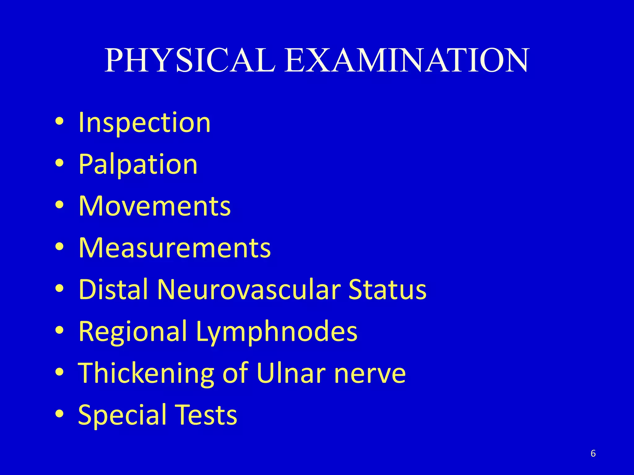
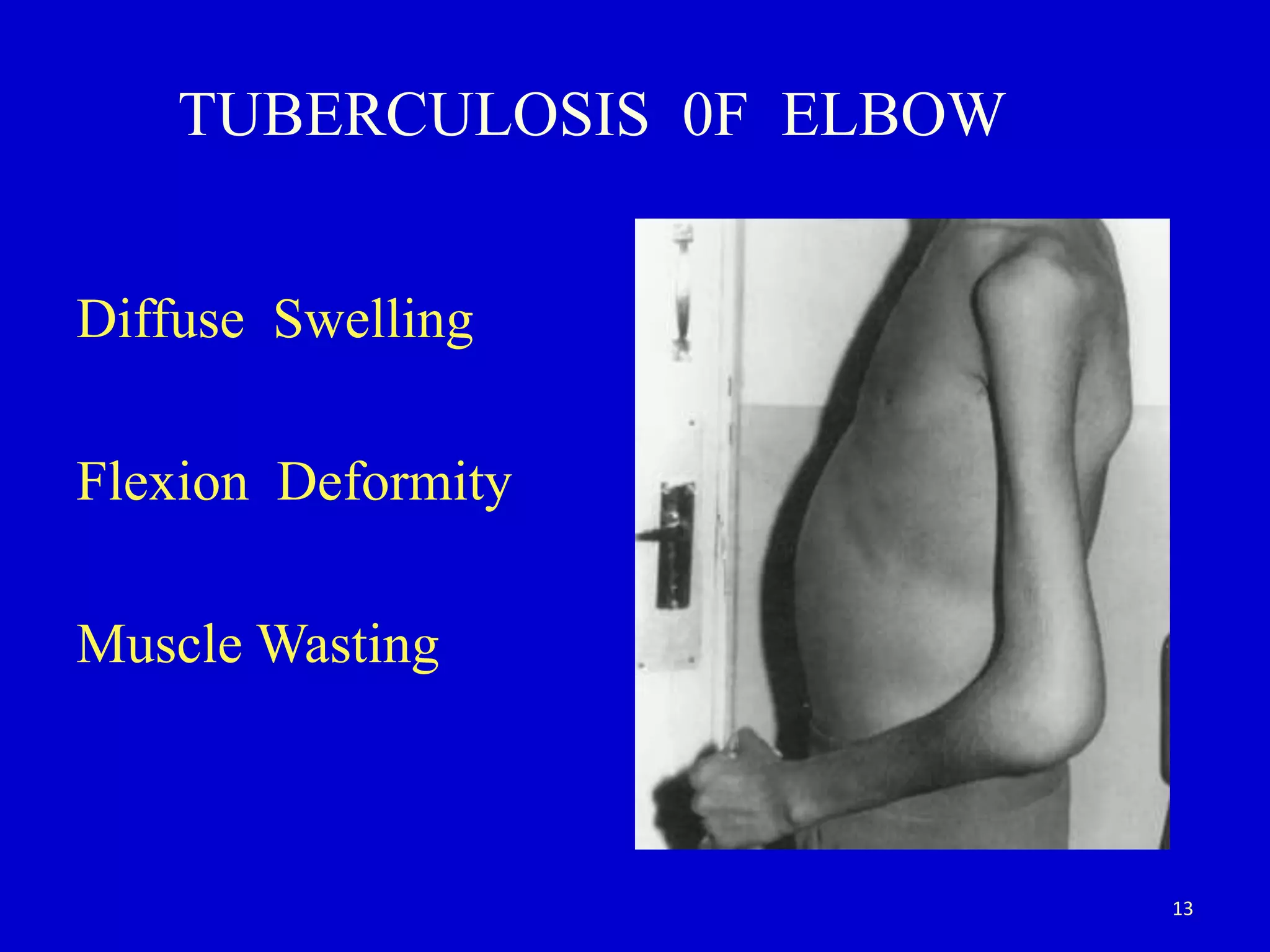
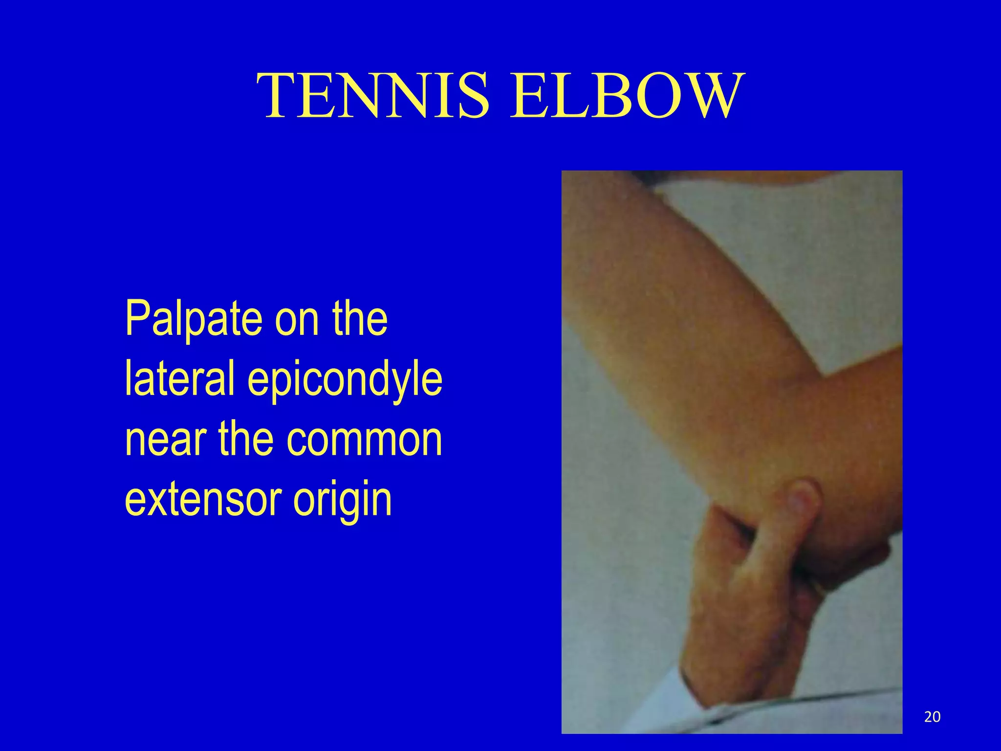
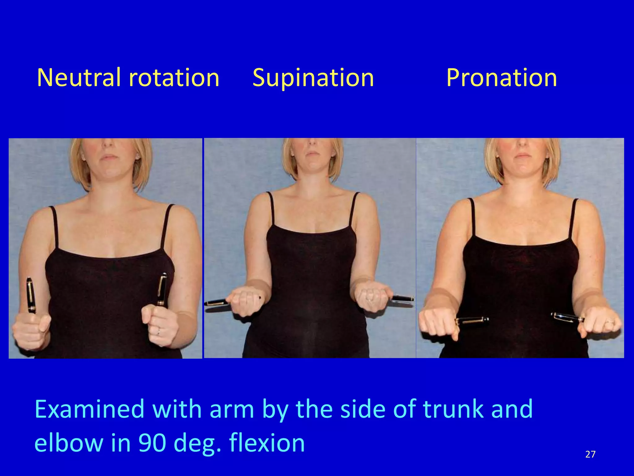
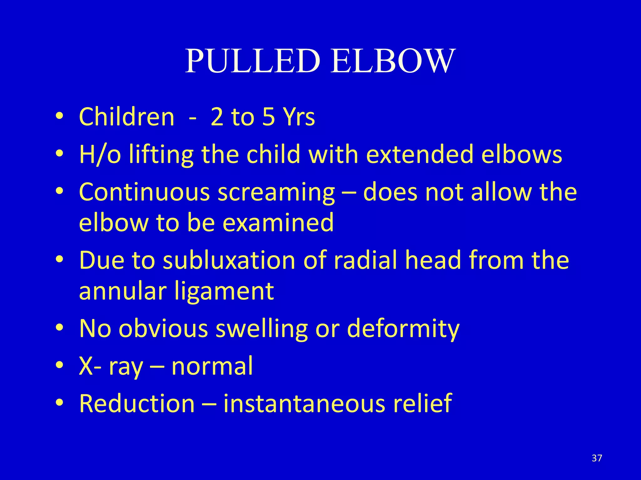
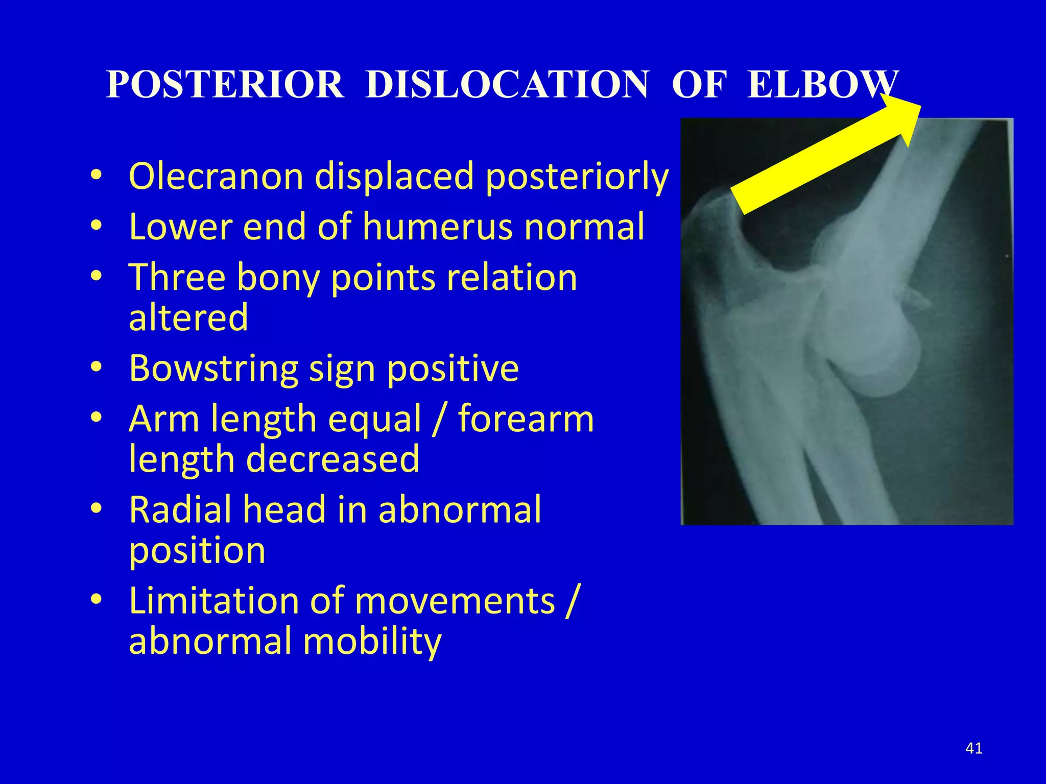
(Rocky) Jaipur Call Girl - 09521753030 Escorts Service 50% Off with Cash ON D...
Clinical examination of elbow joint
1. CLINICAL EXAMINATION
OF ELBOW JOINT
Dr K. Anjaneyulu
Prof & HOD of Orthopaedics
Gandhi Medical College / gandhi hospital
Secunderabad
1
2. The approach for clinical examination of
trauma cases differs from non traumatic
conditions
It also differs - acute injuries examination from
old neglected cases
2
3. • Hinge joint (Humero ulnar, Radiohumeral
Sup.Radioulnar)
• Common - childhood injuries
• Easily prone for stiffness
• Often neglected & inappropiately Rx
• Functional position - different - R – L
3
5. HISTORY
• Duration
• Dominant Limb - Profession
• H/O injury / consti. sympt.
• H/o polyarthralgia / UTI
• Rx History
• H/o massage
• Limitation of ADL
• Referred pain from neck / shoulder
5
6. PHYSICAL EXAMINATION
• Inspection
• Palpation
• Movements
• Measurements
• Distal Neurovascular Status
• Regional Lymphnodes
• Thickening of Ulnar nerve
• Special Tests
6
7. ELBOW FRACTURES IN CHILDREN
• Neuro-motor exam may be limited by the
child’s ability to cooperate because of age,
pain, or fear.
• Thumb extension - EPL (radial – PIN branch)
• Thumb flexion - FPL (median – AIN branch)
• Cross fingers - Interossei (ulnar)
7
14. PALPATION
• Local rise of temperature
• Tenderness
• Bony components
• Soft Tissue components
• Ulnar nerve thickening
• Supratrochlear lymph node
14
15. LOCAL RISE OF TEMPERATURE
Infective - Pyogenic
Tubercular
Inflammatory - Polyarthritis
Acute Myositis
Traumatic - Fresh injury - haematoma
oedema
15
16. TENDERNESS
Maximum point of tenderness
Lat. Epicondyle - Tennis elbow
Med. Epicondyle - Golfer’s elbow
Lower end of Humerus - S/C # Humerus
Radial head - # Radial head
Upper end of Ulna - Olecranon #
16
17. PALPATION
BONY COMPONENTS
irregularity, bowing, thickening
and steps
Medial epicondyle
Lateral epicondyle
Olecranon
Supracondylar ridges
Radial head and capitellum (springing of forearm)
Soft Tissue
Medial aspect
Lateral aspect
Posterior aspect
Anterior aspect 17
18. THREE BONY POINT RELATIONSHIP
COMPARE WITH OPPOSITE NORMAL ELBOW
• Medial epicondyle Extension
• Lateral epicondyle
• Olecranon
F
L
E
X
I
O
N
18
19. PALPATION OF SUPRATROCHLEAR NODE
• Flex the Elbow to right angle to relax
surrounding structures
• Palpated on anterior surface of medial
intermuscular septum 1 cm above the
medial epicondyle
• Not Palpable: Normal elbow, Traumatic causes
• Palpable : Unilateral or Bil (systemic)
19
25. CRITICAL ANGLE OF FLEXION
The arc of flexion 30 – 110 deg
Inspite of some degree of morbidity with
partial limitation of motion a person will be
able to perform the day to day activities with
out much difficulty
25
36. CONCLUDE BY FOLLOWING
• NEUROLOGICAL EXAMINATION
Motor
Sensory
Thickening of ulnar nerve
• EXAMINATION of DISTAL PULSES
Brachial
Radial
• EXAMINATION OF CERVICAL SPINE
• EXAMINATION OF SHOULDER , WRIST,
OPPOSITE ELBOW
36
37. PULLED ELBOW
• Children - 2 to 5 Yrs
• H/o lifting the child with extended elbows
• Continuous screaming – does not allow the
elbow to be examined
• Due to subluxation of radial head from the
annular ligament
• No obvious swelling or deformity
• X- ray – normal
• Reduction – instantaneous relief
37
38. COMMON EXAM CASES
• Cubitus varus - malunited Supracondylar #
• Cubitus Valgus - Non Union of Lateral condyle
+/- Tardy Ulnar nerve Palsy
• Neglected Posterior Dislocation Elbow
• Ankylosed / Stiff Elbow
Trauma/Infection
Myositis Ossificans
38
39. CUBITUS VARUS
• Gunstock deformity
• Medial deviation of forearm
• Thickening / irregularity of L/E of humerus
• Sometimes difficult to identify lat.epicondyle
• Relation of three bony points maintained
• Downward Tilting of the triangle medially
• Shortening of arm – forearm length equal
• Hyperextension / limitation of flexion
39
41. POSTERIOR DISLOCATION OF ELBOW
• Olecranon displaced posteriorly
• Lower end of humerus normal
• Three bony points relation
altered
• Bowstring sign positive
• Arm length equal / forearm
length decreased
• Radial head in abnormal
position
• Limitation of movements /
abnormal mobility
41
42. # LATERAL CONDYLE HUMERUS
• Cubitus valgus deformity – increased carrying
• Lateral supracondylar ridge irregular/ stepping
• Medial supracondylar ridge normal
• Widening of interepicondylar distance
• Distance bet.lat.epicondyle and tip of
olecranon increased
• Abnormal mobility of lat.cond - nonunion
42
45. MYOSITIS OSSIFICANS
• H/o injury (can form with or without fracture)
• H/o massage
• Irregular bony mass infront & behind of elbow
• Limitation of movts of elbow / ankylosis
45