The document details the specifications and functionalities of a Toshiba dual head gamma camera (model GCA-7200A) used in nuclear medicine, capable of scanning the whole body in anterior and posterior views simultaneously. It includes components such as a collimator, photomultiplier tubes, and amplifiers, explaining their roles in detecting gamma rays and converting them into electrical signals for image processing. The gamma camera is employed for various medical applications including locating tumors and assessing organ function using radioactive tracers like iodine-131 and technetium-99.
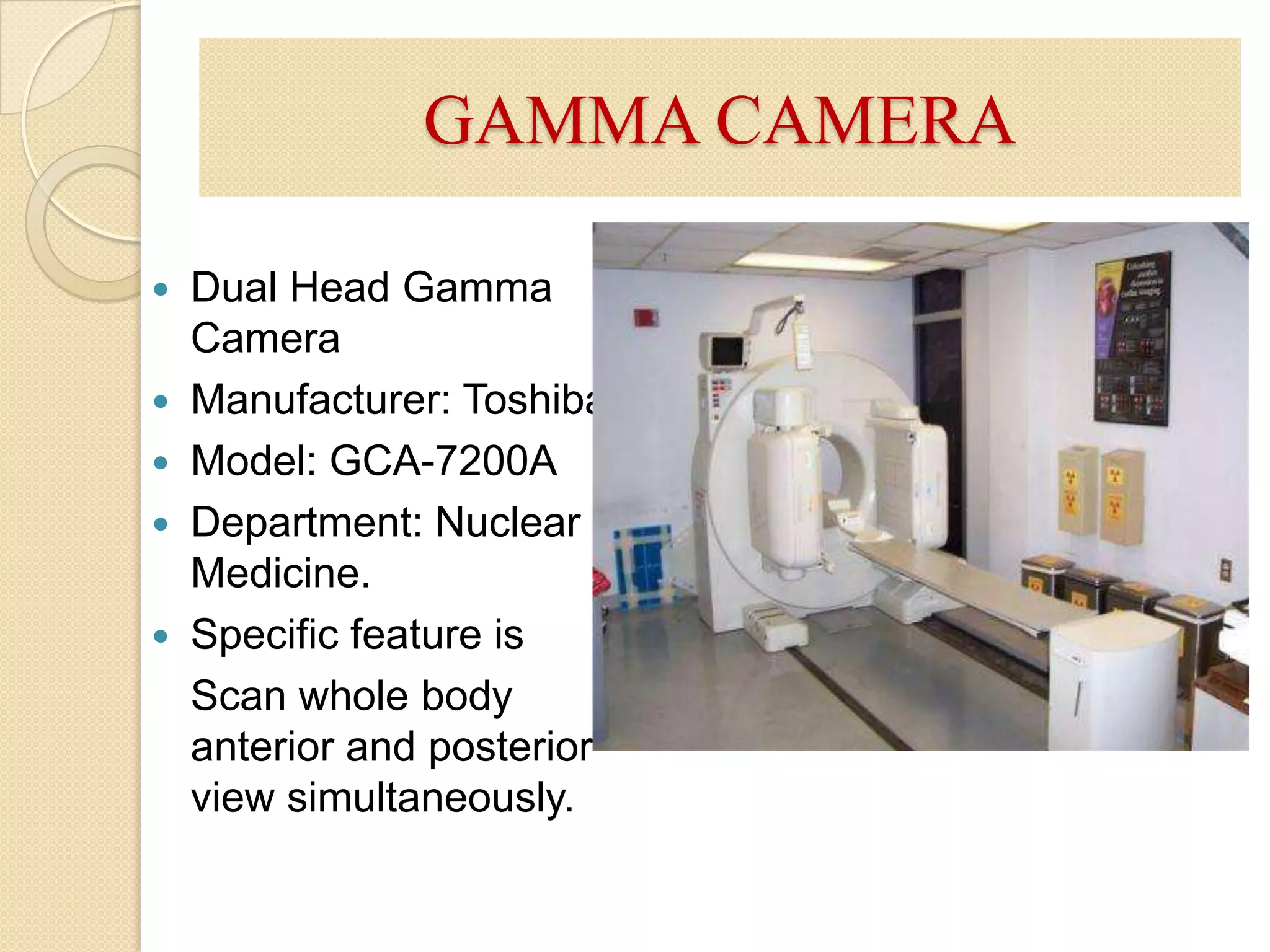
GAMMA CAMERADual HeadGamma CameraManufacturer: ToshibaModel: GCA-7200ADepartment: Nuclear Medicine.Specific feature is Scan whole body anterior and posterior view simultaneously.
3.
GAMMA CAMERADeveloped byHal Anger at Berkeley in 1957 therefore also called Anger cameraAn electronic device that detects gamma rays emitted by radio pharmaceautical (e.gtechnetium 99m (Tc-99m)that have been introduced into the body as tracers. The position of the source of the radioactivity can be plotted and displayed on a TV monitor or photographic film.
4.
COMPONENTS OF GAMMACAMERACollimatorNaI(Tl) crystal.Photomultiplier Tubes(PMT)Pre-amplifierPosition logic circuitsAmplifierPulse height analyzerData Analysis ComputerDisplay (Cathode Ray Tube etc).Gantry
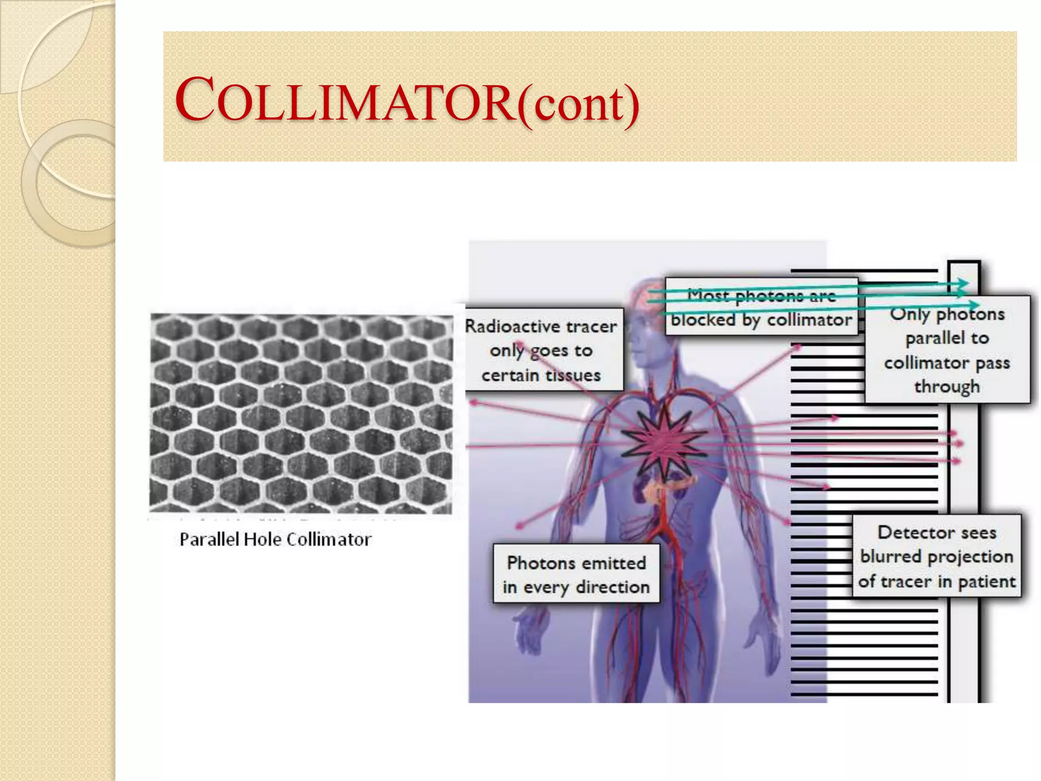
COLLIMATORCollimator is madefrom lead. Maintains the quality of imageSpaces between holes known as septaCollimator consisting of a series of holes in a lead plate can be used to select the direction of the rays falling on the crystal. There are 4 types of collimator.Parallel-hole collimatorPin-hole collimatorDivergingConvergingMost collimators in use are parallel hole collimators. A parallel hole collimator is shown schematically in Figure.
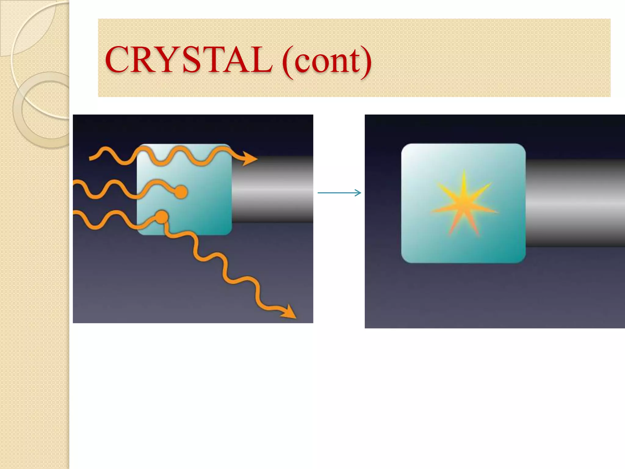
SCINTILLATOR (CRYSTAL)Sodium iodidewith thallium NaI( Tl )The main function of crystal is convert gamma ray to photons of visible light process called scintillation.Amount of light proportional to deposited energy.
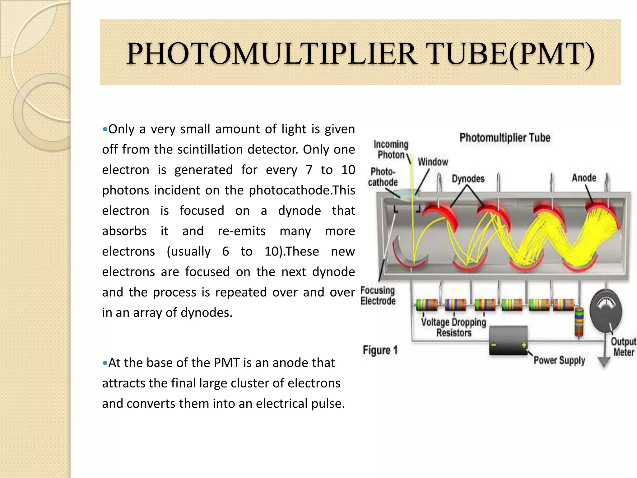
It detects andamplifies the electrons that are produced by the photocathode. The photocathode, when stimulated by light photons, ejects electrons. The PMT is attached to the back of the crystal. PHOTOMULTIPLIER TUBE(PMT)Only a very small amount of light is given off from the scintillation detector. Only one electron is generated for every 7 to 10 photons incident on the photocathode.This electron is focused on a dynode that absorbs it and re-emits many more electrons (usually 6 to 10).These new electrons are focused on the next dynode and the process is repeated over and over in an array of dynodes. At the base of the PMT is an anode that attracts the final large cluster of electrons and converts them into an electrical pulse.
13.
PRE AMPLIFIER ANDAMPLIFIERPreamps attach above the PMT.The amount of charge given by PMT is very small. Even though we have used a sophisticated photodetector like a PMT we still end up with quite a small electrical signal.A very sensitive amplifier is therefore needed to amplify this signal. This type of amplifier is generally called a pre-amplifier.Afte that use amlifier to amlify the signal as need.
14.
POSITION CIRCUITARY &PULSE HEIGHT ANALYSERPosition circuitary receive the electrical impulses from the tubes in the summing matrix circuit (SMC). This allows the position circuits to determine where each scintillation event occurred in the detector crystal.The amplitude of each electrical pulse from the amplifiers is measured in the electrical circuits of the pulse-height analyzerPeak height analyzer and a computer convert the light into a useful anatomical image
15.
DATA ANALYSIS COMPUTERFinally,a processing computer is used to deal with the incoming projection data and processes it into a readable image of the 3D spatial distribution of activity within the patient. The computer may use various methods to reconstruct an image, such as filtered back projection or iterative reconstruction.
16.
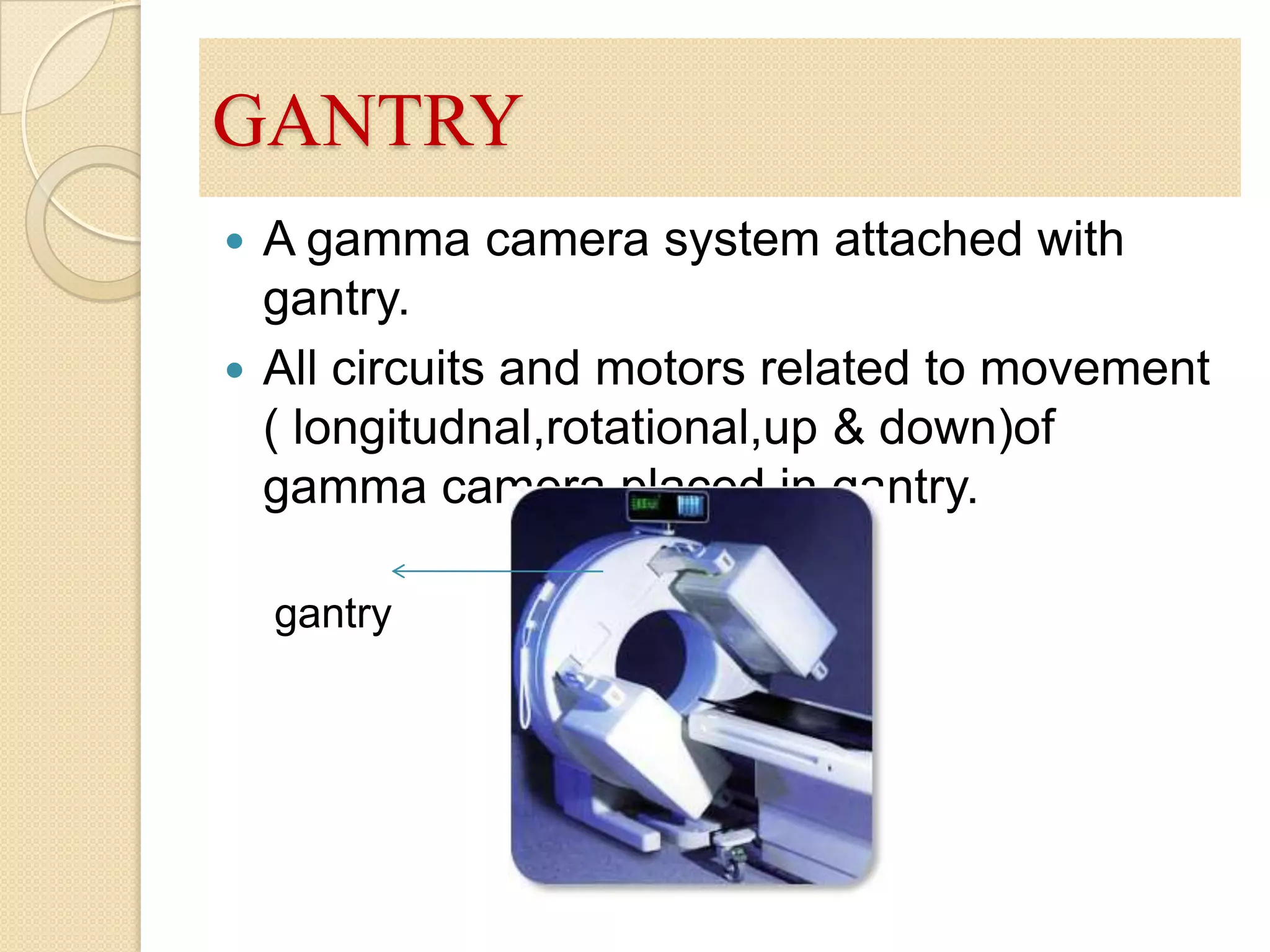
GANTRYA gamma camerasystem attached with gantry.All circuits and motors related to movement ( longitudnal,rotational,up & down)of gamma camera placed in gantry. gantry
APPLICATION OF GAMMACAMERAGAMMA CAMERA used to locate cancerous tumours,minor bone fractures,abnormal functioning of organs and other medical problems .Iodine-131 is used to detect thyroid (a gland that absorbs Iodine) problems. Technetium-99 is used to find tumours in the body. Gamma camera give structural and functional image of body organs.Bone scan. Myocardial Perfusion Lungs scan. Kidney function. Thyroid uptakeWhole body scan.


![Vibe Coding vs. Spec-Driven Development [Free Meetup]](https://cdn.slidesharecdn.com/ss_thumbnails/vibecodingvsspecdrivendevelopment-251209105622-43f455e7-thumbnail.jpg?width=640&height=640&fit=bounds)


