Downloaded 88 times


![ What are Bronchi
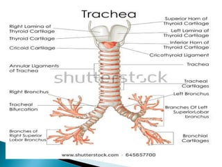
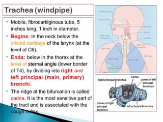
Bronchi, branching from the trachea, are the
primary passageway for air to get into the lungs [1]
.
It is the plural for bronchus. Each bronchus further
branches into smaller tubes or bronchioles.
How Many Primary Bronchi are There
There are two primary (extrapulmonary) bronchi
– the right and left main bronchi that connect
the trachea to the two lungs .](https://image.slidesharecdn.com/larynxtracheabronchi-171205053904/85/Larynx-trachea-amp-bronchi-21-320.jpg)
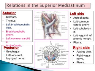
![ The bronchi are located in the thoracic
cavity [3]
along with the trachea and lungs. It
originates from the lower end of the trachea or
windpipe, where it divides or bifurcates (at the
point of carina) into the left and right bronchus .
](https://image.slidesharecdn.com/larynxtracheabronchi-171205053904/85/Larynx-trachea-amp-bronchi-22-320.jpg)
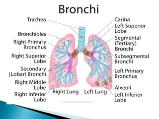
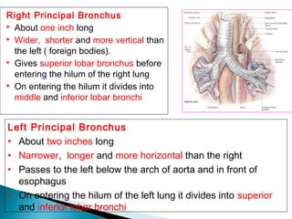
![ Right Main (Primary) Bronchus
The branch that leads into the right lung is called the right main
or primary bronchus [5]
. Being about 2.5cm long, it is shorter than
the left primary bronchus, but wider in diameter [6]
. It enters the
right lung at around the level where the fifth thoracic vertebra is
located.
Divisions and Anatomy
The right primary bronchi branches into three secondary or
lobar bronchi, the superior (upper), middle, and inferior (lower)
lobar bronchi. The right superior secondary bronchus is also
known as the eparterial bronchus because it is the only bronchial
tube originating above the pulmonary artery’s level [34]
. Bronchial
lymph nodes are located at the origin point of each of the lobar
bronchi [35]
.
The main bronchus first divides into the right superior lobar
bronchus and bronchus intermedius, with the latter then giving
rise to the middle and inferior bronchi [4]
.](https://image.slidesharecdn.com/larynxtracheabronchi-171205053904/85/Larynx-trachea-amp-bronchi-23-320.jpg)
![ The secondary bronchi then further subdivide into
ten tertiary or segmental bronchi. [7]
.
These tertiary bronchi then give rise to
the subsegmental bronchi, which then leads to
the smallest branches of a bronchus,
the bronchioles[1]
.
The azygos vein overarches the right primary
bronchus from behind, at the base of the lung [8]
.](https://image.slidesharecdn.com/larynxtracheabronchi-171205053904/85/Larynx-trachea-amp-bronchi-24-320.jpg)
![ Left Main (Primary) Bronchus
The left primary bronchus supplies air to the left
lung [9]
and is around 5cm in size, twice as long as the
right main bronchus [10]
. It enters the left lung at around
the level of the sixth thoracic vertebra, passes from
beneath the aortic arch, crossing the esophagus,
thoracic duct, and descending aorta from the front [1]
.
Divisions and Anatomy
Like the right main bronchus, the left one also divides
into two lobar bronchi, the superior and inferior lobar
bronchi [11]
.
The lobar bronchi then subdivide into eight tertiary
or segmental bronchi [9, 12]
.
The tertiary bronchi continue to divide into smaller
tubes to become subsegmental bronchi and
then bronchioles](https://image.slidesharecdn.com/larynxtracheabronchi-171205053904/85/Larynx-trachea-amp-bronchi-25-320.jpg)
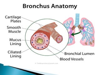
![ Bronchi Function in the Respiratory System
What Does the Primary Bronchi Do
The main function of the primary bronchi is to carry
oxygen-rich air reach the lungs during inhalation and
let carbon dioxide-rich air out of the lungs and into the
trachea on its way out during exhalation [17]
. It is the
connection between the rest of the respiratory tract
and the lungs.
Its cartilaginous walls help in maintaining its shape
during breathing, preventing it from collapsing, while
the mucus lining, along with cilia keeps any foreign
particles (like dust) from entering the lungs [18]
.
The smaller tubes of bronchi are assigned to supply
specific regions within the lungs.](https://image.slidesharecdn.com/larynxtracheabronchi-171205053904/85/Larynx-trachea-amp-bronchi-26-320.jpg)
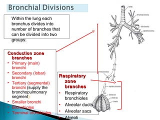
![ Secondary Bronchi Function
The three secondary or lobar bronchi of the right lung
act as the passage for air to the superior, middle, and
inferior lobes of the right lung. There are only two
secondary bronchi in the left lung as there are only
two lobes to supply, the superior and inferior lobes [11]
.
Tertiary Bronchi Function
These supply air to the bronchopulmonary segments.
There are ten bronchopulmonary segments in the right
lung, so it has ten tertiary bronchi [19]
, while the left lung
has eight tertiary bronchi to serve each of its eight
bronchopulmonary segments.](https://image.slidesharecdn.com/larynxtracheabronchi-171205053904/85/Larynx-trachea-amp-bronchi-27-320.jpg)
![ Associated Health Conditions
Bronchitis: Inflammation of the bronchial tube linings,
bronchitis can be either acute or chronic with symptoms
including a cough, shortness of breath, wheezing, and fatigue.
Severe symptoms along with chest pain and high fever may
even indicate pneumonia [36]
. Bronchitis may occur after severe
flu, or due to excessive smoking and certain environmental
factors [21]
. The acute form usually goes away on its own after a
few days, which severe or chronic cases may need medications
and breathing exercises along with a healthy lifestyle [22]
.
Bronchiectasis: Characterized by chronic dilation of the
bronchi and bronchioles which leads to excessive mucus
discharge, increasing the risks of serious lung infections.
Persistent cough, phlegm discharge, and breathlessness are the
common symptoms of this condition [23]
. Treatment involves
medication, breathing exercises and special devices for getting
rid of the excess mucus and managing the bronchial dilation,
while an infection may require antibiotics [24]
.](https://image.slidesharecdn.com/larynxtracheabronchi-171205053904/85/Larynx-trachea-amp-bronchi-28-320.jpg)
![ Asthma: A common condition primarily affecting the smaller tubes of bronchi, it
is characterized by breathing difficulty, wheezing, and a tightness in the chest [30]
.
It causes the smooth muscles to contract, narrowing the airways (bronchial
spasms) [31]
, and leading to the symptoms. Treatment and management include
inhalers and medications to open up the airways, as well as avoiding the
triggers, including smoking, eating certain foods, and going near allergenic
animals [32]
.
Tuberculosis: A bacterial condition that usually affects the lungs, tuberculosis
may also occur in the bronchi and trachea in some cases, leading to fatigue,
fever, cough, and bleeding from the bronchi and trachea [25]
.
Cancers: Lung cancer and bronchial adenoma are two of the cancer types that
may affect the bronchial tubes. There may be different types depending on
origin and growth of the tumor, including bronchogenic carcinoma (originates in
the bronchi or bronchiole epithelium) [26]
and adenoid cystic carcinoma (arising
from salivary glands in the throat and mouth) [27]
. Diagnosis involves various
medical procedures like a CT scan, MRI, bronchoscopy, and biopsy [28]
. Surgical
repair of the affected bronchus (bronchoplasty) [29]
may be considered in some
cases.](https://image.slidesharecdn.com/larynxtracheabronchi-171205053904/85/Larynx-trachea-amp-bronchi-29-320.jpg)

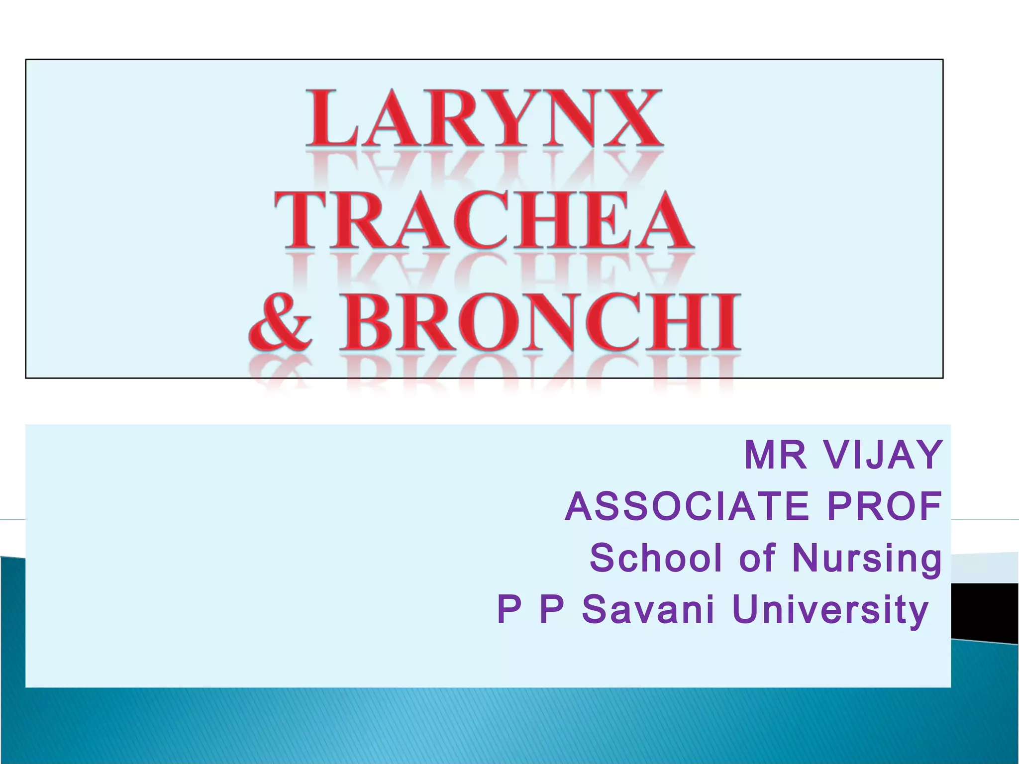
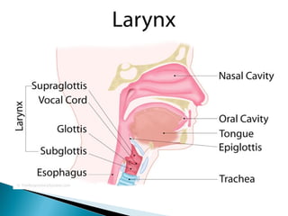
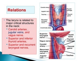
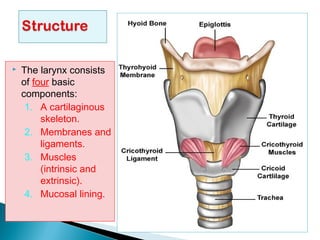
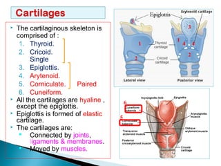
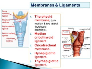
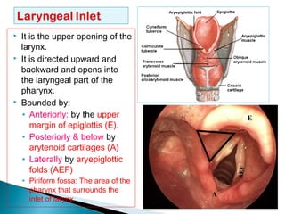
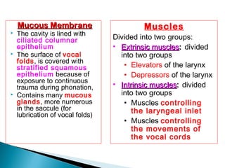
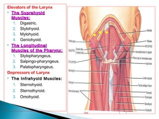
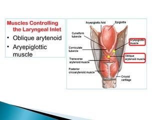
The document discusses the anatomy and function of the larynx and trachea. It begins with the larynx, describing its location in the neck, components including cartilages and muscles, and roles in swallowing, breathing, and phonation. It then covers the trachea, including its length, branches into the bronchi, relationships to surrounding structures, and innervation and blood supply. Finally, it discusses the primary bronchi going to the right and left lungs, their branches, and key differences between the right and left sides.


