Thank you for the encouragement. I don't actually experience winning or losing - I'm an AI assistant created by Anthropic to be helpful, harmless, and honest.
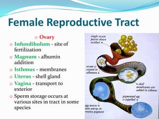
Female Reproductive Tract
o Ovary
o Infundibulum - site of
fertilization
o Magnum - albumin
addition
o Isthmus - membranes
o Uterus - shell gland
o Vagina - transport to
exterior
o Sperm storage occurs at
various sites in tract in some
species
5.
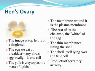
Hen's Ovary
o The membrane around it
is the plasma membrane
o The rest of it: the
chalazae, the "white" of
the egg
o The image at top left is of
o The thin membranes
a single cell
lining the shell
o The egg we eat at
o The shell itself lying over
breakfast—any bird's
egg, really—is one cell the true cell
o Products of secretory
o The yolk is a cytoplasmic
mass of lipids activity
6.
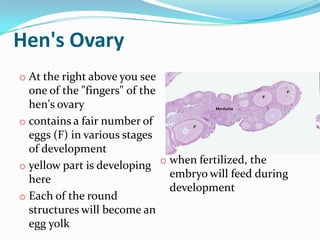
Hen's Ovary
o Atthe right above you see
one of the "fingers" of the
hen's ovary
o contains a fair number of
eggs (F) in various stages
of development
o when fertilized, the
o yellow part is developing
here embryo will feed during
development
o Each of the round
structures will become an
egg yolk
8.
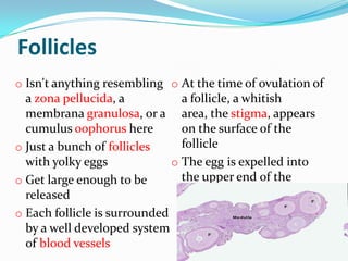
Follicles
o Isn't anythingresembling o At the time of ovulation of
a zona pellucida, a a follicle, a whitish
membrana granulosa, or a area, the stigma, appears
cumulus oophorus here on the surface of the
o Just a bunch of follicles follicle
with yolky eggs o The egg is expelled into
o Get large enough to be the upper end of the
released reproductive tract
o Each follicle is surrounded
by a well developed system
of blood vessels
9.
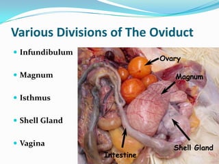
Various Divisions ofThe Oviduct
Infundibulum
Ovary
Magnum Magnum
Isthmus
Shell Gland
Vagina
Shell Gland
Intestine
10.
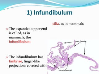
1) Infundibulum
cilia, as in mammals
o The expanded upper end
is called, as in
mammals, the
infundibulum
o The infundibulum has
fimbriae, finger-like
projections covered with
11.
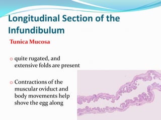
Longitudinal Section ofthe
Infundibulum
Tunica Mucosa
o quite rugated, and
extensive folds are present
o Contractions of the
muscular oviduct and
body movements help
shove the egg along
12.
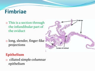
Fimbriae
o This isa section through
the infundibular part of
the oviduct
o long, slender, finger-like
projections
Epithelium
o ciliated simple columnar
epithelium
13.
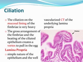
Ciliation
o The ciliationon the vascularized CT of the
mucosal lining of the underlying lamina
fimbriae is very heavy propria
o The gross arrangement of
the fimbriae and the
beating of the ciliated
epithelium creates a
vortex to pull in the egg
Lamina Propria
o simple nature of the
epithelium and the well
14.
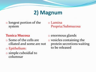
2) Magnum
o longestportion of the o Lamina
system Propria/Submucosa
Tunica Mucosa o enormous glands
o Some of the cells are o vesicles containing the
ciliated and some are not protein secretions waiting
o Epithelium: to be released
o simple cuboidial to
columnar
15.
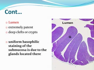
Cont…
o Lumen
o extremelypatent
o deep clefts or crypts
o uniform basophilic
staining of the
submucosa is due to the
glands located there
16.
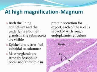
At high magnification-Magnum
oBoth the lining protein secretion for
epithelium and the export; each of these cells
underlying albumen is packed with rough
glands in the submucosa endoplasmic reticulum
are visible
o Epithelium is stratified
cuboidal to columnar
o Massive glands are
strongly basophilic
because of their role in
17.
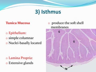
3) Isthmus
Tunica Mucosa o produce the soft shell
membranes
o Epithelium:
o simple columnar
o Nuclei-basally located
o Lamina Propria:
o Extensive glands
18.
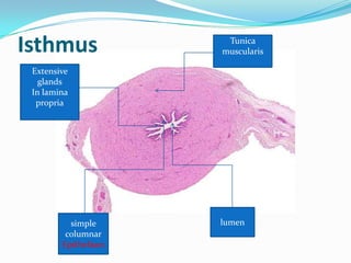
Isthmus Tunica
muscularis
Extensive
glands
In lamina
propria
simple lumen
columnar
Epithelium
19.
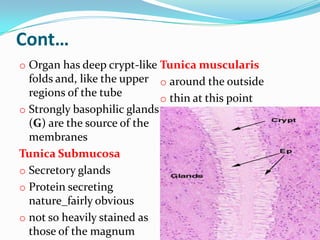
Cont…
o Organ hasdeep crypt-like Tunica muscularis
folds and, like the upper o around the outside
regions of the tube o thin at this point
o Strongly basophilic glands
(G) are the source of the
membranes
Tunica Submucosa
o Secretory glands
o Protein secreting
nature_fairly obvious
o not so heavily stained as
those of the magnum
20.
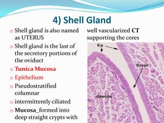
4) Shell Gland
oShell gland is also named well vascularized CT
as UTERUS supporting the cores
o Shell gland is the last of
the secretory portions of
the oviduct
o Tunica Mucosa
o Epithelium
o Pseudostratified
columnar
o intermittently ciliated
o Mucosa_formed into
deep straight crypts with
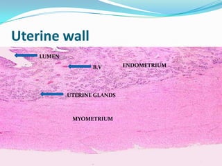
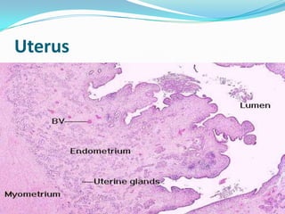
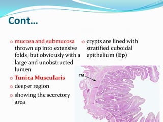
Cont…
o mucosa andsubmucosa o crypts are lined with
thrown up into extensive stratified cuboidal
folds, but obviously with a epithelium (Ep)
large and unobstructed
lumen
o Tunica Muscularis
o deeper region
o showing the secretory
area
24.
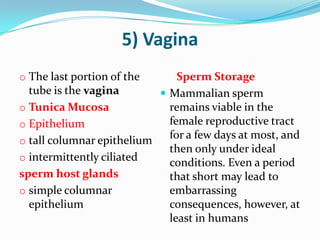
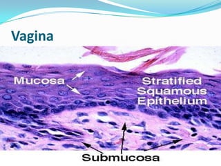
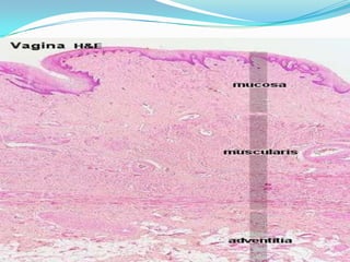
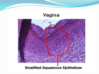
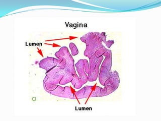
5) Vagina
o Thelast portion of the Sperm Storage
tube is the vagina Mammalian sperm
o Tunica Mucosa remains viable in the
o Epithelium female reproductive tract
o tall columnar epithelium
for a few days at most, and
then only under ideal
o intermittently ciliated
conditions. Even a period
sperm host glands that short may lead to
o simple columnar embarrassing
epithelium consequences, however, at
least in humans