
Recommended
More Related Content
What's hot
What's hot (20)
Similar to REGULATION OF VENTILATION
Similar to REGULATION OF VENTILATION (20)
Recently uploaded
Recently uploaded (20)
REGULATION OF VENTILATION
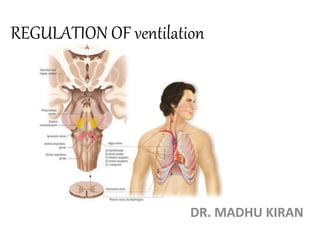
- 1. REGULATION OF ventilation DR. MADHU KIRAN
- 2. Breathing is regulated by two mechanisms: • Nervous or neural mechanism • Chemical mechanism
- 3. Nervous Mechanism: • It involves respiratory centers, afferent and efferent nerves. • Respiratory centers: The centres in the medulla oblongata and pons that collects sensory information about the level of oxygen and carbon dioxide in the blood and determines the signals to be sent to the respiratory muscles. • Stimulation of these respiratory muscles provide respiratory movements which leads to alveolar ventilation. • Respiratory centers are situated in the reticular formation of the brainstem and depending upon the situation in brainstem, the respiratory centers are classified into two groups: 1. Medullary centers 2. Pontine centers
- 4. There are two centers in each group: • Medullary Centers: A. Dorsal respiratory group B. Ventral respiratory group • Pontine Centers: A. Pneumotaxic center B. Apneustic center
- 5. Pre- Botzinger complex: • Spontaneous automatic rhythmic respiration is initiated by small group of synaptically coupled pace maker cells in the pre-botzinger complex situated on either side of medulla between nucleus ambiguous and lateral reticular nucleus. • Pre-BOTC neurons discharge rhythmically, producing rhythmic discharge in phrenic motor neurons • Pre-BOTC contact hypoglossal nuclei, and the tongue is involved in the regulation of airway resistance. • DRG & VRG involved in generation of respiratory pattern project to pre-BOTC.
- 6. Dorsal respiratory group • Center is situated in upper part of medulla oblongata • This center is previously called inspiratory centre. • It is formed by nucleus of tractus solitarius. • DRG constitute the initial intra cranial processing station for afferent inputs from 9th & 10th cranial nerves, which originate from peripheral chemoreceptors, baroreceptors, and several other receptors of airways and lungs. • Function: it is concerned with inspiration and cause rhythmic inspiratory discharge. • Action potential in inspiratory neurons begin weakly and increase steadily in a ramp like manner for about 2 sec. then it ceases abruptly for 3 sec, which turns off inspiratory muscles(diaphragm) and allows elastic recoil of lung and chest wall to cause expiration. .
- 7. Ventral respiratory group: • It is situated in medulla oblongata anterior and lateral to the DRG • Contains both inspiratory and expiratory neurons. • It is formed by neurons of nucleus retro fascialis, nucleus paraambiguous and nucleus retro ambiguous • Function: this center is inactive during quiet breathing and inspiratory center is the active center, but during forced breathing or when the inspiratory center is inhibited it becomes active
- 8. Pneumotaxic center: • It is situated in upper Pons. • It is formed by nucleus parabrachialis. • Function: it controls medullary respiratory centers, particularly the inspiratory center through apneustic center. It always controls the activity of inspiratory center so that duration of inspiration is controlled. Stimulation leads to shallow and rapid breathing. Apnuestic center: • It is situated in lower Pons. • Normally inhibited by pneumotaxic centre and vagus. • Function: this center increases depth of inspiration by acting directly on the inspiratory center.
- 9. Nervous connections of respiratory centers: Afferent pathway: • Respiratory center receive afferent impulses from different parts of the body according to movements of thoracic cage and lungs. • From peripheral, chemoreceptor and baroreceptor impulses are carried by glossopharyngeal and vagus nerves to respiratory center. Efferent pathway: • Nerve fiber from respiratory center leaves the brain and descend in anterior part of lateral column of spinal cord. • These nerve fibers terminate in the motor neurons in the anterior horn cells of the cervical and thoracic segments of spinal cord. • From motor neurons two sets of nerve fiber arise which supplies particular muscle: 1. Phrenic nerve fibers: supplies diaphragm 2. The intercostal nerve fibers: supplies intercostal muscles.
- 10. Effect of lesion/ Transection • Above pons: loss of voluntary control Respiration remains normal • At upper/mid pons: slow and deep breathing (vagi intact) Apneustic breathing (or prolonged respiratory spasm) when vagi are also cut. • At inferior border of pons & upper border of medulla: spontaneous respiration continues some what irregular and gasping occurs • Below medulla: Respiration stops.
- 11. Factors affecting respiratory centers: 1) Impulses from higher centers: impulses from higher center can stimulate or inhibit respiratory centers directly. 2) Impulses from stretch receptors of lung:
- 12. 3) Impulses from ‘J’ receptors of lungs: • ‘J’ receptors are juxtacapillary receptors which are present in wall of the alveoli and have close contact with the pulmonary capillaries. • These receptors get stimulated during conditions like pulmonary edema, pulmonary congestion, pneumonia as well as due to exposure of exogenous and endogenous chemicals like histamine, serotonin. • Stimulation of ‘J’ receptor produces a reflex response called apnea. 4) Impulses from irritant receptors of lungs: • Irritant receptors are situated on the wall of bronchi and bronchioles of lungs. • They got stimulated by harmful chemicals like ammonia and sulfur dioxide. • Stimulation of irritant receptors produces reflex hyperventilation along with bronchospasm which prevents entry of harmful chemicals into the alveoli.
- 13. 5) Impulses from Proprioceptors: • Proprioceptors are the receptors which give response to the change in the position of different parts of the body. • This receptors are situated in joints, muscles and tendons. They get stimulated during exercise and sends impulses to the cerebral cortex. • Cerebral cortex in turn by activating medullary respiratory centres causes hyperventilation. 6) Impulses from Thermoreceptors: • Thermoreceptors give response to change in the body temperature. • They are cutaneous receptors namely cold and warmth • When this receptors get stimulated they send signals to cerebral cortex • Cerebral cortex in turn stimulates respiratory centres and causes hyperventilation.
- 14. 7) Impulses from pain receptors: • Pain receptors give response to pain stimulus. • Like other receptors this receptors also send impulses to the cerebral cortex. • Cerebral cortex in turn stimulates the respiratory centers ad causes hyperventilation. 8) Cough reflex: • This is a protective reflex caused by irritation of parts of the respiratory tract beyond nose like larynx, trachea and bronchi. • Irritation of any of this part causes stimulation of vagus nerve and cough occurs. • Cough begins with deep inspiration followed by forceful expiration with closed glottis. • So the intrapleural pressure rises above 100 mm Hg. • Then, glottis is suddenly opened with explosive outflow of air at a higher velocity. So the irritants may be expelled out of the respiratory tract.
- 15. 9) Sneezing reflex: • It is also a protective reflex which occurs due to the irritation of nasal mucus membrane. • During irritation of nasal mucus membrane, the olfactory receptors and trigeminal nerve endings present in the nasal mucosa are stimulated leading to sneezing. • Sneezing starts with deep inspiration, followed by forceful expiratory effort with opened glottis and the irritants are expelled out of the respiratory tract. 10) Deglutition reflex: • During swallowing of the food, the respiration is arrested for a while. • Temporary arrest of the respiration is called apnea and apnea which occurs during swallowing called swallowing apnea or deglutition apnea. • This prevents entry of the food particles into the respiratory tract.
- 16. Chemical Mechanism: • The chemical mechanism of the respiration is operated through the chemoreceptors. Chemoreceptors: • They are the receptors which give response to change in the chemical constituents of blood like.. A. Hypoxia B. Hypercapnea C. Increased hydrogen ions concentration (decreased blood pH)
- 17. • Chemoreceptors are classified into two groups: 1. Central chemoreceptors 2. Peripheral chemoreceptors
- 18. Central chemoreceptors Situation: • They are situated in deeper part of medulla oblongata, close to the dorsal group of neurons. • This area is known as chemosensitive area and neurons are called as chemoreceptors. • They are in close contact with blood and CSF. Action: • They are very sensitive to increase in hydrogen ion concentration. • Hydrogen ion cannot cross the blood brain barrier and blood cerebrospinal fluid barrier. • On the other hand if carbon dioxide increases in the blood as it is a gas it can cross both the barrier easily and after entering the brain it combines with water to form carbonic acid. • As carbonic acid is unstable, it immediately dissociates into hydrogen and bicarbonate ions. • The hydrogen ion now stimulates the central cemoreceptors which stimulates dorsal group of respiratory center (inspiratory group) and increase rate and force of breathing. The chemoreceptors present in the brain are called central chemoreceptors.
- 19. Peripheral chemoreceptors: Situation: Action: • They are very sensitive to reduction in partial pressure of oxygen. • Whenever, the partial pressure of oxygen decreases these chemoreceptors become activated and send impulses to inspiratory center and stimulate them. • Thereby increases rate and force of respiration and rectifies the lack of oxygen. The receptors are present in peripheral portions of the body that’s why called as peripheral chemoreceptors.
- 20. Thank you