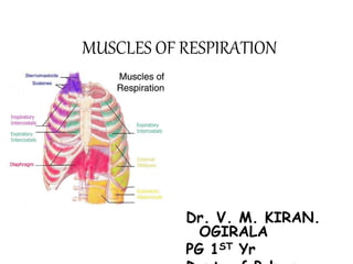
1. The diaphragm and external intercostal muscles are responsible for quiet inspiration by contracting to increase the thoracic cavity volume.
2. Quiet expiration is a passive process where these muscles relax and the lungs and chest wall recoil, decreasing thoracic cavity volume and expelling air.
3. Forced expiration involves contraction of the internal intercostals and abdominal muscles like the rectus abdominis to compress the abdomen and further increase intrathoracic pressure, expelling more air from the lungs.
2. Muscles of Ventilation
Ventilation is the movement of air into and out
of the lungs.
Respiration is the cellular process of breaking
down chemical energy (substrates) to make
ATP
C6H12O6 + 6 O2 6 CO2 + 6 H20 + E
3. Ventilation
• Inspiration (Inhalation): active process.
– Requires muscular action
– Change in thoracic volume change in pressure.
(Boyle’s Law)
• Change in pressure drives air into lungs
• Expiration (exhalation): passive process
– Occurs when muscles of breathing relax
– Forced exhalation: muscular activity required
4. Resting Ventilation
• Two muscles of resting ventilation
1. Diaphragm
• A musculotendinous partition between thoracic &
abdominal cavity
• Convex toward thoracic & concave toward abdominal
cavity
• It consists of two functionally distinct parts, costal and
crural parts.
– Origin: Xiphoid, ribs 7-12 and their costal cartilage, anterior
surface of lumbar vertebrae.
– Insertion: Central tendinous sheet
– Nerve supply: phrenic nerve (C3,4,5), penetrates diaphragm &
innervates it from abdominal surface
– Action: contraction (descent) of diaphragm increase vertical
diameter of thoracic cavity (essential for normal breathing)
5.
6. Resting Ventilation
2. External intercostals
– Origin: Inferior border of each rib
– Insertion: Superior border of the
more inferior rib
(the rib below the superior one)
-Nerve supply: intercostal nerves
• Action: Lifts the lower rib to
expand the thoracic cavity
7. Resting Exhalation
• This is a passive process in which the
diaphragm and external intercostals relax and
the thoracic cavity recoils back to its resting
volume (gets smaller) forcing air from the
lungs due to the increase in intrathoracic
pressure.
8. Muscles of Inspiration during
Exercise
• These muscles lift the clavicle and ribs
• These are known as Accessory Inspiratory
muscles.
These muscles include:
• Scalene muscles
• Sternocleidomastoid
• Trapezius
• Pectoralis major and minor
10. Scalene:
Work in conjunction with SC
Origin: Transverse processes
of C2 – C7
Insertion: First two ribs
Action: Ant &Middle scalene
lift rib 1
Post Scalene
lifts rib 2
11. Muscles of Inspiration during
Exercise
• Diaphragm
• External intercostals – raises the ribs
– Other Accessory Muscles
• Trapezius (upper fibers) – stabilizes head and
raises the clavicle
• scalene– raises the ribs 1 & 2
• Pec minor – raises ribs when scapula is fixed
18. Anterior abdominal wall
• Is formed of 3 layers of muscles of fibers running in
different directions (to increase strength of anterior
abdominal wall)
• The 3 muscles form a sheath in which a fourth muscles
lies (rectus abdominis)
• Muscles are attached to: sternum, costal cartilages and
ribs + hip bones
• The aponeurosis of the 3 muscles on both sides fuse in
the midline to form linea alba
• Action (during forced expiration): Compression of
abdominal viscera to help in ascent of diaphragm
(during forced expiration)
• Nerve supply: lower intercostal nerves (T7 – T11),
subcostal nerve (T12) and first lumbar nerve
19. RESPIRATORY MOVEMENTS
A- MOVEMENTS OF DIAPHRAGM
Contraction (descent)
of diaphragm
Increase of vertical diameter
of thoracic cavity
Inspiration
Expiration
Relaxation (ascent)
of diaphragm)
20. RESPIRATORY MOVEMENTS
B - MOVEMENTS OF RIBS
PUMP HANDLE MOVEMENT
Elevation of ribs
Increase in antero-posterior diameter
of thoracic cavity
BUCKET HANDLE MOVEMENT
Elevation of ribs
Increase in lateral diameter of thoracic
cavity
21. SUMMARY OF RESPIRATORY MOVEMENTS
Inspiration
Quiet Inspiration (active)
Expiration
Quiet Expiration (passive)
1. Elastic recoil of lung
2. Relaxation of diaphragm & external
intercostal
Forced Expiration (active):
Contraction of anterior Depression of ribs
abdominal wall muscles (rest of intercostal
muscles)
Compression of abdominal
viscera
Ascent of diaphragm
Contraction (Descent) Elevation of ribs
of diaphragm (external intercostal)
Increase in vertical Increase in:
diameter - anteroposterior
diameter
- lateral diameter
Forced Inspiration (active)
Accessory muscles of inspiration:
1. Pectoralis major
2. Scalene muscles