Downloaded 356 times


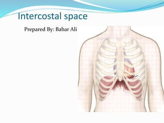
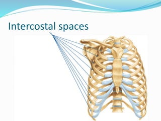
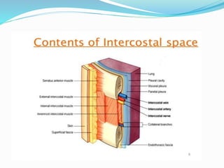
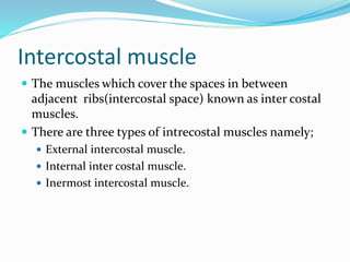
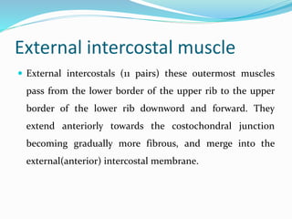
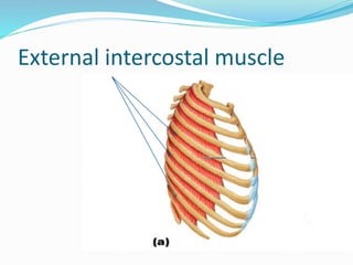
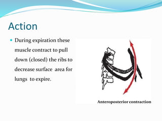
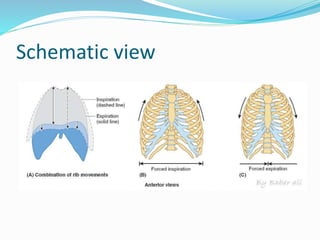
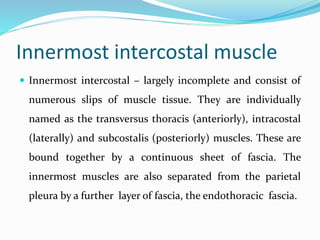
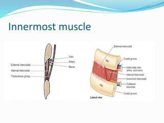
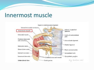
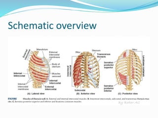
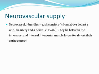
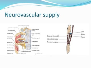


This document summarizes the anatomy of the intercostal spaces. It describes that the intercostal spaces are located between adjacent ribs, and contain intercostal muscles and neurovascular bundles. The three types of intercostal muscles - external, internal, and innermost - are discussed, along with their actions and nerve/blood supply. The importance of the intercostal spaces in breathing, ECG placement, auscultation, and surgical access is also noted.


