SlideShare a Scribd company logo
1 of 22
Albugo(Cystopus)
V.S.Patil
Assosiate Professor
Department of Botany
Shri Shivaji College of Arts, Commerce & Science
Akola
Albugo (Cystopus)
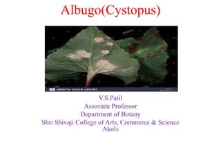
White rust or white blister
diseases
Kingdom- Mycota (Fungi)
Division- Eumycota
Sub-Division- Mastigomycotina
Class- Oomycetes
Order- Albuginales
Family- Albuginaceae
Genus- Albugo sp.
• This organism causes white rust or white blister diseases in above-
ground plant tissues.
• While these organisms affect many types of plants, the destructive
aspect of infection is limited to a few agricultural crops, including :
beets, cabbages, cauliflower, mustards, radish, spinach, sweet
potatoes, turnips etc.
• Symptoms of white rust caused by Albugo typically include yellow
lesions on the upper leaf surface and white pustules on the underside
of the leaf.
• The pathogen is spread by wind, water, and insects.
• Management includes use of resistant cultivars,
proper irrigation practices, crop rotation, sanitation, and chemical
control. White rust is an important economic disease, causing severe
crop losses if not controlled.
Figure 1.Symptoms on leaves, branches, flower parts
A B C
D E F
• White rust pathogens create chlorotic (yellowed)
lesions and sometimes galls on the upper leaf
surface and there are corresponding white blister-
like dispersal pustules of sporangia on the underside
of the leaf.
• Species of the Albuginaceae deform the branches
and flower parts of many host species. Increase in
the size of the cells (hypertrophy) and organs takes
place.
• Proliferation of the lateral buds, discoloration of
flowers, malformation of floral parts and sterile
gynoecium.
1.White rust is an obligate parasite.(needs a living host
to grow and reproduce).
2.The Albuginaceae reproduce by producing both sexual
spores (called oospores) and asexual spores
(called sporangia) in a many-stage (polycyclic) disease
cycle.
3.White rust is an economically important foliar disease,
causing substantial yield losses and eventual death of
various crops.
Structure-
Thallus is eucarpic and mycelial. Hyphae are intercellular, coenocytic,
aseptate and profusely branched(Fig. 2 B). Cell wall is composed of
fungal cellulose. The protoplasm contains a large number of nuclei
distributed in the cytoplasm.
• Reserve food material is in the form of oil drops and glycogen bodies.
Some mycelium is intracellular in the form of knob-like haustoria for
the absorption of food material from the host cells.
Haustoria
Reproduction-
1)Asexual Reproduction:
• The asexual reproduction takes place by conidia, condiosporangia or
zoosporangia.
• They are produced on the sporangiophores. Under suitable conditions
the mycelium grows and branches rapidly. After attaining a certain age
of maturity, it produces a dense mat like growth just beneath the
epidermis of the host. (Fig. 2 D). These hyphae produce, at right
angles to the epidermis are short, thick walled, un-branched and club
shaped. These are the sporangiophores or conidiophores.
• They form a solid, palisade like layer beneath the epidermis (Fig. 2 A-
D).They are thick walled on lateral sides and thin walled at tip. The
sporangiophores contain dense cytoplasm and about a dozen nuclei.
After reaching a certain stage of maturity, the apical portion of
sporangiophore gets swollen and is ready to cut off a sporangium or
conidium (Fig. 2E).
The sporangia are produced at the tip by abstraction method. A
Deeping constriction appears below the swollen end (fig 2. F)
and results in the formation of first sporangium. A second
sporangium is similarly formed from the tip just beneath the
previous one (Fig. 2 G).
This process is repeated several times. The new nuclei
migrates from mycelium to cytoplasm and are used in the
formation of another sporangium or conidium. Thus along
chain of sporangia or conidia is formed above each
sporangiophore in basipetal succession. (youngest at the base
and oldest at the tip) (Fig. 2 H). The sporangia or conidia are
spherical, smooth, hyaline and multinucleate structures. The
walls between them fuse to form a gelatinous disc-like
structure called disjunctor or separation disc or intercalary
disc. (Fig. 2 G).
It tends to hold the sporangia together. The continued growth
and production of sporangia exerts a pressure upon the
enveloping epidermis. Which is firstly raised up but finally
ruptured exposing the underlying sours containing white
powdery dust of multinucleate sporangia or conidia (Fig. 2 A,
3).
The separation discs are dissolved by water, and the sporangia
are set free. They are blown away in the air by wind or washed
away by rain water under suitable environmental conditions
and falling on a suitable host, sporangia germinates with in 2
or 3 hours. The sporangia germinate directly or indirectly
depending on temperature conditions.
1. Direct Germination:At high temperature and comparative
dry conditions the sporangium germinates directly. It gives
rise to a germ tube which in-fact the host tissue through stoma
or through an injury in the epidermis (Fig. 2 I, P).
2. Indirect Germination:
In the presence of moisture and low temperature (10°C) the sporangium
germinates indirectly i.e., it behaves like zoosporangium and produces
zoospores. It absorbs water, swells up, and its contents divide by
cleaving into 5-8 polyhedral parts (Fig. 2 J) depending upon the nuclei
present in it. Each part later on rounds up and metamorphoses into
zoospore (Fig. 2 K, L). A papilla is developed on one side which later
burst and liberates the zoospores.
Zoospore: The zoospores are uninucleate, slightly concavo-convex and
biflagellate. The flagella are attached laterally near the vacuole. Of the
two flagella one is of whiplash type and the other tinsel type (Fig. 2M).
After swimming for some time in water, they settle down on the host.
They retract their flagella, secrete a wall and undergo a period of
encystment (Fig. 2 N). On germination, they put out a short germ tube
which enters the host through stomata (Fig. 2 O, P) or again infects the
healthy plants.
Sexual Reproduction:
It takes place when the growing season comes to an end. The
mycelium penetrates into the deeper tissues of the host. The
sexual reproduction is highly oogamous type. The antheridium
and oogonium develops deeper in the host tissue in close
association within the intercellular spaces.
Its formation is externally indicated by hypertrophy. The
antheridium and oogonium are formed near each other on
hyphal branches. They are terminal in position, however,
intercalary oogonia also occur, though rarely.
Antheridium:
It is elongated and club shaped structure. It is multinucleate
(6-12 nuclei) but only one nucleus remains functional at the
time of fertilization in C. candidus. It is paragynous i.e.,
laterally attached to the oogonium (Fig. 4 A-C). It is separated
by a cross wall from the rest of the male hyphae.
Oogonium:
It is spherical and multinucleate containing as many as 65 to 115 nuclei.
All nuclei are evenly distributed throughout the cytoplasm (Fig. 4 A-C).
As the oogonium reaches towards the maturity the contents of the
oogonium get organised into an outer peripheral region of periplasm and
the inner dense central region of ooplasm or oosphere or the egg (Fig. 4
D-G). The ooplasm and periplasm are separated by a plasma membrane.
The nuclei in the oogonium divides mitotically. The first mitotic division
takes place before the organization of the periplasm and oosplasm (Fig. 4
E). After the organization, all the nuclei of the ooplasm, except one,
migrate to the periplasm forming a ring and undergo second mitotic
division. They divide in such a manner that one pole of each spindle is in
ooplasm and the other in the periplasm (Fig. 4 E).
At the end of the division one daughter nucleus of each spindle goes to
the oosplasm and other in periplasm (Fig. 4 F). However, at the time of
maturity, all nuclei disintegrate, except single functional nucleus (Fig. 4
G). On the basis of functional nuclei in ooplasm,” Albugo is divided into
following groups:
Group I:
The number of functional egg nucleus in ooplasm is
one. It is represented by C. Candidus.
Group II:
The number of functional eggs in ooplasm is many. It
is in many species.
It carries a single male nucleus. Its tip ruptures to
discharge the male nucleus near the female nucleus.
Ultimately the male nucleus fuses with the female
nucleus (karyogamy).
Fertilization:
Before fertilization a deeply staining mass of
cytoplasm, (Fig. 4 H) appears almost in the centre of
the ooplasm. This is called coenocentrum. It persists
only up to the time of fertilization. The functional
female nucleus attracted towards it and becomes
attached to a point near it.
The oogonium develops a papilla like out growth at
the point of contact with the antheridium. This is
called as receptive papilla (Fig. 4 G). Soon it
disappears, and the antheridium develops a
fertilization tube.It penetrates through receptive
papilla, oogonial wall and periplasm and finally
reaches upto the ooplasm (Fig. 4 H, L).
Oospore:
The oospore alongwith the fusion nucleus is called
oospore (Fig. 4 J). In C. candidus, one male functional
nucleus fuses with one female functional nucleus. So,
the oospore is uninucleate..
The same number of functional male nuclei are
discharged by the fertilization tube. Both male and
female nuclei fuse, and the oospore produced in these
species in multinucleate. Such oospore is called a
compound oospore.
The oospore on maturity secretes a two to three
layered wall (Fig. 4 J-L). The outer layer is thick,
warty or tuberculated and represents the exospore. The
inner layer is thin and culled the endospore.
Germination of oospore:
With the secretion of the wall, the zygotic nucleus divides
repeatedly to form about 32 nuclei. The first division is
meiotic. At this stage the oospore undergoes a long period of
rest until unfavorable conditions are over. Meanwhile its host
tissues disintegrate leaving the oospore free. After a long
period of rest the oospore germinates. Its nuclei divide
mitotically and large number of nuclei are produced.
A small amount of cytoplasm gathers around each nucleus.
Protoplasm undergoes segmentation and each segment later on
rounds up and metamorphoses into a zoomeiospre or zoospore
(Fig. 4 O). The exospore is ruptured and the endospore comes
out as a thin vesicle (Fig. 4 M). The zoospores move out into
the thin vesicle which soon perishes to liberate the zoospores.
However, Vanterpool (1959) reported that oospore forms a
short exit or germ tube which ends in a thin vesicle. According
to Stevens (1899), Sansome and Sansome (1974), the thallus
of Albugo is diploid and the meiosis occurs in gametangla i.e.,
Antheridia and oogomia. Zygotic nucleus divides only
mitotically and not meiotically.
Germination of Zoospore:
The zoospores are reniform (kidney shaped) and biflagellate.
Of the two Safe flagella, long one is of whiplash type and
short one is of tinsel type (Fig. 4 O). The zoospores after
swimming for sometime encyst and germinate by a germ tube
which reinfects the host plant (Fig. 4 O, P, Q).
Albugo
Albugo
Albugo

More Related Content

What's hot (20)

Anthoceros.ppt
Anthoceros.pptAnthoceros.ppt
Anthoceros.ppt
 
Kingdom fungi (ustilago)
Kingdom fungi (ustilago)Kingdom fungi (ustilago)
Kingdom fungi (ustilago)
 
Pellia
PelliaPellia
Pellia
 
Riccia ppt
Riccia ppt Riccia ppt
Riccia ppt
 
Ectocarpus
EctocarpusEctocarpus
Ectocarpus
 
Structure, reproduction, life history and systematic position of Lycopodium
Structure, reproduction, life history and systematic position of LycopodiumStructure, reproduction, life history and systematic position of Lycopodium
Structure, reproduction, life history and systematic position of Lycopodium
 
Marchantia
MarchantiaMarchantia
Marchantia
 
Basidiomycotina
BasidiomycotinaBasidiomycotina
Basidiomycotina
 
Algae thallus
Algae thallusAlgae thallus
Algae thallus
 
Pteris ppt
Pteris pptPteris ppt
Pteris ppt
 
Stelar evolution in Pteridophytes-BOTANY
Stelar evolution in Pteridophytes-BOTANYStelar evolution in Pteridophytes-BOTANY
Stelar evolution in Pteridophytes-BOTANY
 
Hydrodictyon
HydrodictyonHydrodictyon
Hydrodictyon
 
Psilotum
PsilotumPsilotum
Psilotum
 
Classification of brayophyta
Classification of brayophytaClassification of brayophyta
Classification of brayophyta
 
Gnetum
 Gnetum Gnetum
Gnetum
 
Chara
CharaChara
Chara
 
STRUCTURE AND EVOLUTION IN GAMETOPHYTES, SPOROPHYTES
STRUCTURE AND EVOLUTION IN GAMETOPHYTES, SPOROPHYTESSTRUCTURE AND EVOLUTION IN GAMETOPHYTES, SPOROPHYTES
STRUCTURE AND EVOLUTION IN GAMETOPHYTES, SPOROPHYTES
 
Life cycle of pinus
Life cycle of pinusLife cycle of pinus
Life cycle of pinus
 
Selaginella ppt
Selaginella pptSelaginella ppt
Selaginella ppt
 
Agaricus
AgaricusAgaricus
Agaricus
 

Similar to Albugo

Mushroom Cultivation Report Shasyendra Yadav
Mushroom Cultivation Report Shasyendra YadavMushroom Cultivation Report Shasyendra Yadav
Mushroom Cultivation Report Shasyendra YadavShasyendra J.B. Yadav
 
Course Name-Biology and Diversity of Algae, Bryophyta and Pteridophyta (BOT 5...
Course Name-Biology and Diversity of Algae, Bryophyta and Pteridophyta (BOT 5...Course Name-Biology and Diversity of Algae, Bryophyta and Pteridophyta (BOT 5...
Course Name-Biology and Diversity of Algae, Bryophyta and Pteridophyta (BOT 5...ShahanasherinA
 
Class 12||Chapter 2|| Sexual Reproduction in flowering plants
Class 12||Chapter 2|| Sexual Reproduction in flowering plantsClass 12||Chapter 2|| Sexual Reproduction in flowering plants
Class 12||Chapter 2|| Sexual Reproduction in flowering plantsPrathamBiology
 
Plant reproduction responses
Plant reproduction responsesPlant reproduction responses
Plant reproduction responsesmartyynyyte
 
Introduction to fungus of all kinds.pptx
Introduction to fungus of all kinds.pptxIntroduction to fungus of all kinds.pptx
Introduction to fungus of all kinds.pptxchandanabinu
 
NCERT 12TH CLASS BIOLOGY CHANGES
NCERT 12TH CLASS BIOLOGY CHANGESNCERT 12TH CLASS BIOLOGY CHANGES
NCERT 12TH CLASS BIOLOGY CHANGESNEETRICKSJEE
 
Ascomycetes. / The Sac Fungi
Ascomycetes. / The Sac FungiAscomycetes. / The Sac Fungi
Ascomycetes. / The Sac FungiMd Azizul Haque
 

Similar to Albugo (20)

Mastigomycotina
MastigomycotinaMastigomycotina
Mastigomycotina
 
Sumbali
SumbaliSumbali
Sumbali
 
Vaucheria ppt.pptx
Vaucheria ppt.pptxVaucheria ppt.pptx
Vaucheria ppt.pptx
 
Mushroom Cultivation Report Shasyendra Yadav
Mushroom Cultivation Report Shasyendra YadavMushroom Cultivation Report Shasyendra Yadav
Mushroom Cultivation Report Shasyendra Yadav
 
Reproduction in chromista
Reproduction in chromistaReproduction in chromista
Reproduction in chromista
 
Course Name-Biology and Diversity of Algae, Bryophyta and Pteridophyta (BOT 5...
Course Name-Biology and Diversity of Algae, Bryophyta and Pteridophyta (BOT 5...Course Name-Biology and Diversity of Algae, Bryophyta and Pteridophyta (BOT 5...
Course Name-Biology and Diversity of Algae, Bryophyta and Pteridophyta (BOT 5...
 
Fungi Penicillium ppt
Fungi Penicillium pptFungi Penicillium ppt
Fungi Penicillium ppt
 
Anthoceros
Anthoceros Anthoceros
Anthoceros
 
Embryonic and post embryonic development in insect
Embryonic and post embryonic development in insectEmbryonic and post embryonic development in insect
Embryonic and post embryonic development in insect
 
Aspergillus niger (FUNGUS)
Aspergillus niger  (FUNGUS)Aspergillus niger  (FUNGUS)
Aspergillus niger (FUNGUS)
 
BOTANY .docx
BOTANY .docxBOTANY .docx
BOTANY .docx
 
Saprolegnia
Saprolegnia Saprolegnia
Saprolegnia
 
Class 12||Chapter 2|| Sexual Reproduction in flowering plants
Class 12||Chapter 2|| Sexual Reproduction in flowering plantsClass 12||Chapter 2|| Sexual Reproduction in flowering plants
Class 12||Chapter 2|| Sexual Reproduction in flowering plants
 
Plasmodiophoromycetes
PlasmodiophoromycetesPlasmodiophoromycetes
Plasmodiophoromycetes
 
Plant reproduction responses
Plant reproduction responsesPlant reproduction responses
Plant reproduction responses
 
Introduction to fungus of all kinds.pptx
Introduction to fungus of all kinds.pptxIntroduction to fungus of all kinds.pptx
Introduction to fungus of all kinds.pptx
 
NCERT 12TH CLASS BIOLOGY CHANGES
NCERT 12TH CLASS BIOLOGY CHANGESNCERT 12TH CLASS BIOLOGY CHANGES
NCERT 12TH CLASS BIOLOGY CHANGES
 
Ascomycetes. / The Sac Fungi
Ascomycetes. / The Sac FungiAscomycetes. / The Sac Fungi
Ascomycetes. / The Sac Fungi
 
Zygomycotina
ZygomycotinaZygomycotina
Zygomycotina
 
Ascomycotina converted
Ascomycotina convertedAscomycotina converted
Ascomycotina converted
 

More from vaishalidandge3

Host parasite relationship ,interaction & mechanism of
Host parasite relationship ,interaction & mechanism ofHost parasite relationship ,interaction & mechanism of
Host parasite relationship ,interaction & mechanism ofvaishalidandge3
 
Economic importance of fungi
Economic importance of fungiEconomic importance of fungi
Economic importance of fungivaishalidandge3
 
Signs and symptoms of plant disease
Signs and symptoms of plant diseaseSigns and symptoms of plant disease
Signs and symptoms of plant diseasevaishalidandge3
 
General account of post harvest diseases of vegetables
General account of post harvest diseases of vegetablesGeneral account of post harvest diseases of vegetables
General account of post harvest diseases of vegetablesvaishalidandge3
 
Classification of fungi proposed by Ainsworth (1971)
Classification of fungi proposed by Ainsworth (1971)Classification of fungi proposed by Ainsworth (1971)
Classification of fungi proposed by Ainsworth (1971)vaishalidandge3
 
Important fungal disease on vegetables-Brinjal
Important fungal disease on vegetables-BrinjalImportant fungal disease on vegetables-Brinjal
Important fungal disease on vegetables-Brinjalvaishalidandge3
 
History,classification & importance of plant pathology
History,classification & importance of plant pathologyHistory,classification & importance of plant pathology
History,classification & importance of plant pathologyvaishalidandge3
 
Koch's postulate –principals & method
Koch's postulate –principals & methodKoch's postulate –principals & method
Koch's postulate –principals & methodvaishalidandge3
 
Industrial fungal metabolites enzymes pectinases
Industrial fungal metabolites enzymes  pectinasesIndustrial fungal metabolites enzymes  pectinases
Industrial fungal metabolites enzymes pectinasesvaishalidandge3
 
Industrial fungal metabolites enzymes lipases
Industrial fungal metabolites enzymes  lipasesIndustrial fungal metabolites enzymes  lipases
Industrial fungal metabolites enzymes lipasesvaishalidandge3
 
Industrial fungal metabolites enzymes proteases
Industrial fungal metabolites enzymes proteasesIndustrial fungal metabolites enzymes proteases
Industrial fungal metabolites enzymes proteasesvaishalidandge3
 
Fungal diseases of vegetables tomato
Fungal diseases of vegetables tomatoFungal diseases of vegetables tomato
Fungal diseases of vegetables tomatovaishalidandge3
 
Fungal diseases of vegetables potato
Fungal diseases of vegetables potatoFungal diseases of vegetables potato
Fungal diseases of vegetables potatovaishalidandge3
 
Fungal diseases of vegetables bhindi
Fungal diseases of vegetables bhindiFungal diseases of vegetables bhindi
Fungal diseases of vegetables bhindivaishalidandge3
 
Fungal diseases of vegetables-Cabbage
Fungal diseases of vegetables-CabbageFungal diseases of vegetables-Cabbage
Fungal diseases of vegetables-Cabbagevaishalidandge3
 
Fungal diseases of fruit crops grapes
Fungal diseases of fruit crops grapesFungal diseases of fruit crops grapes
Fungal diseases of fruit crops grapesvaishalidandge3
 
Fruiting bodies of Deauteromycetes & Ascomycetes
Fruiting bodies of Deauteromycetes & AscomycetesFruiting bodies of Deauteromycetes & Ascomycetes
Fruiting bodies of Deauteromycetes & Ascomycetesvaishalidandge3
 

More from vaishalidandge3 (20)

Host parasite relationship ,interaction & mechanism of
Host parasite relationship ,interaction & mechanism ofHost parasite relationship ,interaction & mechanism of
Host parasite relationship ,interaction & mechanism of
 
Oomycetes
OomycetesOomycetes
Oomycetes
 
Myxomycotina
MyxomycotinaMyxomycotina
Myxomycotina
 
Economic importance of fungi
Economic importance of fungiEconomic importance of fungi
Economic importance of fungi
 
Signs and symptoms of plant disease
Signs and symptoms of plant diseaseSigns and symptoms of plant disease
Signs and symptoms of plant disease
 
General account of post harvest diseases of vegetables
General account of post harvest diseases of vegetablesGeneral account of post harvest diseases of vegetables
General account of post harvest diseases of vegetables
 
Classification of fungi proposed by Ainsworth (1971)
Classification of fungi proposed by Ainsworth (1971)Classification of fungi proposed by Ainsworth (1971)
Classification of fungi proposed by Ainsworth (1971)
 
Important fungal disease on vegetables-Brinjal
Important fungal disease on vegetables-BrinjalImportant fungal disease on vegetables-Brinjal
Important fungal disease on vegetables-Brinjal
 
History,classification & importance of plant pathology
History,classification & importance of plant pathologyHistory,classification & importance of plant pathology
History,classification & importance of plant pathology
 
Koch's postulate –principals & method
Koch's postulate –principals & methodKoch's postulate –principals & method
Koch's postulate –principals & method
 
Industrial fungal metabolites enzymes pectinases
Industrial fungal metabolites enzymes  pectinasesIndustrial fungal metabolites enzymes  pectinases
Industrial fungal metabolites enzymes pectinases
 
Industrial fungal metabolites enzymes lipases
Industrial fungal metabolites enzymes  lipasesIndustrial fungal metabolites enzymes  lipases
Industrial fungal metabolites enzymes lipases
 
Industrial fungal metabolites enzymes proteases
Industrial fungal metabolites enzymes proteasesIndustrial fungal metabolites enzymes proteases
Industrial fungal metabolites enzymes proteases
 
Fungal diseases of vegetables tomato
Fungal diseases of vegetables tomatoFungal diseases of vegetables tomato
Fungal diseases of vegetables tomato
 
Fungal diseases of vegetables potato
Fungal diseases of vegetables potatoFungal diseases of vegetables potato
Fungal diseases of vegetables potato
 
Fungal diseases of vegetables bhindi
Fungal diseases of vegetables bhindiFungal diseases of vegetables bhindi
Fungal diseases of vegetables bhindi
 
Fungal diseases of vegetables-Cabbage
Fungal diseases of vegetables-CabbageFungal diseases of vegetables-Cabbage
Fungal diseases of vegetables-Cabbage
 
Fungal diseases of fruit crops grapes
Fungal diseases of fruit crops grapesFungal diseases of fruit crops grapes
Fungal diseases of fruit crops grapes
 
Fruiting bodies of Deauteromycetes & Ascomycetes
Fruiting bodies of Deauteromycetes & AscomycetesFruiting bodies of Deauteromycetes & Ascomycetes
Fruiting bodies of Deauteromycetes & Ascomycetes
 
Enzymes cellulases
Enzymes cellulasesEnzymes cellulases
Enzymes cellulases
 

Recently uploaded

Module 4: Mendelian Genetics and Punnett Square
Module 4:  Mendelian Genetics and Punnett SquareModule 4:  Mendelian Genetics and Punnett Square
Module 4: Mendelian Genetics and Punnett SquareIsiahStephanRadaza
 
Spermiogenesis or Spermateleosis or metamorphosis of spermatid
Spermiogenesis or Spermateleosis or metamorphosis of spermatidSpermiogenesis or Spermateleosis or metamorphosis of spermatid
Spermiogenesis or Spermateleosis or metamorphosis of spermatidSarthak Sekhar Mondal
 
Call Us ≽ 9953322196 ≼ Call Girls In Lajpat Nagar (Delhi) |
Call Us ≽ 9953322196 ≼ Call Girls In Lajpat Nagar (Delhi) |Call Us ≽ 9953322196 ≼ Call Girls In Lajpat Nagar (Delhi) |
Call Us ≽ 9953322196 ≼ Call Girls In Lajpat Nagar (Delhi) |aasikanpl
 
Call Us ≽ 9953322196 ≼ Call Girls In Mukherjee Nagar(Delhi) |
Call Us ≽ 9953322196 ≼ Call Girls In Mukherjee Nagar(Delhi) |Call Us ≽ 9953322196 ≼ Call Girls In Mukherjee Nagar(Delhi) |
Call Us ≽ 9953322196 ≼ Call Girls In Mukherjee Nagar(Delhi) |aasikanpl
 
‏‏VIRUS - 123455555555555555555555555555555555555555
‏‏VIRUS -  123455555555555555555555555555555555555555‏‏VIRUS -  123455555555555555555555555555555555555555
‏‏VIRUS - 123455555555555555555555555555555555555555kikilily0909
 
Microphone- characteristics,carbon microphone, dynamic microphone.pptx
Microphone- characteristics,carbon microphone, dynamic microphone.pptxMicrophone- characteristics,carbon microphone, dynamic microphone.pptx
Microphone- characteristics,carbon microphone, dynamic microphone.pptxpriyankatabhane
 
TOTAL CHOLESTEROL (lipid profile test).pptx
TOTAL CHOLESTEROL (lipid profile test).pptxTOTAL CHOLESTEROL (lipid profile test).pptx
TOTAL CHOLESTEROL (lipid profile test).pptxdharshini369nike
 
Evidences of Evolution General Biology 2
Evidences of Evolution General Biology 2Evidences of Evolution General Biology 2
Evidences of Evolution General Biology 2John Carlo Rollon
 
insect anatomy and insect body wall and their physiology
insect anatomy and insect body wall and their  physiologyinsect anatomy and insect body wall and their  physiology
insect anatomy and insect body wall and their physiologyDrAnita Sharma
 
Manassas R - Parkside Middle School 🌎🏫
Manassas R - Parkside Middle School 🌎🏫Manassas R - Parkside Middle School 🌎🏫
Manassas R - Parkside Middle School 🌎🏫qfactory1
 
LIGHT-PHENOMENA-BY-CABUALDIONALDOPANOGANCADIENTE-CONDEZA (1).pptx
LIGHT-PHENOMENA-BY-CABUALDIONALDOPANOGANCADIENTE-CONDEZA (1).pptxLIGHT-PHENOMENA-BY-CABUALDIONALDOPANOGANCADIENTE-CONDEZA (1).pptx
LIGHT-PHENOMENA-BY-CABUALDIONALDOPANOGANCADIENTE-CONDEZA (1).pptxmalonesandreagweneth
 
Welcome to GFDL for Take Your Child To Work Day
Welcome to GFDL for Take Your Child To Work DayWelcome to GFDL for Take Your Child To Work Day
Welcome to GFDL for Take Your Child To Work DayZachary Labe
 
Temporomandibular joint Muscles of Mastication
Temporomandibular joint Muscles of MasticationTemporomandibular joint Muscles of Mastication
Temporomandibular joint Muscles of Masticationvidulajaib
 
Grafana in space: Monitoring Japan's SLIM moon lander in real time
Grafana in space: Monitoring Japan's SLIM moon lander  in real timeGrafana in space: Monitoring Japan's SLIM moon lander  in real time
Grafana in space: Monitoring Japan's SLIM moon lander in real timeSatoshi NAKAHIRA
 
Speech, hearing, noise, intelligibility.pptx
Speech, hearing, noise, intelligibility.pptxSpeech, hearing, noise, intelligibility.pptx
Speech, hearing, noise, intelligibility.pptxpriyankatabhane
 
Best Call Girls In Sector 29 Gurgaon❤️8860477959 EscorTs Service In 24/7 Delh...
Best Call Girls In Sector 29 Gurgaon❤️8860477959 EscorTs Service In 24/7 Delh...Best Call Girls In Sector 29 Gurgaon❤️8860477959 EscorTs Service In 24/7 Delh...
Best Call Girls In Sector 29 Gurgaon❤️8860477959 EscorTs Service In 24/7 Delh...lizamodels9
 
Call Girls in Munirka Delhi 💯Call Us 🔝9953322196🔝 💯Escort.
Call Girls in Munirka Delhi 💯Call Us 🔝9953322196🔝 💯Escort.Call Girls in Munirka Delhi 💯Call Us 🔝9953322196🔝 💯Escort.
Call Girls in Munirka Delhi 💯Call Us 🔝9953322196🔝 💯Escort.aasikanpl
 
BIOETHICS IN RECOMBINANT DNA TECHNOLOGY.
BIOETHICS IN RECOMBINANT DNA TECHNOLOGY.BIOETHICS IN RECOMBINANT DNA TECHNOLOGY.
BIOETHICS IN RECOMBINANT DNA TECHNOLOGY.PraveenaKalaiselvan1
 
RESPIRATORY ADAPTATIONS TO HYPOXIA IN HUMNAS.pptx
RESPIRATORY ADAPTATIONS TO HYPOXIA IN HUMNAS.pptxRESPIRATORY ADAPTATIONS TO HYPOXIA IN HUMNAS.pptx
RESPIRATORY ADAPTATIONS TO HYPOXIA IN HUMNAS.pptxFarihaAbdulRasheed
 

Recently uploaded (20)

Module 4: Mendelian Genetics and Punnett Square
Module 4:  Mendelian Genetics and Punnett SquareModule 4:  Mendelian Genetics and Punnett Square
Module 4: Mendelian Genetics and Punnett Square
 
Spermiogenesis or Spermateleosis or metamorphosis of spermatid
Spermiogenesis or Spermateleosis or metamorphosis of spermatidSpermiogenesis or Spermateleosis or metamorphosis of spermatid
Spermiogenesis or Spermateleosis or metamorphosis of spermatid
 
Call Us ≽ 9953322196 ≼ Call Girls In Lajpat Nagar (Delhi) |
Call Us ≽ 9953322196 ≼ Call Girls In Lajpat Nagar (Delhi) |Call Us ≽ 9953322196 ≼ Call Girls In Lajpat Nagar (Delhi) |
Call Us ≽ 9953322196 ≼ Call Girls In Lajpat Nagar (Delhi) |
 
Engler and Prantl system of classification in plant taxonomy
Engler and Prantl system of classification in plant taxonomyEngler and Prantl system of classification in plant taxonomy
Engler and Prantl system of classification in plant taxonomy
 
Call Us ≽ 9953322196 ≼ Call Girls In Mukherjee Nagar(Delhi) |
Call Us ≽ 9953322196 ≼ Call Girls In Mukherjee Nagar(Delhi) |Call Us ≽ 9953322196 ≼ Call Girls In Mukherjee Nagar(Delhi) |
Call Us ≽ 9953322196 ≼ Call Girls In Mukherjee Nagar(Delhi) |
 
‏‏VIRUS - 123455555555555555555555555555555555555555
‏‏VIRUS -  123455555555555555555555555555555555555555‏‏VIRUS -  123455555555555555555555555555555555555555
‏‏VIRUS - 123455555555555555555555555555555555555555
 
Microphone- characteristics,carbon microphone, dynamic microphone.pptx
Microphone- characteristics,carbon microphone, dynamic microphone.pptxMicrophone- characteristics,carbon microphone, dynamic microphone.pptx
Microphone- characteristics,carbon microphone, dynamic microphone.pptx
 
TOTAL CHOLESTEROL (lipid profile test).pptx
TOTAL CHOLESTEROL (lipid profile test).pptxTOTAL CHOLESTEROL (lipid profile test).pptx
TOTAL CHOLESTEROL (lipid profile test).pptx
 
Evidences of Evolution General Biology 2
Evidences of Evolution General Biology 2Evidences of Evolution General Biology 2
Evidences of Evolution General Biology 2
 
insect anatomy and insect body wall and their physiology
insect anatomy and insect body wall and their  physiologyinsect anatomy and insect body wall and their  physiology
insect anatomy and insect body wall and their physiology
 
Manassas R - Parkside Middle School 🌎🏫
Manassas R - Parkside Middle School 🌎🏫Manassas R - Parkside Middle School 🌎🏫
Manassas R - Parkside Middle School 🌎🏫
 
LIGHT-PHENOMENA-BY-CABUALDIONALDOPANOGANCADIENTE-CONDEZA (1).pptx
LIGHT-PHENOMENA-BY-CABUALDIONALDOPANOGANCADIENTE-CONDEZA (1).pptxLIGHT-PHENOMENA-BY-CABUALDIONALDOPANOGANCADIENTE-CONDEZA (1).pptx
LIGHT-PHENOMENA-BY-CABUALDIONALDOPANOGANCADIENTE-CONDEZA (1).pptx
 
Welcome to GFDL for Take Your Child To Work Day
Welcome to GFDL for Take Your Child To Work DayWelcome to GFDL for Take Your Child To Work Day
Welcome to GFDL for Take Your Child To Work Day
 
Temporomandibular joint Muscles of Mastication
Temporomandibular joint Muscles of MasticationTemporomandibular joint Muscles of Mastication
Temporomandibular joint Muscles of Mastication
 
Grafana in space: Monitoring Japan's SLIM moon lander in real time
Grafana in space: Monitoring Japan's SLIM moon lander  in real timeGrafana in space: Monitoring Japan's SLIM moon lander  in real time
Grafana in space: Monitoring Japan's SLIM moon lander in real time
 
Speech, hearing, noise, intelligibility.pptx
Speech, hearing, noise, intelligibility.pptxSpeech, hearing, noise, intelligibility.pptx
Speech, hearing, noise, intelligibility.pptx
 
Best Call Girls In Sector 29 Gurgaon❤️8860477959 EscorTs Service In 24/7 Delh...
Best Call Girls In Sector 29 Gurgaon❤️8860477959 EscorTs Service In 24/7 Delh...Best Call Girls In Sector 29 Gurgaon❤️8860477959 EscorTs Service In 24/7 Delh...
Best Call Girls In Sector 29 Gurgaon❤️8860477959 EscorTs Service In 24/7 Delh...
 
Call Girls in Munirka Delhi 💯Call Us 🔝9953322196🔝 💯Escort.
Call Girls in Munirka Delhi 💯Call Us 🔝9953322196🔝 💯Escort.Call Girls in Munirka Delhi 💯Call Us 🔝9953322196🔝 💯Escort.
Call Girls in Munirka Delhi 💯Call Us 🔝9953322196🔝 💯Escort.
 
BIOETHICS IN RECOMBINANT DNA TECHNOLOGY.
BIOETHICS IN RECOMBINANT DNA TECHNOLOGY.BIOETHICS IN RECOMBINANT DNA TECHNOLOGY.
BIOETHICS IN RECOMBINANT DNA TECHNOLOGY.
 
RESPIRATORY ADAPTATIONS TO HYPOXIA IN HUMNAS.pptx
RESPIRATORY ADAPTATIONS TO HYPOXIA IN HUMNAS.pptxRESPIRATORY ADAPTATIONS TO HYPOXIA IN HUMNAS.pptx
RESPIRATORY ADAPTATIONS TO HYPOXIA IN HUMNAS.pptx
 

Albugo

  • 1. Albugo(Cystopus) V.S.Patil Assosiate Professor Department of Botany Shri Shivaji College of Arts, Commerce & Science Akola
  • 2. Albugo (Cystopus) White rust or white blister diseases Kingdom- Mycota (Fungi) Division- Eumycota Sub-Division- Mastigomycotina Class- Oomycetes Order- Albuginales Family- Albuginaceae Genus- Albugo sp.
  • 3. • This organism causes white rust or white blister diseases in above- ground plant tissues. • While these organisms affect many types of plants, the destructive aspect of infection is limited to a few agricultural crops, including : beets, cabbages, cauliflower, mustards, radish, spinach, sweet potatoes, turnips etc. • Symptoms of white rust caused by Albugo typically include yellow lesions on the upper leaf surface and white pustules on the underside of the leaf. • The pathogen is spread by wind, water, and insects. • Management includes use of resistant cultivars, proper irrigation practices, crop rotation, sanitation, and chemical control. White rust is an important economic disease, causing severe crop losses if not controlled.
  • 4. Figure 1.Symptoms on leaves, branches, flower parts A B C D E F
  • 5. • White rust pathogens create chlorotic (yellowed) lesions and sometimes galls on the upper leaf surface and there are corresponding white blister- like dispersal pustules of sporangia on the underside of the leaf. • Species of the Albuginaceae deform the branches and flower parts of many host species. Increase in the size of the cells (hypertrophy) and organs takes place. • Proliferation of the lateral buds, discoloration of flowers, malformation of floral parts and sterile gynoecium.
  • 6. 1.White rust is an obligate parasite.(needs a living host to grow and reproduce). 2.The Albuginaceae reproduce by producing both sexual spores (called oospores) and asexual spores (called sporangia) in a many-stage (polycyclic) disease cycle. 3.White rust is an economically important foliar disease, causing substantial yield losses and eventual death of various crops.
  • 7. Structure- Thallus is eucarpic and mycelial. Hyphae are intercellular, coenocytic, aseptate and profusely branched(Fig. 2 B). Cell wall is composed of fungal cellulose. The protoplasm contains a large number of nuclei distributed in the cytoplasm. • Reserve food material is in the form of oil drops and glycogen bodies. Some mycelium is intracellular in the form of knob-like haustoria for the absorption of food material from the host cells. Haustoria
  • 8. Reproduction- 1)Asexual Reproduction: • The asexual reproduction takes place by conidia, condiosporangia or zoosporangia. • They are produced on the sporangiophores. Under suitable conditions the mycelium grows and branches rapidly. After attaining a certain age of maturity, it produces a dense mat like growth just beneath the epidermis of the host. (Fig. 2 D). These hyphae produce, at right angles to the epidermis are short, thick walled, un-branched and club shaped. These are the sporangiophores or conidiophores. • They form a solid, palisade like layer beneath the epidermis (Fig. 2 A- D).They are thick walled on lateral sides and thin walled at tip. The sporangiophores contain dense cytoplasm and about a dozen nuclei. After reaching a certain stage of maturity, the apical portion of sporangiophore gets swollen and is ready to cut off a sporangium or conidium (Fig. 2E).
  • 9. The sporangia are produced at the tip by abstraction method. A Deeping constriction appears below the swollen end (fig 2. F) and results in the formation of first sporangium. A second sporangium is similarly formed from the tip just beneath the previous one (Fig. 2 G). This process is repeated several times. The new nuclei migrates from mycelium to cytoplasm and are used in the formation of another sporangium or conidium. Thus along chain of sporangia or conidia is formed above each sporangiophore in basipetal succession. (youngest at the base and oldest at the tip) (Fig. 2 H). The sporangia or conidia are spherical, smooth, hyaline and multinucleate structures. The walls between them fuse to form a gelatinous disc-like structure called disjunctor or separation disc or intercalary disc. (Fig. 2 G).
  • 10. It tends to hold the sporangia together. The continued growth and production of sporangia exerts a pressure upon the enveloping epidermis. Which is firstly raised up but finally ruptured exposing the underlying sours containing white powdery dust of multinucleate sporangia or conidia (Fig. 2 A, 3). The separation discs are dissolved by water, and the sporangia are set free. They are blown away in the air by wind or washed away by rain water under suitable environmental conditions and falling on a suitable host, sporangia germinates with in 2 or 3 hours. The sporangia germinate directly or indirectly depending on temperature conditions. 1. Direct Germination:At high temperature and comparative dry conditions the sporangium germinates directly. It gives rise to a germ tube which in-fact the host tissue through stoma or through an injury in the epidermis (Fig. 2 I, P).
  • 11. 2. Indirect Germination: In the presence of moisture and low temperature (10°C) the sporangium germinates indirectly i.e., it behaves like zoosporangium and produces zoospores. It absorbs water, swells up, and its contents divide by cleaving into 5-8 polyhedral parts (Fig. 2 J) depending upon the nuclei present in it. Each part later on rounds up and metamorphoses into zoospore (Fig. 2 K, L). A papilla is developed on one side which later burst and liberates the zoospores. Zoospore: The zoospores are uninucleate, slightly concavo-convex and biflagellate. The flagella are attached laterally near the vacuole. Of the two flagella one is of whiplash type and the other tinsel type (Fig. 2M). After swimming for some time in water, they settle down on the host. They retract their flagella, secrete a wall and undergo a period of encystment (Fig. 2 N). On germination, they put out a short germ tube which enters the host through stomata (Fig. 2 O, P) or again infects the healthy plants.
  • 12.
  • 13. Sexual Reproduction: It takes place when the growing season comes to an end. The mycelium penetrates into the deeper tissues of the host. The sexual reproduction is highly oogamous type. The antheridium and oogonium develops deeper in the host tissue in close association within the intercellular spaces. Its formation is externally indicated by hypertrophy. The antheridium and oogonium are formed near each other on hyphal branches. They are terminal in position, however, intercalary oogonia also occur, though rarely. Antheridium: It is elongated and club shaped structure. It is multinucleate (6-12 nuclei) but only one nucleus remains functional at the time of fertilization in C. candidus. It is paragynous i.e., laterally attached to the oogonium (Fig. 4 A-C). It is separated by a cross wall from the rest of the male hyphae.
  • 14. Oogonium: It is spherical and multinucleate containing as many as 65 to 115 nuclei. All nuclei are evenly distributed throughout the cytoplasm (Fig. 4 A-C). As the oogonium reaches towards the maturity the contents of the oogonium get organised into an outer peripheral region of periplasm and the inner dense central region of ooplasm or oosphere or the egg (Fig. 4 D-G). The ooplasm and periplasm are separated by a plasma membrane. The nuclei in the oogonium divides mitotically. The first mitotic division takes place before the organization of the periplasm and oosplasm (Fig. 4 E). After the organization, all the nuclei of the ooplasm, except one, migrate to the periplasm forming a ring and undergo second mitotic division. They divide in such a manner that one pole of each spindle is in ooplasm and the other in the periplasm (Fig. 4 E). At the end of the division one daughter nucleus of each spindle goes to the oosplasm and other in periplasm (Fig. 4 F). However, at the time of maturity, all nuclei disintegrate, except single functional nucleus (Fig. 4 G). On the basis of functional nuclei in ooplasm,” Albugo is divided into following groups:
  • 15. Group I: The number of functional egg nucleus in ooplasm is one. It is represented by C. Candidus. Group II: The number of functional eggs in ooplasm is many. It is in many species. It carries a single male nucleus. Its tip ruptures to discharge the male nucleus near the female nucleus. Ultimately the male nucleus fuses with the female nucleus (karyogamy).
  • 16. Fertilization: Before fertilization a deeply staining mass of cytoplasm, (Fig. 4 H) appears almost in the centre of the ooplasm. This is called coenocentrum. It persists only up to the time of fertilization. The functional female nucleus attracted towards it and becomes attached to a point near it. The oogonium develops a papilla like out growth at the point of contact with the antheridium. This is called as receptive papilla (Fig. 4 G). Soon it disappears, and the antheridium develops a fertilization tube.It penetrates through receptive papilla, oogonial wall and periplasm and finally reaches upto the ooplasm (Fig. 4 H, L).
  • 17. Oospore: The oospore alongwith the fusion nucleus is called oospore (Fig. 4 J). In C. candidus, one male functional nucleus fuses with one female functional nucleus. So, the oospore is uninucleate.. The same number of functional male nuclei are discharged by the fertilization tube. Both male and female nuclei fuse, and the oospore produced in these species in multinucleate. Such oospore is called a compound oospore. The oospore on maturity secretes a two to three layered wall (Fig. 4 J-L). The outer layer is thick, warty or tuberculated and represents the exospore. The inner layer is thin and culled the endospore.
  • 18. Germination of oospore: With the secretion of the wall, the zygotic nucleus divides repeatedly to form about 32 nuclei. The first division is meiotic. At this stage the oospore undergoes a long period of rest until unfavorable conditions are over. Meanwhile its host tissues disintegrate leaving the oospore free. After a long period of rest the oospore germinates. Its nuclei divide mitotically and large number of nuclei are produced. A small amount of cytoplasm gathers around each nucleus. Protoplasm undergoes segmentation and each segment later on rounds up and metamorphoses into a zoomeiospre or zoospore (Fig. 4 O). The exospore is ruptured and the endospore comes out as a thin vesicle (Fig. 4 M). The zoospores move out into the thin vesicle which soon perishes to liberate the zoospores.
  • 19. However, Vanterpool (1959) reported that oospore forms a short exit or germ tube which ends in a thin vesicle. According to Stevens (1899), Sansome and Sansome (1974), the thallus of Albugo is diploid and the meiosis occurs in gametangla i.e., Antheridia and oogomia. Zygotic nucleus divides only mitotically and not meiotically. Germination of Zoospore: The zoospores are reniform (kidney shaped) and biflagellate. Of the two Safe flagella, long one is of whiplash type and short one is of tinsel type (Fig. 4 O). The zoospores after swimming for sometime encyst and germinate by a germ tube which reinfects the host plant (Fig. 4 O, P, Q).