
Recommended
More Related Content
What's hot
What's hot (20)
Similar to Aspergillus niger (FUNGUS)
Similar to Aspergillus niger (FUNGUS) (20)
Recently uploaded
Recently uploaded (20)
Aspergillus niger (FUNGUS)
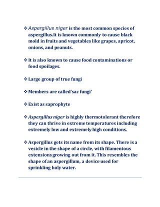
- 1. Aspergillus niger is the most common species of aspergillus.It is known commonly to cause black mold in fruits and vegetables like grapes, apricot, onions, and peanuts. It is also known to cause food contaminations or food spoilages. Large group of true fungi Members are called’sac fungi’ Exist as saprophyte Aspergillus niger is highly thermotolerant therefore they can thrive in extreme temperatures including extremely low and extremely high conditions. Aspergillus gets its name from its shape. There is a vesicle in the shape of a circle, with filamentous extensions growing out from it. This resembles the shape of an aspergillum, a device used for sprinkling holy water.
- 2. MORPHOLOGY OF Aspergillus niger Aspergillus niger is a haploid filamentous fungi Plant body is mycelia Slender, tubular, pale yellow coloured, branched, thin wall hyphae . Each cell is multinucleate and is filled with granular cytoplasm, mitochondria, endoplasmic reticulum, ribosomes and vacuoles. The cross walls between the cells have a simple pore through which the cytoplasm of the adjacent cells remain continuous. Oil globules are reserve food material A niger produce colonies that are composed of white or yellow felt that is covered by dark asexually produced fungal spores. Mycelial, or threadlike, hyphae are divided by a septum and transparent. Conidiophores. Conidiophores are smooth and haline
- 3. each conidia is spherical, echinulate and multinucleate conidial heads are present Compared to the other types, A. niger produces dark or dark brown spores from their conidial heads (biserite). This is a characteristic that has only been seen on A. niger and none of the others. The conidial head to be globose the carbon black/dark brown color of the spores (as well as the conidia) is used to distinguish A. niger from other species in the same genus. Each globose vesicle is completely covered with biseriate phialides which are projections from the conidiophore of A. niger. These phialides come out from brown metulae, which is the site where a conidiogenous cell is created. The phialides go through a process of blastic basipetal conidiogenesis to create globose mitospores. Reproducesby vegetative, asexual and sexual methods:- (i) Vegetative Reproduction: It takes place by the following methods:
- 4. (a) Fragmentation: The vegetative mycelium breaks up into small pieces (fragments) and each fragment grows independently into a new thallus under favourable conditions. (b) Sclerotia: Some species e.g., A niger, A. terreus produce sclerotia. It is more a means of keeping the fungus alive than of propagation. Sclerotia are described as resting structures that remain quiescent in the presence of adverse environmental conditions and are able to germinate when the conditions improve ii) Asexual Reproduction: Asexual reproduction takes place by the hyphae called conidiophores. and are known as foot cells . Each foot cell produces a special erect branch as an outgrowth. It is the young conidiophore. The tip of the conidiophore swells up into on elliptical or globular multinucleate head called vesicle. It forms many radially arranged tubular outgrowths called sterigmata or phialides . In some species primary sterigmata (uniseriate) bear secondary sterigmata. (bi-seriate)
- 5. Conidia, arise exogenously from the sterigmata or phialides (therefore, conidia are also called phialospores or phialoconidia) by abstraction method. They are arranged in basipetal succession (i.e., the youngest conidium is at its base and the oldest at the tip)
- 6. The sterigmata elongate at the tip to form a tube. The conidia are formed inside this. The sterigmata are uninucleate. At the time of formation of conidia the single nucleus of the phialide divide mitotically into two daughter nuclei. One of the daughter nuclei passes into the tube . It is the first conidium. As the first conidium is formed the upper broken wall of the phialide serves as a cap around
- 7. it . The second conidium is formed by phialide just below the first . The cytoplasm of both the conidia is confluent through a narrow cellular link called isthmus . The continuity of the cytoplasm is stopped by the formation of the inner conidial wall. The isthmus becomes empty and now it is called connective.
- 8. Structure and Germination of Conidia conidia are small, globose, unicellular, uninucleate or multinucleate, black, brown or yellow green in colour. They have two layered wall. Outer wall layer is thick spiny, pigmented and known as epispore, whereas the inner one is thin, delicate and is called endospore. Conidia are dispersed by wind. They germinate on suitable substratum by giving out a germ tube. The germ tube becomes septate, branched and forms a mycelium. iii) Sexual Reproduction: The sexual reproduction is of rare occurrence.Majority of the species of Aspergillus are homothallic. However,a few species are heterothallic e.g., A. heterothallicus. It takes place by the formation of male and female sex organs. Male sex organ is known as antheridium and the male branch is called Pollinodium. Female sex organ is called ascogonium and female branch is called as archicarp.
- 9. Ascogonium:Archicarp develops on the mycelium in the form of septate, loosely coiled structure. The young archicarp can be differentiated into three parts : (i) The basal multicellular, multinucleate stalk. (ii) Middle unicellular, multinucleate ascogonium (gametangium). (iii) Apical unicellular, multinucleate receptive organ called trichogyne. At first archicarp is loosely coiled but later on the coil approaches nearer and nearer and finally touch each other to form a cork screw like structure . Antheridium: Pollinodium grows up beside the archicarp on the same or adjacent hyphae . It gets spirally coiled around the archicarp and arches over the apex of ascogonium. It can be differentiated into two parts: (i) Upper part, slightly broader, unicellular, multinucleate and behaves as antheridium. (ii) Lower unicellular and multinucleate part called stalk. Fertilization: The tip of the archegonium arches over the trichogyne and fuses with it. The wall at the point of contact dissolves, thus making a continuous passage. It is plasmogamy. The contents of the antheridium pass into the ascogonium. The
- 10. pairing of male and female nuclei takes place in ascogonium . Development of Ascocarp: the ascogonium develops into a fruiting body called ascocarp . After the pairing of the nuclei, the ascogonium becomes septate. Each segment consists of one male and one female nucleus (dikaryon). From these dikaryotic segments arise ascogenous hyphae. Each ascogenous hypha is multicellular with a pair of nuclei and produces asci by crozier formation. Crozier Formation: The terminal bi-nucleate cell of the ascogenous hyphae elongate and then been itself to form a hook like structure known as crozier . Both nuclei divide in such a way thin spindle apparatus are oriented parallely in vertical direction. Now the separation takes place in crozier and it is differentiated into three cells
- 11. (i) The terminal or ultimate uninucleate cell. (ii) Sub-terminal or penultimate bi-nucleate (‘+’ and nuclei) cell, occurs at the curve position. (iii) A basal uninucleate, anti-penultimate cell. The penultimate bi-nucleate cell acts as ascus mother cell. The nuclei in these cells fuse to form diploid nucleus (karyogamy). The diploid nucleus first divides meiotically and forms four haploid nuclei. Each haploid nucleus divides meitotically and thus 8 haploid nuclei are formed
- 12. Each nucleus later on gets surrounded by cytoplasm and develops a wall. Thus, 8 haploid ascospores are formed in each ascus. The cytoplasm left over in each ascus is known as epiplasm. The asci may be globose or pear shaped. As the asci develop, a large number of sterile hyphae grow around them and form a protective covering called peridium. The entire structure is known as ascocarp. It encloses many asci. It is spherical and has no opening. Such an ascocarp is known as cleistothecium .
- 13. Structure of Ascospore: As the asci mature, the ascospores are set free by dissolution of the wall of the asci in the ascocarp. They are liberated only after the decay of the ascocarp wall. Each ascospore is pulley wheel shaped, unicellular, uninucleate . Spore wall is differentiated into two layers, the outer thick, sculpturous epispore and inner thin endospore . After falling on a suitable substratum each ascospore germinates to give rise to a germ tube which develops into a new haploid mycelium .