07 heart
•Download as PPT, PDF•
5 likes•368 views

For D.Pharm students

Recommended
More Related Content
What's hot
What's hot (20)
Similar to 07 heart
Similar to 07 heart (20)
More from Prin.K.M.Kundnani Pharmacy Polytechnic
More from Prin.K.M.Kundnani Pharmacy Polytechnic (18)
Recently uploaded
Recently uploaded (20)
07 heart
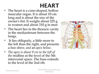
- 1. Prof.Sunil Chavan Prin.K.M.Kundnani Pharmacy Polytechnic HEART • The heart is a cone-shaped, hollow muscular organ. It is about 10 cm long and is about the size of the owner's fist. It weighs about 225 g in women and about 310 g in men • The heart lies in the thoracic cavity in the mediastinum between the lungs. • It lies obliquely, a little more to the left than the right, and presents a base above, and an apex below. • The apex is about 9 cm to the left of the midline at the level of the 5th intercostal space. The base extends to the level of the 2nd rib.
- 2. Prof.Sunil Chavan Prin.K.M.Kundnani Pharmacy Polytechnic Structure • The heart is composed of three layers of tissue: pericardium, myocardium and endocardium • Pericardium: is made up of two sacs. The outer sac consists of fibrous tissue and the inner of a continuous double layer of serous membrane. Its inelastic, fibrous nature prevents overdistension of the heart. • The outer layer of the serous membrane, the parietal pericardium. The inner layer, the visceral pericardium, or epicardium, is adherent to the heart muscle. The serous membrane consists of flattened epithelial cells. It secretes serous fluid into the space between the visceral and parietal layers which allows smooth movement between them when the heart beats. The space between the parietal and visceral pericardium is only a potential space.
- 5. Prof.Sunil Chavan Prin.K.M.Kundnani Pharmacy Polytechnic Myocardium: The myocardium is composed of specialised cardiac muscle found only in the heart .. Each fibre (cell) has a nucleus and one or more branches. The ends of the cells and their branches are in very close contact with the ends and branches of adjacent cells.
- 7. Prof.Sunil Chavan Prin.K.M.Kundnani Pharmacy Polytechnic • Microscopically these 'joints', or intercalated discs, can be seen as thicker, darker lines than the ordinary cross-stripes. This arrangement gives cardiac muscle the appearance of being a sheet of muscle rather than a very large number of individual cells. Because of the end-to-end continuity of the fibres, each one does not need to have a separate nerve supply. When an impulse is initiated it spreads from cell to cell via the branches and intercalated discs over the whole 'sheet’ of muscle, causing contraction.
- 8. Prof.Sunil Chavan Prin.K.M.Kundnani Pharmacy Polytechnic The 'sheet' arrangement of the myocardium enables the atria and ventricles to contract in a coordinated and efficient manner. The myocardium is thickest at the apex and thins out towards the base . It is thickest in the left ventricle. Endocardium: This forms the lining of the myocardium and the heart valves. It is a thin, smooth, glistening membrane which permits smooth flow of blood inside the heart. It consists of flattened epithelial cells, continuous with the endothelium that lines the blood vessels.
- 9. Prof.Sunil Chavan Prin.K.M.Kundnani Pharmacy Polytechnic Interior of the heart • The heart is divided into a right and left side by the septum, a partition consisting of myocardium covered by endocardium. • Each side is divided by an atrioventricular valve into an upper chamber, the atrium, and a lower chamber, the ventricle. • The atrioventricular valves are formed by double folds of endocardium strengthened by a little fibrous tissue. The right atrioventricular valve (tricuspid valve) has three flaps or cusps and the left atrioventricular valve (mitral valve) has two cusps.
- 11. Prof.Sunil Chavan Prin.K.M.Kundnani Pharmacy Polytechnic • During ventricular systole (contraction) the pressure in the ventricles rises above that in the atria and the valves snap shut preventing backward flow of blood. The valves are prevented from opening upwards into the atria by threads called chordae tendineae, which extend from the inferior surface of the cusps to little projections of myocardium covered with endothelium, called papillary muscles • Pulmonary valve is present between right atrium & pulmonary artery; Arotic valve is between left ventricle & arota
- 12. Prof.Sunil Chavan Prin.K.M.Kundnani Pharmacy Polytechnic Conducting system of the heart
- 13. Conducting system of heart • The heart has inbuilt system of cardiac muscle contraction. Small groups of specialized neuromuscular cells are present in myocardium. These cells conduct impulses, causing co-ordinated and synchronized contraction of the heart muscle. Prof.Sunil Chavan Prin.K.M.Kundnani Pharmacy Polytechnic
- 14. • Sinoatrial Node (SA node): • This small mass of specialised cells is in the wall of the right atrium near the opening of the superior vena cava. This node initiates impulses and spread through both atrial walls, that causes contraction of atria. • Atrioventricular node (AV node): • This small mass of neuromuscular tissue. It is situated in the wall of the atrial septum near the atrioventricular valves. AV node conducts impulses that arrive via atria. If there is problem with SA node, then AV node generates impulses. Prof.Sunil Chavan Prin.K.M.Kundnani Pharmacy Polytechnic
- 15. • Atrioventricular bundle, bundle branches, and Purkinje fibers: • This is a mass of specialized fibres that originate from the AV node. The AV bundle at the upper end of the ventricular septum, it divides into right and left bundle branches. Within the ventricular myocardium the branches break up into fine fibres, called the Purkinje fibres. • The AV bundle, bundle branches and Purkinje fibers convey electrical impulses from AV node to the apex of ventricles. It causes forceful ventricular contraction. Prof.Sunil Chavan Prin.K.M.Kundnani Pharmacy Polytechnic
- 16. Prof.Sunil Chavan Prin.K.M.Kundnani Pharmacy Polytechnic The cardiac cycle • The function of the heart is to maintain a constant circulation of blood throughout the body. The heart acts as a pump and its action consists of a series of events known as the cardiac cycle • During each heartbeat, or cardiac cycle, the heart contracts and then relaxes. The period of contraction is called systole and that of relaxation, diastole.
- 17. Prof.Sunil Chavan Prin.K.M.Kundnani Pharmacy Polytechnic Stages of the cardiac cycle • The normal number of cardiac cycles per minute ranges from 60 to 80. Taking 75 as an example each cycle lasts about 0.8 of a second and consists of: • atrial systole — contraction of the atria(0.1s) • ventricular systole — contraction of the ventricles(0.3s) • complete cardiac diastole — relaxation of the atria and ventricles(0.4s)
- 18. Prof.Sunil Chavan Prin.K.M.Kundnani Pharmacy Polytechnic •The superior vena cava and the inferior vena cava transport deoxygenated blood into the right atrium at the same time as the four pulmonary veins convey oxygenated blood into the left atrium. •The atrioventricular valves are open and blood flows through to the ventricles. The SA node triggers a wave of contraction that spreads over the myocardium of both atria, emptying the atria and completing ventricular filling (atrial systole 0.1 s).
- 19. Prof.Sunil Chavan Prin.K.M.Kundnani Pharmacy Polytechnic •When the wave of contraction reaches the AV node it is stimulated to emit an impulse which quickly spreads to the ventricular muscle via the AV bundle, the bundle branches and Purkinje fibres. This results in a wave of contraction which sweeps upwards from the apex of the heart and across the walls of both ventricles pumping the blood into the pulmonary artery and the aorta (ventricular systole 0.3 s).
- 20. Prof.Sunil Chavan Prin.K.M.Kundnani Pharmacy Polytechnic •The high pressure generated during ventricular contraction is greater than that in the aorta and forces the atrioventricular valves to close, preventing backflow of blood into the atria. • After contraction of the ventricles there is complete cardiac diastole, a period of 0.4 seconds, when atria and ventricles are relaxed. During this time the myocardium recovers until it is able to contract again, and the atria refill in preparation for the next cycle.
- 22. Prof.Sunil Chavan Prin.K.M.Kundnani Pharmacy Polytechnic Heart sounds (Auscultation): •Two sounds, separated by a short pause, can be clearly distinguished. They are described in words as 'lub dup'. •The first sound, 'lub', is fairly loud and is due to the closure of the atrioventricular valves. This corresponds with ventricular systole. •The second sound, 'dup', is softer and is due to the closure of the aortic and pulmonary valves. This corresponds with atrial systole.
- 23. Prof.Sunil Chavan Prin.K.M.Kundnani Pharmacy Polytechnic Electrical changes in the heart: • The electrical activity within the heart can be detected by attaching electrodes to the surface of the body. The pattern of electrical activity may be displayed on an oscilloscope screen or traced on paper. The apparatus used is an electrocardiograph and the tracing is an electrocardiogram (ECG). • The normal ECG tracing shows five waves which, by convention, have been named P, Q, R, S and T.
- 24. Prof.Sunil Chavan Prin.K.M.Kundnani Pharmacy Polytechnic •The P wave arises when the impulse from the SA node sweeps over the atria. •The QRS complex represents the very rapid spread of the impulse from the AV node through the AV bundle and the Purkinje fibres and the electrical activity of the ventricular muscle. •The T wave represents the relaxation of the ventricular muscle.
- 25. Prof.Sunil Chavan Prin.K.M.Kundnani Pharmacy Polytechnic •The ECG described originates from the SA node and is known as sinus rhythm. The rate of sinus rhythm is 60 to 100 beats per minute. •By examining the pattern of waves and the time interval between cycles and parts of cycles, information about the state of the myocardium and the cardiac conduction system is obtained.
- 26. Prof.Sunil Chavan Prin.K.M.Kundnani Pharmacy Polytechnic Cardiac output Cardiac output = Stroke volume x Heart rate • stroke volume: The amount of blood expelled by each contraction of the ventricles. • Cardiac output is expressed in liters per minute (l/min) • In a healthy adult at rest, the stroke volume is approximately 70 ml and if the heart rate is 72 per minute, the cardiac output is 5 l/minute. • This can be greatly increased to meet the demands of exercise by 5 – 7 times.
- 28. Prof.Sunil Chavan Prin.K.M.Kundnani Pharmacy Polytechnic Factors affecting Stroke volume • Ventricular end-diastolic volume: The volume of blood in the ventricles immediately before they contract, i.e. the (VEDV), sometimes called preload. This depends on the amount of blood returning to the heart through the superior and inferior venae cavae (the venous return). Increased VEDV leads to stronger myocardial contraction, and more blood is expelled.
- 29. Prof.Sunil Chavan Prin.K.M.Kundnani Pharmacy Polytechnic •Venous return: • the major determinant of cardiac output • The force of contraction of the left ventricle ejecting blood into the aorta is not sufficient to return the blood through the veins and back to the heart. Other factors are involved. The position of the body: Gravity assists the venous return from the head and neck when standing or sitting and offers less resistance to venous return from the lower parts of the body when an individual is lying flat.
- 30. Prof.Sunil Chavan Prin.K.M.Kundnani Pharmacy Polytechnic Muscular contraction: Back flow of blood in veins of the limbs, especially when standing, is prevented by valves. The contraction of skeletal muscles surrounding the deep veins puts pressure on them, pushing blood towards the heart. In the lower limbs, this is called the skeletal muscle pump. When the pressure in deep veins is lowered during muscle relaxation, blood flows into them from superficial veins through communicating veins.
- 31. Prof.Sunil Chavan Prin.K.M.Kundnani Pharmacy Polytechnic The respiratory pump: During inspiration the expansion of the chest creates a negative pressure within the thorax, assisting flow of blood towards the heart. In addition, when the diaphragm descends during inspiration, the increased intra-abdominal pressure pushes blood towards the heart. •Strength of myocardial contraction: factors that increase myocardial contraction include: • increased stimulation of the sympathetic nerves innervating the heart • hormones, e.g. adrenaline, noradrenaline, thyroxine •Blood volume: This is normally kept constant by the kidneys and if deficient the stroke volume, cardiac output and venous return decrease.
- 32. Prof.Sunil Chavan Prin.K.M.Kundnani Pharmacy Polytechnic Factors affecting heart rate • Autonomic nervous system: The rate at which the heart beats is a balance of sympathetic and parasympathetic activity and this is the most important factor in determining heart rate. • Circulating chemicals: The hormones adrenaline and noradrenaline, secreted by the adrenal medulla, have the same effect as sympathetic stimulation, i.e. they increase the heart rate. Other hormones including thyroxine increase heart rate by their metabolic effect. Some drugs, dissolved gases and electrolytes in the blood may either increase or decrease the heart rate.
- 33. Prof.Sunil Chavan Prin.K.M.Kundnani Pharmacy Polytechnic Position: When the person is upright, the heart rate is usually faster than when lying down. Exercise: Active muscles need more blood than resting muscles and this is achieved by an increased heart rate and selective vasodilatation. Emotional states: During excitement, fear or anxiety the heart rate is increased. Other effects mediated by the sympathetic nervous system may be present. Gender: The heart rate is faster in women than men. Age: In babies and small children the heart rate is more rapid than in older children and adults Temperature: The heart rate rises and falls with body temperature
- 34. Prof.Sunil Chavan Prin.K.M.Kundnani Pharmacy Polytechnic Baroreceptor reflex: • Baroreceptors are nerve endings sensitive to pressure changes (stretch) within the vessel, situated in the arch of the aorta and in the carotid sinuses. • A rise in blood pressure in these arteries stimulates the baroreceptors, increasing their input to the CVC. The CVC responds by increasing parasympathetic nerve activity to the heart; this slows the heart down. At the same time, sympathetic stimulation to the blood vessels is inhibited, causing vasodilatation. The net result is a fall in systemic blood pressure.
- 35. Prof.Sunil Chavan Prin.K.M.Kundnani Pharmacy Polytechnic Conversely, if pressure within the aortic arch and carotid sinuses falls, the rate of baroreceptor discharge also falls. The CVC responds by increasing sympathetic drive to the heart to speed it up. Sympathetic activity in blood vessels is also increased, leading to vasoconstriction. Both these measures counteract the falling blood pressure.