SlideShare a Scribd company logo
1 of 66
Prof.Sunil Chavan Prin.K.M.Kundnani Pharmacy
Polytechnic
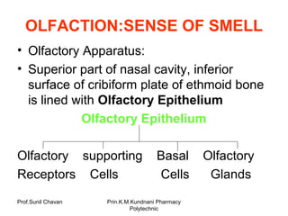
OLFACTION:SENSE OF SMELL
• Olfactory Apparatus:
• Superior part of nasal cavity, inferior
surface of cribiform plate of ethmoid bone
is lined with Olfactory Epithelium
Olfactory Epithelium
Olfactory supporting Basal Olfactory
Receptors Cells Cells Glands
Prof.Sunil Chavan Prin.K.M.Kundnani Pharmacy
Polytechnic
Prof.Sunil Chavan Prin.K.M.Kundnani Pharmacy
Polytechnic
Olfactory Receptors:
• These are first order neurons of Olfactory
Pathway
• Each Olfactory receptor is a bipolar
neuron
• Knob shaped dendrites have olfactory
hairs, cilia, these causes transduction.
• Axons project through cribiform plate &
end in Olfactory bulb
• Olfactory receptors respond to chemical
stimulation of an odorant molecule by
producing nerve impulse.
Prof.Sunil Chavan Prin.K.M.Kundnani Pharmacy
Polytechnic
Prof.Sunil Chavan Prin.K.M.Kundnani Pharmacy
Polytechnic
Supporting Cells:
• These are columnar epithelium cells of
mucus lining of nose.
• Provide physical support, nourishment &
electrical insulation for olfactory receptors.
Basal Cells:
• These are stem cells, located between
bases of supporting cells.
• These continually undergo cell division to
produce new olfactory receptors.
Prof.Sunil Chavan Prin.K.M.Kundnani Pharmacy
Polytechnic
Olfactory Gland (Bowman’s):
• These are present within connective
tissue that support olfactory epithelium.
• These produce mucus that is carried to
surface to epithelium.
• Secretion moisten surface & dissolves
odorants, so that transduction can occur.
Prof.Sunil Chavan Prin.K.M.Kundnani Pharmacy
Polytechnic
Physiology of Olfaction
Odorant molecule binds to olfactory hair
It causes transduction
Propagation of impulse along axon of
olfactory receptor
In some cases odorant binds to G-Protein
in Plasma membrane of olfactory receptor
activates enzyme adenylate cyclase
Production of cAMP Influx of Na+
Generation of impulse & propagation of
impulse
Prof.Sunil Chavan Prin.K.M.Kundnani Pharmacy
Polytechnic
Olfactory Pathway
• On each side of nose, bundles of axons of
olfactory receptors extend through 20 olfactory
foramina in cribiform plate
• These bundles collectively form Right & Left
Olfactory Nerve (I).
• These nerve terminate in olfactory bulb. Here
axons of receptors form synapses with dendrites
of second order neurons in olfactory
pathway.
• Axons of olfactory bulb extend posteriorly
& form olfactory Tract
Prof.Sunil Chavan Prin.K.M.Kundnani Pharmacy
Polytechnic
Olfactory Tract
Some axons project to primary olfactory
area conscious awareness of smell
begins
Other axons projected to Limbic system &
Hypothalamus Responsible for
emotional & memory evoked responses to
odors
Prof.Sunil Chavan Prin.K.M.Kundnani Pharmacy
Polytechnic
GUSTATION:SENSE OF
TASTE
• Taste-Chemical sense
• Five primary tastes: Sour, Bitter, Sweet, Salty, &
U….
• Anatomy of Taste Buds & Papillae
• Vallate: Circular, large, 8-12, form inverted V
shape. Each papilla has 100-300 taste buds.
• Fungiform: Mushroom shapr, entire surface,
each papilla has about 5 taste buds.
• Foliate: Located in small trenches on lateral
margins. These degenerate in early childhood
Prof.Sunil Chavan Prin.K.M.Kundnani Pharmacy
Polytechnic
Prof.Sunil Chavan Prin.K.M.Kundnani Pharmacy
Polytechnic
Taste Buds: Receptors for
sensation of taste
• About 10,000 TB in young adult
• Most of them on tongue, some of them on
soft palate, pharynx & epiglottis.
• No. of TB declines with age.
• Oval body-consist of 3 kinds of epithelial
cells: Supporting cell, Gustatory receptor
cell Basal cell
• Supporting cell: Surround about 50
gustatory receptor cells in each taste bud.
Prof.Sunil Chavan Prin.K.M.Kundnani Pharmacy
Polytechnic
• Gustatory Receptor Cell: Each cell
projects single long microvillus-Gustatory
hair.
• Basal Cell; Present at periphery, produce
supporting cells Gustatory receptor
cell. Each has 10 days life span.
• At the base gustatory receptor cell
synapses with dendrites of first-order
neuron. This neuron forms the first part of
gustatory pathway.
Prof.Sunil Chavan Prin.K.M.Kundnani Pharmacy
Polytechnic
Physiology of Gustation
Testant Dissolved in Saliva
Gustatory hair
Transduction
Receptor Potential
Exocytosis of Synaptic vesicles
Liberation of Neurotransmitter
Trigger nerve impulse
Prof.Sunil Chavan Prin.K.M.Kundnani Pharmacy
Polytechnic
SWEET, BITTER, UMAMI TESTANTS
• Binds to receptors on plasma membrane
linked to G-proteins
Activates several different chemicals
(secondary
messengers)
Release of Neurotransmitter
Prof.Sunil Chavan Prin.K.M.Kundnani Pharmacy
Polytechnic
Salty food: Na +
from testants
Na+
Gustatory receptor Cell
Accumulation of Na+
inside
Opening of Ca2+
channels
Influx of Ca2+
Exocytosis of Synaptic Vesicles
Sour Food: H+
from testants
Prof.Sunil Chavan Prin.K.M.Kundnani Pharmacy
Polytechnic
Gustatory Pathway
Taste Buds Taste Buds Taste buds in
In Ant.2/3 in post. 1/3 throat, epiglottis
Facial Glossopharyngeal Vagus
Nerve(VII) Nerve(IX) Nerve(X)
Medulla Oblongata
Limbic System Thalamus
& Hypothalamus
Primary Gustatory Area
(Parietal lobe)
Prof.Sunil Chavan Prin.K.M.Kundnani Pharmacy
Polytechnic
EYE : SENSE OF VISION (SIGHT)
Vision: important to human
survival
More than half sensory receptors
Larger part of cerebral cortex
Prof.Sunil Chavan Prin.K.M.Kundnani Pharmacy
Polytechnic
Anatomy of Eye Ball
Wall of Eyeball
Fibrous Vascular Retina
layer layer Neuronal layer
-Cornea -Choroid
-Sclera -Ciliary body
-Iris
Prof.Sunil Chavan Prin.K.M.Kundnani Pharmacy
Polytechnic
Prof.Sunil Chavan Prin.K.M.Kundnani Pharmacy
Polytechnic
Interior of Eye ball
Aqueous Vitreous Lens
Humour body
Prof.Sunil Chavan Prin.K.M.Kundnani Pharmacy
Polytechnic
Fibrous Layer:
• Outer coat of eyeball
• Consist of anterior cornea, posterior sclera
CORNEA:
-Transparent epithelial coat, covers iris
-Curved shape helps focus light into retina
SCLERA (white of eye):
-Layer of dense connective tissue, mostly collagen
fibers & fibroblasts
-Covers eyeball except cornea
-Gives shape to eyeball, make it more rigid,
protects inner parts
Prof.Sunil Chavan Prin.K.M.Kundnani Pharmacy
Polytechnic
Canal of Schlemm:
• An opening at junction of sclera & cornea
• It drains aqueous humour from anterior
chamber
Prof.Sunil Chavan Prin.K.M.Kundnani Pharmacy
Polytechnic
Vascular Layer:
• Middle layer of the eyeball
• Three parts: Choroid, Ciliary body, Iris
CHOROID:
-Posterior portion of meddle layer
-Lines most of internal surface of sclera
-Rich in blood vessels, provides nutrients to retina
-Contains melanocytes, produces melanin
-Melanin absorbs stray light rays, prevent
reflection & scattering of light within eyeball
Sharp & clear image
Prof.Sunil Chavan Prin.K.M.Kundnani Pharmacy
Polytechnic
CILIARY BODY:
-Anterior portion of choroid
-Consists of Ciliary Processes & Ciliary Muscle
Ciliary processes are folds on internal side,
contain blood capillaries, secrete aqueous
humour
Suspensory Ligaments (Zonular fibers):
-Extends from Processes to lens
Ciliary muscle- circular band of smooth muscle.
Its contraction/relaxation Tightness of zonular
fibers alters shape of lens
Prof.Sunil Chavan Prin.K.M.Kundnani Pharmacy
Polytechnic
Prof.Sunil Chavan Prin.K.M.Kundnani Pharmacy
Polytechnic
IRIS:
-Coloured portion of eyeball
-Suspended between Cornea & Lens
-Melanocytes, Circular & Radial smooth
muscle fibers
Amount of melanin Colour of eye
-Regulate amount of light entering
through pupil
Prof.Sunil Chavan Prin.K.M.Kundnani Pharmacy
Polytechnic
Prof.Sunil Chavan Prin.K.M.Kundnani Pharmacy
Polytechnic
Retina:
-Innermost layer of eyeball
-Lines about ¾ of eyeball
Thickest at the back, thins out interiorly,
ends just behind ciliary body
-Consists of Pigmented layer & Neural layer
Pigmented Layer:
-Made up of melanocytes
-Melanin helps to absorb stray light
Prof.Sunil Chavan Prin.K.M.Kundnani Pharmacy
Polytechnic
Prof.Sunil Chavan Prin.K.M.Kundnani Pharmacy
Polytechnic
NEURAL LAYER:
Three distinct layers of retinal neurons:
Photoreceptor Layer, Bipolar layer &
Ganglion Cell Layer
-Separated by two zones: Outer & Inner
Synaptic layer
PHOTORECEPTORS:
Specialized cells, begin process of
conversion of light rays to nerve impulse
Prof.Sunil Chavan Prin.K.M.Kundnani Pharmacy
Polytechnic
Prof.Sunil Chavan Prin.K.M.Kundnani Pharmacy
Polytechnic
-Cells: two types;
ROD- outer segment is cylindrical
CONE- outer segment cone shaped
-Each retina has about 6 million cones &
120 million rods
-Rods allow us to see in dim light
-Cones produce colour vision
Axons of ganglion cells extend posteriorly
exit at a site called OPTIC DISC (Blind
Spot) as Optic Nerve.
Prof.Sunil Chavan Prin.K.M.Kundnani Pharmacy
Polytechnic
INTERIOR OF EYEBALL
LENS:
-Highly elastic circular biconvex transparent
body
-Located behind pupil & iris, enclosed in
connective tissue capsule
-Held in position by zonular fibers, attached to
ciliary processes
-Consists of protein Crystallins
-Lens help focus image on retina by refraction,
facilitates clear vision
-Vary its refracting power by changing its
thickness
Prof.Sunil Chavan Prin.K.M.Kundnani Pharmacy
Polytechnic
LENS DIVIDES interior of eyeball into two
cavities: Anterior Cavity & Vitreous
Chamber
ANTERIOR CAVITY: Consists of two
chambers:
Anterior Chamber- between cornea & Iris
Posterior Chamber-behind Iris & infront of
lens & zonular fibers
Both chamber are filled with aqueous
humour-watery fluid nourishes lens &
cornea.
Prof.Sunil Chavan Prin.K.M.Kundnani Pharmacy
Polytechnic
Aqueous humour filters out of blood
capillaries in ciliary processes & enters
posterior chamber
-Flows forward between iris & lens, through
pupil into anterior chamber
-From ant. Chamber it drains into Canal of
Schlemm, and then into blood.
-it is completely replaced about every 90
minutes
Prof.Sunil Chavan Prin.K.M.Kundnani Pharmacy
Polytechnic
VITREOUS CHAMBER:
-lies between lens & retina
-Contains viscous substance:Vitreous Body
-Formed during embryonic development not
replaced thereafter.
-It contains 99% water, mucoprotein, salts
and phagocytic cells
-It prevents collapsing of eye walls
-It keeps this part clear for unobstructed
vision
Prof.Sunil Chavan Prin.K.M.Kundnani Pharmacy
Polytechnic
INTRA-OCULAR PRESSURE:
-Produced mainly by Aq. Humour & partly by
Vitreous body
-normal 16 mm Hg (15 – 20 mm Hg)
-maintains shape of Eyeball & prevents it
from collapsing
GLAUCOMA:
Condition in which there is increased
intraocular pressure due to defective
drainage of aqueous humour through
canal of Schlemm.
Prof.Sunil Chavan Prin.K.M.Kundnani Pharmacy
Polytechnic
Accessory structures of Eye
Eyelids, Eyelashes, Eyebrows, Lacrimal
Apparatus & Extrinsic eye Muscles
Superior rectusrotates eyeball upwards
Inferior rectus rotates eyeball downwards
Lateral rectusrotates eyeball outwards
Medial rectusrotates eyeball inwards
Superior obliquerotates eyeball so that
cornea turns in downwards & outwards
direction
Inferior obliquecornea turns upward &
outward direction
Prof.Sunil Chavan Prin.K.M.Kundnani Pharmacy
Polytechnic
PHYSIOLOGY OF VISION
• ISOMERIZATION OF RETINAL:
Retinal in photoreceptor cells present in cis- form
in darkness. This absorbs light (photon) and
converts into trans- form
• RELEASE OF NEUROTRANSMITTER:
Isomerization activates enzyme that breaks down
cyclic Guanosine Monophosphate (cGMP)
closure of cGMP gated Na+ channels Na+
influx Membrane potential more negative
affects release of Glutamate
(Hyperpolarisation)
Prof.Sunil Chavan Prin.K.M.Kundnani Pharmacy
Polytechnic
DIM LIGHT-small & brief receptor potential
that partially turnoff glutamate release
BRIGHT LIGHT-larger & longer receptor
potential that completely shut down
glutamate release
Excites bipolar cell & subsequently
stimulates ganglion cells to generate
nerve impulse in their axons
Axons of all retinal ganglion cells exit
eyeball at Optic disc and form Optic Nerve
(II)
Prof.Sunil Chavan Prin.K.M.Kundnani Pharmacy
Polytechnic
Visual Pathway
• At the optic chaism axons from temporal half of
each retina do not cross, continue directly to
thalamus on same side
• Axons from nasal half of each retina cross optic
chaism and continue to opposite thalamus
• Each optic tract consists of crossed & uncrossed axons
• Branches of axon project to midbrain that
govern constriction of pupils in response to light
& co-ordination of head & Eye movements
• Axons of thalamic neurons project to primary
visual area in occipital lobe of cortex image is
perceived
Prof.Sunil Chavan Prin.K.M.Kundnani Pharmacy
Polytechnic
Prof.Sunil Chavan Prin.K.M.Kundnani Pharmacy
Polytechnic
EAR:SENSE OF SOUND
(HEARING)
Ear- an engineering marvel,
contains receptors for Hearing &
Equilibrium
Transmits 1000 times faster
than photoreceptors
Prof.Sunil Chavan Prin.K.M.Kundnani Pharmacy
Polytechnic
ANATOMY OF EAR
External Ear Middle Ear Internal ear
-Auricle (tympanic cavity) (Labyrinth)
-Auditory canal -Auditory Ossicles -Bony
(acoustic 1.Malleus
-Membranous
meatus) 2.Incus
3.Stapes
-Eustachian Tube
Prof.Sunil Chavan Prin.K.M.Kundnani Pharmacy
Polytechnic
Prof.Sunil Chavan Prin.K.M.Kundnani Pharmacy
Polytechnic
External Ear
AURICLE (Pinna):
• Expanded portion from side of head
• Composed of fibroelastic cartilage
• Deeply grooved & ridged
• Prominent outer ridge-Helix
• Soft inferior portion-Lobule, composed of
fibrous & adipose tissue, richly supplied
with blood capillaries
Prof.Sunil Chavan Prin.K.M.Kundnani Pharmacy
Polytechnic
EXTERNAL AUDITORY CANAL
(ACOUSTIC MEATUS):
• Slightly curved tube, about 2.5cm long,
lies in temporal bone, extended from
auricle to tympanic membrane
• Lined with hairy skin, continues with auricle
• Contains numerous specialized sweat glands-
Ceruminus glands, secret earwax, sticky
material containing lysozome & immunoglobins
• Prevent foreign material like dust, insects,
microbes reaching eardrum.
Function:Collection of sound waves &
channel them inward  eardrum
Prof.Sunil Chavan Prin.K.M.Kundnani Pharmacy
Polytechnic
TYMPANIC MEMBRANE/EARDRUM
• Thin partition between auditory canal &
tympanic cavity
• Oval shaped, slightly broader edge
upwards
• Composed of three layers of tissues-
-outer covering of hairless skin
-middle layer of fibrous tissue
-inner lining of mucus membrane
FUNCTION: production of vibrations, as
sound waves strike it.
Prof.Sunil Chavan Prin.K.M.Kundnani Pharmacy
Polytechnic
Prof.Sunil Chavan Prin.K.M.Kundnani Pharmacy
Polytechnic
Middle Ear/Tympanic Cavity
• Small, irregular, air-filled cavity in temporal bone
• Cavity & its contents are lined with either simple
squamous or cuboidal epithelium
• Extend till oval window & round window of
internal ear
AUDITORY OSSICLES:
• Three very small bones extend across cavity-
Malleus, Incus, Stapes
• Attached to cavity by ligaments
• Connected by synovial joints
Prof.Sunil Chavan Prin.K.M.Kundnani Pharmacy
Polytechnic
Prof.Sunil Chavan Prin.K.M.Kundnani Pharmacy
Polytechnic
MALLEUS:
• Handle is in contact with eardrum
• Head forms joint with incus
INCUS:
• Body articulates with malleus, long
process with stapes, stabilized by short
process
STAPES:
• Head articulates with incus, base or
footplate fits into oval window
Prof.Sunil Chavan Prin.K.M.Kundnani Pharmacy
Polytechnic
EUSTACHIAN TUBE (AUDITORY)
• About 4 cm long tube, connects middle
ear with nasopharynx, lined with ciliated
epithelium
• Normally closed, opens during swallowing,
yawning, sneezing
• Balanced pressure allows eardrum
vibrates freely as sound waves strike it
FUNCTION: Transmission of vibrations till
oval window, work as piston, its action add
force
Prof.Sunil Chavan Prin.K.M.Kundnani Pharmacy
Polytechnic
Prof.Sunil Chavan Prin.K.M.Kundnani Pharmacy
Polytechnic
Inner Ear (Labyrinth)
• Contains the organs of hearing and
balance.
• Described in two parts, the bony labyrinth
and the membranous labyrinth.
BONY LABYRINTH:
• This is a cavity within the temporal bone
lined with periosteum.
• The bony labyrinth consists of:
1 vestibule , 1 cochlea , 3 semicircular
canals
Prof.Sunil Chavan Prin.K.M.Kundnani Pharmacy
Polytechnic
VESTIBULE:
• Expanded part nearest the middle ear
• Contains the oval and round windows in
its
lateral wall.
COCHLEA:
• Has a broad base where it is continuous with the
vestibule and a narrow apex, and it spirals round
a central bony column.
THE SEMICIRCULAR CANALS:
• These are three tubes arranged so that one is
situated in each of the three planes of space.
They are continuous with the vestibule.
Prof.Sunil Chavan Prin.K.M.Kundnani Pharmacy
Polytechnic
MEMBARNOUS LABYRINTH
• Contains endolymph
• It comprises of: vestibule, cochlea & 3
semicircular canals
COCHLEA: contains three compartments:
-the scala vestibuli
-the scala media, or cochlear duct
-the scala tympani.
Prof.Sunil Chavan Prin.K.M.Kundnani Pharmacy
Polytechnic
Prof.Sunil Chavan Prin.K.M.Kundnani Pharmacy
Polytechnic
Cochlear duct:
• It is triangular in shape
• On the base of triangle there are
supporting cells and specialised cochlear
hair cells containing auditory receptors.
• These cells for Spiral organ (of Corti) that
responds to vibrations
• Auditory receptors are dendrites of
efferent nerves that form cohlear nevre,
part of 8th
cranial nerve
Prof.Sunil Chavan Prin.K.M.Kundnani Pharmacy
Polytechnic
Prof.Sunil Chavan Prin.K.M.Kundnani Pharmacy
Polytechnic
PHYSIOLOGY OF HEARING
1. The auricle because of it shape
concentrates sound waves and direct
them along auditory canal
2. Soundwaves stike eardrumproduces
vibrations
3. Transmission of vibrations across middle
ear via auditory ossicles
4. As stapes moves back & froth it pushes
membrane of oval window in & out
Prof.Sunil Chavan Prin.K.M.Kundnani Pharmacy
Polytechnic
5. Movement of oval window sets up fluid
pressure waves in the perilymph of
cochlea. It pushes perilymph of scala
vestibuli
6. Transmission of pressure from scala
vestibuli to scala tympani to roun window
7. Pressure in scala vestibuli & scala
tympani transmits pressure waves in
endolymph inside cochlear duct
Prof.Sunil Chavan Prin.K.M.Kundnani Pharmacy
Polytechnic
8. Pressure waves in endolymph causes
basilar membrane to vibrate, it moves
hair cells of spiral organ against tectorial
membrane, bending of hair cells of spiral
organ against tectorial membrane
9. Bending of hair cell Stereocilia produces
receptor potentialgeneration of nerve
imp[ulse
Prof.Sunil Chavan Prin.K.M.Kundnani Pharmacy
Polytechnic
Prof.Sunil Chavan Prin.K.M.Kundnani Pharmacy
Polytechnic
AUDITORY PATHWAY
First order sensory neuron (cochlear
branch) Cochlear nuvlei in medulla
oblongataOlivery nuclei in
ponsInferior colicullus of midbrain
Geniculate nucleus of Thalamus
Primary Auditory Area in superior
temporal gyrus of Cerebral cortex

More Related Content

What's hot

Sensory organs ; ears and nose
Sensory organs ; ears and noseSensory organs ; ears and nose
Sensory organs ; ears and noseBasraJaswinder
 
15 inst guide
15 inst guide15 inst guide
15 inst guideTheSlaps
 
Physiology of nose, olfaction and pathway
Physiology of nose, olfaction and pathwayPhysiology of nose, olfaction and pathway
Physiology of nose, olfaction and pathwayanunya reddy
 
Sense organs: Nose (The Sense of Smell)
Sense organs: Nose (The Sense of Smell)Sense organs: Nose (The Sense of Smell)
Sense organs: Nose (The Sense of Smell)PradeepKumarPoudel
 
Physiology of olfaction
Physiology of olfactionPhysiology of olfaction
Physiology of olfactionKarthik Raja
 
Special senses ear ppt
Special senses  ear pptSpecial senses  ear ppt
Special senses ear pptHemant Vanjari
 
Physiology and theories of olfaction
Physiology and theories of olfactionPhysiology and theories of olfaction
Physiology and theories of olfactionLakhan M S
 
Physiology of hearing,smell and taste
Physiology of hearing,smell and tastePhysiology of hearing,smell and taste
Physiology of hearing,smell and tastedina merzeban
 
Special senses olfaction
Special senses   olfactionSpecial senses   olfaction
Special senses olfactionAmbika Jawalkar
 

What's hot (13)

Physio ear
Physio   earPhysio   ear
Physio ear
 
Sensory organs ; ears and nose
Sensory organs ; ears and noseSensory organs ; ears and nose
Sensory organs ; ears and nose
 
15 inst guide
15 inst guide15 inst guide
15 inst guide
 
Physiology of nose, olfaction and pathway
Physiology of nose, olfaction and pathwayPhysiology of nose, olfaction and pathway
Physiology of nose, olfaction and pathway
 
Olfaction
OlfactionOlfaction
Olfaction
 
Sense organs: Nose (The Sense of Smell)
Sense organs: Nose (The Sense of Smell)Sense organs: Nose (The Sense of Smell)
Sense organs: Nose (The Sense of Smell)
 
Sensory organs
Sensory organsSensory organs
Sensory organs
 
Physiology of olfaction
Physiology of olfactionPhysiology of olfaction
Physiology of olfaction
 
Sensory Disorders
Sensory DisordersSensory Disorders
Sensory Disorders
 
Special senses ear ppt
Special senses  ear pptSpecial senses  ear ppt
Special senses ear ppt
 
Physiology and theories of olfaction
Physiology and theories of olfactionPhysiology and theories of olfaction
Physiology and theories of olfaction
 
Physiology of hearing,smell and taste
Physiology of hearing,smell and tastePhysiology of hearing,smell and taste
Physiology of hearing,smell and taste
 
Special senses olfaction
Special senses   olfactionSpecial senses   olfaction
Special senses olfaction
 

Similar to The Anatomy and Physiology of Vision and Olfaction

special sense organs (anatomy and physiology) - a brief discussion
special sense organs (anatomy and physiology) - a brief discussion special sense organs (anatomy and physiology) - a brief discussion
special sense organs (anatomy and physiology) - a brief discussion Pallab Nath
 
Lecture 7 special sense
Lecture 7   special senseLecture 7   special sense
Lecture 7 special sensemissazyaziz
 
160456 water-template-16x9
160456 water-template-16x9160456 water-template-16x9
160456 water-template-16x9bennardnicholas1
 
BIOL 2304 Chapter 17 - regarding sensory organs
BIOL 2304 Chapter 17 - regarding sensory organsBIOL 2304 Chapter 17 - regarding sensory organs
BIOL 2304 Chapter 17 - regarding sensory organsjdbsfz
 
Special senses
Special sensesSpecial senses
Special sensesdrjopogs
 
Somatic and special senses
Somatic and special sensesSomatic and special senses
Somatic and special sensesSoneeshah
 
Chapter16 specialsensesmarieb
Chapter16 specialsensesmariebChapter16 specialsensesmarieb
Chapter16 specialsensesmariebLawrence James
 
lecture 6 special senses.ppt
lecture 6 special senses.pptlecture 6 special senses.ppt
lecture 6 special senses.pptWILLIAMSADU1
 
lecture 6 special senses.ppt
lecture 6 special senses.pptlecture 6 special senses.ppt
lecture 6 special senses.pptWILLIAMSADU1
 
Cranial nerve examination / dental implant courses
Cranial nerve examination / dental implant coursesCranial nerve examination / dental implant courses
Cranial nerve examination / dental implant coursesIndian dental academy
 
Physioloy of olfaction and olfactory dysfunctions
Physioloy of olfaction and olfactory dysfunctionsPhysioloy of olfaction and olfactory dysfunctions
Physioloy of olfaction and olfactory dysfunctionsYousuf Choudhury
 

Similar to The Anatomy and Physiology of Vision and Olfaction (20)

special sense organs (anatomy and physiology) - a brief discussion
special sense organs (anatomy and physiology) - a brief discussion special sense organs (anatomy and physiology) - a brief discussion
special sense organs (anatomy and physiology) - a brief discussion
 
Secial senses (1)
Secial senses (1)Secial senses (1)
Secial senses (1)
 
Lecture 7 special sense
Lecture 7   special senseLecture 7   special sense
Lecture 7 special sense
 
13 digestive system
13 digestive system13 digestive system
13 digestive system
 
160456 water-template-16x9
160456 water-template-16x9160456 water-template-16x9
160456 water-template-16x9
 
BIOL 2304 Chapter 17 - regarding sensory organs
BIOL 2304 Chapter 17 - regarding sensory organsBIOL 2304 Chapter 17 - regarding sensory organs
BIOL 2304 Chapter 17 - regarding sensory organs
 
Special senses
Special sensesSpecial senses
Special senses
 
Chapter16 specialsensesmarieb
Chapter16 specialsensesmariebChapter16 specialsensesmarieb
Chapter16 specialsensesmarieb
 
Special senses.ppsx
Special senses.ppsxSpecial senses.ppsx
Special senses.ppsx
 
Somatic and special senses
Somatic and special sensesSomatic and special senses
Somatic and special senses
 
Chapter16 specialsensesmarieb
Chapter16 specialsensesmariebChapter16 specialsensesmarieb
Chapter16 specialsensesmarieb
 
Bio 166 presentation-A
Bio 166 presentation-ABio 166 presentation-A
Bio 166 presentation-A
 
Receptors
ReceptorsReceptors
Receptors
 
lecture 6 special senses.ppt
lecture 6 special senses.pptlecture 6 special senses.ppt
lecture 6 special senses.ppt
 
lecture 6 special senses.ppt
lecture 6 special senses.pptlecture 6 special senses.ppt
lecture 6 special senses.ppt
 
TASTE & SMELL
TASTE & SMELLTASTE & SMELL
TASTE & SMELL
 
Cranial nerve examination / dental implant courses
Cranial nerve examination / dental implant coursesCranial nerve examination / dental implant courses
Cranial nerve examination / dental implant courses
 
Physiology of smell
Physiology of smellPhysiology of smell
Physiology of smell
 
Physioloy of olfaction and olfactory dysfunctions
Physioloy of olfaction and olfactory dysfunctionsPhysioloy of olfaction and olfactory dysfunctions
Physioloy of olfaction and olfactory dysfunctions
 
Sense organs
Sense organsSense organs
Sense organs
 

More from Prin.K.M.Kundnani Pharmacy Polytechnic (17)

15 reproductive system
15 reproductive system15 reproductive system
15 reproductive system
 
14 endocrine system
14 endocrine system14 endocrine system
14 endocrine system
 
11 nerous system1
11 nerous system111 nerous system1
11 nerous system1
 
11 nervous tissue
11 nervous tissue11 nervous tissue
11 nervous tissue
 
10 muscular tissue
10 muscular tissue10 muscular tissue
10 muscular tissue
 
09 urinary system
09 urinary system09 urinary system
09 urinary system
 
08 respiratory system
08 respiratory system08 respiratory system
08 respiratory system
 
07 heart
07 heart07 heart
07 heart
 
07 general circulation
07 general circulation07 general circulation
07 general circulation
 
06 lymphatic system
06 lymphatic system06 lymphatic system
06 lymphatic system
 
05 blood liquid connective tissue
05 blood liquid connective tissue05 blood liquid connective tissue
05 blood liquid connective tissue
 
04 joints
04 joints04 joints
04 joints
 
04 bone
04 bone04 bone
04 bone
 
04 skeleton
04 skeleton04 skeleton
04 skeleton
 
03 tissues 1
03 tissues 103 tissues 1
03 tissues 1
 
02 cell
02 cell02 cell
02 cell
 
01 hap anatomical terms
01 hap anatomical terms01 hap anatomical terms
01 hap anatomical terms
 

Recently uploaded

call girls in Kamla Market (DELHI) 🔝 >༒9953330565🔝 genuine Escort Service 🔝✔️✔️
call girls in Kamla Market (DELHI) 🔝 >༒9953330565🔝 genuine Escort Service 🔝✔️✔️call girls in Kamla Market (DELHI) 🔝 >༒9953330565🔝 genuine Escort Service 🔝✔️✔️
call girls in Kamla Market (DELHI) 🔝 >༒9953330565🔝 genuine Escort Service 🔝✔️✔️9953056974 Low Rate Call Girls In Saket, Delhi NCR
 
BASLIQ CURRENT LOOKBOOK LOOKBOOK(1) (1).pdf
BASLIQ CURRENT LOOKBOOK  LOOKBOOK(1) (1).pdfBASLIQ CURRENT LOOKBOOK  LOOKBOOK(1) (1).pdf
BASLIQ CURRENT LOOKBOOK LOOKBOOK(1) (1).pdfSoniaTolstoy
 
ENGLISH5 QUARTER4 MODULE1 WEEK1-3 How Visual and Multimedia Elements.pptx
ENGLISH5 QUARTER4 MODULE1 WEEK1-3 How Visual and Multimedia Elements.pptxENGLISH5 QUARTER4 MODULE1 WEEK1-3 How Visual and Multimedia Elements.pptx
ENGLISH5 QUARTER4 MODULE1 WEEK1-3 How Visual and Multimedia Elements.pptxAnaBeatriceAblay2
 
Science lesson Moon for 4th quarter lesson
Science lesson Moon for 4th quarter lessonScience lesson Moon for 4th quarter lesson
Science lesson Moon for 4th quarter lessonJericReyAuditor
 
Computed Fields and api Depends in the Odoo 17
Computed Fields and api Depends in the Odoo 17Computed Fields and api Depends in the Odoo 17
Computed Fields and api Depends in the Odoo 17Celine George
 
Blooming Together_ Growing a Community Garden Worksheet.docx
Blooming Together_ Growing a Community Garden Worksheet.docxBlooming Together_ Growing a Community Garden Worksheet.docx
Blooming Together_ Growing a Community Garden Worksheet.docxUnboundStockton
 
Introduction to ArtificiaI Intelligence in Higher Education
Introduction to ArtificiaI Intelligence in Higher EducationIntroduction to ArtificiaI Intelligence in Higher Education
Introduction to ArtificiaI Intelligence in Higher Educationpboyjonauth
 
Employee wellbeing at the workplace.pptx
Employee wellbeing at the workplace.pptxEmployee wellbeing at the workplace.pptx
Employee wellbeing at the workplace.pptxNirmalaLoungPoorunde1
 
Organic Name Reactions for the students and aspirants of Chemistry12th.pptx
Organic Name Reactions  for the students and aspirants of Chemistry12th.pptxOrganic Name Reactions  for the students and aspirants of Chemistry12th.pptx
Organic Name Reactions for the students and aspirants of Chemistry12th.pptxVS Mahajan Coaching Centre
 
Science 7 - LAND and SEA BREEZE and its Characteristics
Science 7 - LAND and SEA BREEZE and its CharacteristicsScience 7 - LAND and SEA BREEZE and its Characteristics
Science 7 - LAND and SEA BREEZE and its CharacteristicsKarinaGenton
 
CARE OF CHILD IN INCUBATOR..........pptx
CARE OF CHILD IN INCUBATOR..........pptxCARE OF CHILD IN INCUBATOR..........pptx
CARE OF CHILD IN INCUBATOR..........pptxGaneshChakor2
 
Alper Gobel In Media Res Media Component
Alper Gobel In Media Res Media ComponentAlper Gobel In Media Res Media Component
Alper Gobel In Media Res Media ComponentInMediaRes1
 
Solving Puzzles Benefits Everyone (English).pptx
Solving Puzzles Benefits Everyone (English).pptxSolving Puzzles Benefits Everyone (English).pptx
Solving Puzzles Benefits Everyone (English).pptxOH TEIK BIN
 
Kisan Call Centre - To harness potential of ICT in Agriculture by answer farm...
Kisan Call Centre - To harness potential of ICT in Agriculture by answer farm...Kisan Call Centre - To harness potential of ICT in Agriculture by answer farm...
Kisan Call Centre - To harness potential of ICT in Agriculture by answer farm...Krashi Coaching
 
The Most Excellent Way | 1 Corinthians 13
The Most Excellent Way | 1 Corinthians 13The Most Excellent Way | 1 Corinthians 13
The Most Excellent Way | 1 Corinthians 13Steve Thomason
 
How to Configure Email Server in Odoo 17
How to Configure Email Server in Odoo 17How to Configure Email Server in Odoo 17
How to Configure Email Server in Odoo 17Celine George
 
History Class XII Ch. 3 Kinship, Caste and Class (1).pptx
History Class XII Ch. 3 Kinship, Caste and Class (1).pptxHistory Class XII Ch. 3 Kinship, Caste and Class (1).pptx
History Class XII Ch. 3 Kinship, Caste and Class (1).pptxsocialsciencegdgrohi
 
Presiding Officer Training module 2024 lok sabha elections
Presiding Officer Training module 2024 lok sabha electionsPresiding Officer Training module 2024 lok sabha elections
Presiding Officer Training module 2024 lok sabha electionsanshu789521
 
Final demo Grade 9 for demo Plan dessert.pptx
Final demo Grade 9 for demo Plan dessert.pptxFinal demo Grade 9 for demo Plan dessert.pptx
Final demo Grade 9 for demo Plan dessert.pptxAvyJaneVismanos
 
18-04-UA_REPORT_MEDIALITERAСY_INDEX-DM_23-1-final-eng.pdf
18-04-UA_REPORT_MEDIALITERAСY_INDEX-DM_23-1-final-eng.pdf18-04-UA_REPORT_MEDIALITERAСY_INDEX-DM_23-1-final-eng.pdf
18-04-UA_REPORT_MEDIALITERAСY_INDEX-DM_23-1-final-eng.pdfssuser54595a
 

Recently uploaded (20)

call girls in Kamla Market (DELHI) 🔝 >༒9953330565🔝 genuine Escort Service 🔝✔️✔️
call girls in Kamla Market (DELHI) 🔝 >༒9953330565🔝 genuine Escort Service 🔝✔️✔️call girls in Kamla Market (DELHI) 🔝 >༒9953330565🔝 genuine Escort Service 🔝✔️✔️
call girls in Kamla Market (DELHI) 🔝 >༒9953330565🔝 genuine Escort Service 🔝✔️✔️
 
BASLIQ CURRENT LOOKBOOK LOOKBOOK(1) (1).pdf
BASLIQ CURRENT LOOKBOOK  LOOKBOOK(1) (1).pdfBASLIQ CURRENT LOOKBOOK  LOOKBOOK(1) (1).pdf
BASLIQ CURRENT LOOKBOOK LOOKBOOK(1) (1).pdf
 
ENGLISH5 QUARTER4 MODULE1 WEEK1-3 How Visual and Multimedia Elements.pptx
ENGLISH5 QUARTER4 MODULE1 WEEK1-3 How Visual and Multimedia Elements.pptxENGLISH5 QUARTER4 MODULE1 WEEK1-3 How Visual and Multimedia Elements.pptx
ENGLISH5 QUARTER4 MODULE1 WEEK1-3 How Visual and Multimedia Elements.pptx
 
Science lesson Moon for 4th quarter lesson
Science lesson Moon for 4th quarter lessonScience lesson Moon for 4th quarter lesson
Science lesson Moon for 4th quarter lesson
 
Computed Fields and api Depends in the Odoo 17
Computed Fields and api Depends in the Odoo 17Computed Fields and api Depends in the Odoo 17
Computed Fields and api Depends in the Odoo 17
 
Blooming Together_ Growing a Community Garden Worksheet.docx
Blooming Together_ Growing a Community Garden Worksheet.docxBlooming Together_ Growing a Community Garden Worksheet.docx
Blooming Together_ Growing a Community Garden Worksheet.docx
 
Introduction to ArtificiaI Intelligence in Higher Education
Introduction to ArtificiaI Intelligence in Higher EducationIntroduction to ArtificiaI Intelligence in Higher Education
Introduction to ArtificiaI Intelligence in Higher Education
 
Employee wellbeing at the workplace.pptx
Employee wellbeing at the workplace.pptxEmployee wellbeing at the workplace.pptx
Employee wellbeing at the workplace.pptx
 
Organic Name Reactions for the students and aspirants of Chemistry12th.pptx
Organic Name Reactions  for the students and aspirants of Chemistry12th.pptxOrganic Name Reactions  for the students and aspirants of Chemistry12th.pptx
Organic Name Reactions for the students and aspirants of Chemistry12th.pptx
 
Science 7 - LAND and SEA BREEZE and its Characteristics
Science 7 - LAND and SEA BREEZE and its CharacteristicsScience 7 - LAND and SEA BREEZE and its Characteristics
Science 7 - LAND and SEA BREEZE and its Characteristics
 
CARE OF CHILD IN INCUBATOR..........pptx
CARE OF CHILD IN INCUBATOR..........pptxCARE OF CHILD IN INCUBATOR..........pptx
CARE OF CHILD IN INCUBATOR..........pptx
 
Alper Gobel In Media Res Media Component
Alper Gobel In Media Res Media ComponentAlper Gobel In Media Res Media Component
Alper Gobel In Media Res Media Component
 
Solving Puzzles Benefits Everyone (English).pptx
Solving Puzzles Benefits Everyone (English).pptxSolving Puzzles Benefits Everyone (English).pptx
Solving Puzzles Benefits Everyone (English).pptx
 
Kisan Call Centre - To harness potential of ICT in Agriculture by answer farm...
Kisan Call Centre - To harness potential of ICT in Agriculture by answer farm...Kisan Call Centre - To harness potential of ICT in Agriculture by answer farm...
Kisan Call Centre - To harness potential of ICT in Agriculture by answer farm...
 
The Most Excellent Way | 1 Corinthians 13
The Most Excellent Way | 1 Corinthians 13The Most Excellent Way | 1 Corinthians 13
The Most Excellent Way | 1 Corinthians 13
 
How to Configure Email Server in Odoo 17
How to Configure Email Server in Odoo 17How to Configure Email Server in Odoo 17
How to Configure Email Server in Odoo 17
 
History Class XII Ch. 3 Kinship, Caste and Class (1).pptx
History Class XII Ch. 3 Kinship, Caste and Class (1).pptxHistory Class XII Ch. 3 Kinship, Caste and Class (1).pptx
History Class XII Ch. 3 Kinship, Caste and Class (1).pptx
 
Presiding Officer Training module 2024 lok sabha elections
Presiding Officer Training module 2024 lok sabha electionsPresiding Officer Training module 2024 lok sabha elections
Presiding Officer Training module 2024 lok sabha elections
 
Final demo Grade 9 for demo Plan dessert.pptx
Final demo Grade 9 for demo Plan dessert.pptxFinal demo Grade 9 for demo Plan dessert.pptx
Final demo Grade 9 for demo Plan dessert.pptx
 
18-04-UA_REPORT_MEDIALITERAСY_INDEX-DM_23-1-final-eng.pdf
18-04-UA_REPORT_MEDIALITERAСY_INDEX-DM_23-1-final-eng.pdf18-04-UA_REPORT_MEDIALITERAСY_INDEX-DM_23-1-final-eng.pdf
18-04-UA_REPORT_MEDIALITERAСY_INDEX-DM_23-1-final-eng.pdf
 

The Anatomy and Physiology of Vision and Olfaction

  • 1. Prof.Sunil Chavan Prin.K.M.Kundnani Pharmacy Polytechnic OLFACTION:SENSE OF SMELL • Olfactory Apparatus: • Superior part of nasal cavity, inferior surface of cribiform plate of ethmoid bone is lined with Olfactory Epithelium Olfactory Epithelium Olfactory supporting Basal Olfactory Receptors Cells Cells Glands
  • 2. Prof.Sunil Chavan Prin.K.M.Kundnani Pharmacy Polytechnic
  • 3. Prof.Sunil Chavan Prin.K.M.Kundnani Pharmacy Polytechnic Olfactory Receptors: • These are first order neurons of Olfactory Pathway • Each Olfactory receptor is a bipolar neuron • Knob shaped dendrites have olfactory hairs, cilia, these causes transduction. • Axons project through cribiform plate & end in Olfactory bulb • Olfactory receptors respond to chemical stimulation of an odorant molecule by producing nerve impulse.
  • 4. Prof.Sunil Chavan Prin.K.M.Kundnani Pharmacy Polytechnic
  • 5. Prof.Sunil Chavan Prin.K.M.Kundnani Pharmacy Polytechnic Supporting Cells: • These are columnar epithelium cells of mucus lining of nose. • Provide physical support, nourishment & electrical insulation for olfactory receptors. Basal Cells: • These are stem cells, located between bases of supporting cells. • These continually undergo cell division to produce new olfactory receptors.
  • 6. Prof.Sunil Chavan Prin.K.M.Kundnani Pharmacy Polytechnic Olfactory Gland (Bowman’s): • These are present within connective tissue that support olfactory epithelium. • These produce mucus that is carried to surface to epithelium. • Secretion moisten surface & dissolves odorants, so that transduction can occur.
  • 7. Prof.Sunil Chavan Prin.K.M.Kundnani Pharmacy Polytechnic Physiology of Olfaction Odorant molecule binds to olfactory hair It causes transduction Propagation of impulse along axon of olfactory receptor In some cases odorant binds to G-Protein in Plasma membrane of olfactory receptor activates enzyme adenylate cyclase Production of cAMP Influx of Na+ Generation of impulse & propagation of impulse
  • 8. Prof.Sunil Chavan Prin.K.M.Kundnani Pharmacy Polytechnic Olfactory Pathway • On each side of nose, bundles of axons of olfactory receptors extend through 20 olfactory foramina in cribiform plate • These bundles collectively form Right & Left Olfactory Nerve (I). • These nerve terminate in olfactory bulb. Here axons of receptors form synapses with dendrites of second order neurons in olfactory pathway. • Axons of olfactory bulb extend posteriorly & form olfactory Tract
  • 9. Prof.Sunil Chavan Prin.K.M.Kundnani Pharmacy Polytechnic Olfactory Tract Some axons project to primary olfactory area conscious awareness of smell begins Other axons projected to Limbic system & Hypothalamus Responsible for emotional & memory evoked responses to odors
  • 10. Prof.Sunil Chavan Prin.K.M.Kundnani Pharmacy Polytechnic GUSTATION:SENSE OF TASTE • Taste-Chemical sense • Five primary tastes: Sour, Bitter, Sweet, Salty, & U…. • Anatomy of Taste Buds & Papillae • Vallate: Circular, large, 8-12, form inverted V shape. Each papilla has 100-300 taste buds. • Fungiform: Mushroom shapr, entire surface, each papilla has about 5 taste buds. • Foliate: Located in small trenches on lateral margins. These degenerate in early childhood
  • 11. Prof.Sunil Chavan Prin.K.M.Kundnani Pharmacy Polytechnic
  • 12. Prof.Sunil Chavan Prin.K.M.Kundnani Pharmacy Polytechnic Taste Buds: Receptors for sensation of taste • About 10,000 TB in young adult • Most of them on tongue, some of them on soft palate, pharynx & epiglottis. • No. of TB declines with age. • Oval body-consist of 3 kinds of epithelial cells: Supporting cell, Gustatory receptor cell Basal cell • Supporting cell: Surround about 50 gustatory receptor cells in each taste bud.
  • 13. Prof.Sunil Chavan Prin.K.M.Kundnani Pharmacy Polytechnic • Gustatory Receptor Cell: Each cell projects single long microvillus-Gustatory hair. • Basal Cell; Present at periphery, produce supporting cells Gustatory receptor cell. Each has 10 days life span. • At the base gustatory receptor cell synapses with dendrites of first-order neuron. This neuron forms the first part of gustatory pathway.
  • 14. Prof.Sunil Chavan Prin.K.M.Kundnani Pharmacy Polytechnic Physiology of Gustation Testant Dissolved in Saliva Gustatory hair Transduction Receptor Potential Exocytosis of Synaptic vesicles Liberation of Neurotransmitter Trigger nerve impulse
  • 15. Prof.Sunil Chavan Prin.K.M.Kundnani Pharmacy Polytechnic SWEET, BITTER, UMAMI TESTANTS • Binds to receptors on plasma membrane linked to G-proteins Activates several different chemicals (secondary messengers) Release of Neurotransmitter
  • 16. Prof.Sunil Chavan Prin.K.M.Kundnani Pharmacy Polytechnic Salty food: Na + from testants Na+ Gustatory receptor Cell Accumulation of Na+ inside Opening of Ca2+ channels Influx of Ca2+ Exocytosis of Synaptic Vesicles Sour Food: H+ from testants
  • 17. Prof.Sunil Chavan Prin.K.M.Kundnani Pharmacy Polytechnic Gustatory Pathway Taste Buds Taste Buds Taste buds in In Ant.2/3 in post. 1/3 throat, epiglottis Facial Glossopharyngeal Vagus Nerve(VII) Nerve(IX) Nerve(X) Medulla Oblongata Limbic System Thalamus & Hypothalamus Primary Gustatory Area (Parietal lobe)
  • 18. Prof.Sunil Chavan Prin.K.M.Kundnani Pharmacy Polytechnic EYE : SENSE OF VISION (SIGHT) Vision: important to human survival More than half sensory receptors Larger part of cerebral cortex
  • 19. Prof.Sunil Chavan Prin.K.M.Kundnani Pharmacy Polytechnic Anatomy of Eye Ball Wall of Eyeball Fibrous Vascular Retina layer layer Neuronal layer -Cornea -Choroid -Sclera -Ciliary body -Iris
  • 20. Prof.Sunil Chavan Prin.K.M.Kundnani Pharmacy Polytechnic
  • 21. Prof.Sunil Chavan Prin.K.M.Kundnani Pharmacy Polytechnic Interior of Eye ball Aqueous Vitreous Lens Humour body
  • 22. Prof.Sunil Chavan Prin.K.M.Kundnani Pharmacy Polytechnic Fibrous Layer: • Outer coat of eyeball • Consist of anterior cornea, posterior sclera CORNEA: -Transparent epithelial coat, covers iris -Curved shape helps focus light into retina SCLERA (white of eye): -Layer of dense connective tissue, mostly collagen fibers & fibroblasts -Covers eyeball except cornea -Gives shape to eyeball, make it more rigid, protects inner parts
  • 23. Prof.Sunil Chavan Prin.K.M.Kundnani Pharmacy Polytechnic Canal of Schlemm: • An opening at junction of sclera & cornea • It drains aqueous humour from anterior chamber
  • 24. Prof.Sunil Chavan Prin.K.M.Kundnani Pharmacy Polytechnic Vascular Layer: • Middle layer of the eyeball • Three parts: Choroid, Ciliary body, Iris CHOROID: -Posterior portion of meddle layer -Lines most of internal surface of sclera -Rich in blood vessels, provides nutrients to retina -Contains melanocytes, produces melanin -Melanin absorbs stray light rays, prevent reflection & scattering of light within eyeball Sharp & clear image
  • 25. Prof.Sunil Chavan Prin.K.M.Kundnani Pharmacy Polytechnic CILIARY BODY: -Anterior portion of choroid -Consists of Ciliary Processes & Ciliary Muscle Ciliary processes are folds on internal side, contain blood capillaries, secrete aqueous humour Suspensory Ligaments (Zonular fibers): -Extends from Processes to lens Ciliary muscle- circular band of smooth muscle. Its contraction/relaxation Tightness of zonular fibers alters shape of lens
  • 26. Prof.Sunil Chavan Prin.K.M.Kundnani Pharmacy Polytechnic
  • 27. Prof.Sunil Chavan Prin.K.M.Kundnani Pharmacy Polytechnic IRIS: -Coloured portion of eyeball -Suspended between Cornea & Lens -Melanocytes, Circular & Radial smooth muscle fibers Amount of melanin Colour of eye -Regulate amount of light entering through pupil
  • 28. Prof.Sunil Chavan Prin.K.M.Kundnani Pharmacy Polytechnic
  • 29. Prof.Sunil Chavan Prin.K.M.Kundnani Pharmacy Polytechnic Retina: -Innermost layer of eyeball -Lines about ¾ of eyeball Thickest at the back, thins out interiorly, ends just behind ciliary body -Consists of Pigmented layer & Neural layer Pigmented Layer: -Made up of melanocytes -Melanin helps to absorb stray light
  • 30. Prof.Sunil Chavan Prin.K.M.Kundnani Pharmacy Polytechnic
  • 31. Prof.Sunil Chavan Prin.K.M.Kundnani Pharmacy Polytechnic NEURAL LAYER: Three distinct layers of retinal neurons: Photoreceptor Layer, Bipolar layer & Ganglion Cell Layer -Separated by two zones: Outer & Inner Synaptic layer PHOTORECEPTORS: Specialized cells, begin process of conversion of light rays to nerve impulse
  • 32. Prof.Sunil Chavan Prin.K.M.Kundnani Pharmacy Polytechnic
  • 33. Prof.Sunil Chavan Prin.K.M.Kundnani Pharmacy Polytechnic -Cells: two types; ROD- outer segment is cylindrical CONE- outer segment cone shaped -Each retina has about 6 million cones & 120 million rods -Rods allow us to see in dim light -Cones produce colour vision Axons of ganglion cells extend posteriorly exit at a site called OPTIC DISC (Blind Spot) as Optic Nerve.
  • 34. Prof.Sunil Chavan Prin.K.M.Kundnani Pharmacy Polytechnic INTERIOR OF EYEBALL LENS: -Highly elastic circular biconvex transparent body -Located behind pupil & iris, enclosed in connective tissue capsule -Held in position by zonular fibers, attached to ciliary processes -Consists of protein Crystallins -Lens help focus image on retina by refraction, facilitates clear vision -Vary its refracting power by changing its thickness
  • 35. Prof.Sunil Chavan Prin.K.M.Kundnani Pharmacy Polytechnic LENS DIVIDES interior of eyeball into two cavities: Anterior Cavity & Vitreous Chamber ANTERIOR CAVITY: Consists of two chambers: Anterior Chamber- between cornea & Iris Posterior Chamber-behind Iris & infront of lens & zonular fibers Both chamber are filled with aqueous humour-watery fluid nourishes lens & cornea.
  • 36. Prof.Sunil Chavan Prin.K.M.Kundnani Pharmacy Polytechnic Aqueous humour filters out of blood capillaries in ciliary processes & enters posterior chamber -Flows forward between iris & lens, through pupil into anterior chamber -From ant. Chamber it drains into Canal of Schlemm, and then into blood. -it is completely replaced about every 90 minutes
  • 37. Prof.Sunil Chavan Prin.K.M.Kundnani Pharmacy Polytechnic VITREOUS CHAMBER: -lies between lens & retina -Contains viscous substance:Vitreous Body -Formed during embryonic development not replaced thereafter. -It contains 99% water, mucoprotein, salts and phagocytic cells -It prevents collapsing of eye walls -It keeps this part clear for unobstructed vision
  • 38. Prof.Sunil Chavan Prin.K.M.Kundnani Pharmacy Polytechnic INTRA-OCULAR PRESSURE: -Produced mainly by Aq. Humour & partly by Vitreous body -normal 16 mm Hg (15 – 20 mm Hg) -maintains shape of Eyeball & prevents it from collapsing GLAUCOMA: Condition in which there is increased intraocular pressure due to defective drainage of aqueous humour through canal of Schlemm.
  • 39. Prof.Sunil Chavan Prin.K.M.Kundnani Pharmacy Polytechnic Accessory structures of Eye Eyelids, Eyelashes, Eyebrows, Lacrimal Apparatus & Extrinsic eye Muscles Superior rectusrotates eyeball upwards Inferior rectus rotates eyeball downwards Lateral rectusrotates eyeball outwards Medial rectusrotates eyeball inwards Superior obliquerotates eyeball so that cornea turns in downwards & outwards direction Inferior obliquecornea turns upward & outward direction
  • 40. Prof.Sunil Chavan Prin.K.M.Kundnani Pharmacy Polytechnic PHYSIOLOGY OF VISION • ISOMERIZATION OF RETINAL: Retinal in photoreceptor cells present in cis- form in darkness. This absorbs light (photon) and converts into trans- form • RELEASE OF NEUROTRANSMITTER: Isomerization activates enzyme that breaks down cyclic Guanosine Monophosphate (cGMP) closure of cGMP gated Na+ channels Na+ influx Membrane potential more negative affects release of Glutamate (Hyperpolarisation)
  • 41. Prof.Sunil Chavan Prin.K.M.Kundnani Pharmacy Polytechnic DIM LIGHT-small & brief receptor potential that partially turnoff glutamate release BRIGHT LIGHT-larger & longer receptor potential that completely shut down glutamate release Excites bipolar cell & subsequently stimulates ganglion cells to generate nerve impulse in their axons Axons of all retinal ganglion cells exit eyeball at Optic disc and form Optic Nerve (II)
  • 42. Prof.Sunil Chavan Prin.K.M.Kundnani Pharmacy Polytechnic Visual Pathway • At the optic chaism axons from temporal half of each retina do not cross, continue directly to thalamus on same side • Axons from nasal half of each retina cross optic chaism and continue to opposite thalamus • Each optic tract consists of crossed & uncrossed axons • Branches of axon project to midbrain that govern constriction of pupils in response to light & co-ordination of head & Eye movements • Axons of thalamic neurons project to primary visual area in occipital lobe of cortex image is perceived
  • 43. Prof.Sunil Chavan Prin.K.M.Kundnani Pharmacy Polytechnic
  • 44. Prof.Sunil Chavan Prin.K.M.Kundnani Pharmacy Polytechnic EAR:SENSE OF SOUND (HEARING) Ear- an engineering marvel, contains receptors for Hearing & Equilibrium Transmits 1000 times faster than photoreceptors
  • 45. Prof.Sunil Chavan Prin.K.M.Kundnani Pharmacy Polytechnic ANATOMY OF EAR External Ear Middle Ear Internal ear -Auricle (tympanic cavity) (Labyrinth) -Auditory canal -Auditory Ossicles -Bony (acoustic 1.Malleus -Membranous meatus) 2.Incus 3.Stapes -Eustachian Tube
  • 46. Prof.Sunil Chavan Prin.K.M.Kundnani Pharmacy Polytechnic
  • 47. Prof.Sunil Chavan Prin.K.M.Kundnani Pharmacy Polytechnic External Ear AURICLE (Pinna): • Expanded portion from side of head • Composed of fibroelastic cartilage • Deeply grooved & ridged • Prominent outer ridge-Helix • Soft inferior portion-Lobule, composed of fibrous & adipose tissue, richly supplied with blood capillaries
  • 48. Prof.Sunil Chavan Prin.K.M.Kundnani Pharmacy Polytechnic EXTERNAL AUDITORY CANAL (ACOUSTIC MEATUS): • Slightly curved tube, about 2.5cm long, lies in temporal bone, extended from auricle to tympanic membrane • Lined with hairy skin, continues with auricle • Contains numerous specialized sweat glands- Ceruminus glands, secret earwax, sticky material containing lysozome & immunoglobins • Prevent foreign material like dust, insects, microbes reaching eardrum. Function:Collection of sound waves & channel them inward  eardrum
  • 49. Prof.Sunil Chavan Prin.K.M.Kundnani Pharmacy Polytechnic TYMPANIC MEMBRANE/EARDRUM • Thin partition between auditory canal & tympanic cavity • Oval shaped, slightly broader edge upwards • Composed of three layers of tissues- -outer covering of hairless skin -middle layer of fibrous tissue -inner lining of mucus membrane FUNCTION: production of vibrations, as sound waves strike it.
  • 50. Prof.Sunil Chavan Prin.K.M.Kundnani Pharmacy Polytechnic
  • 51. Prof.Sunil Chavan Prin.K.M.Kundnani Pharmacy Polytechnic Middle Ear/Tympanic Cavity • Small, irregular, air-filled cavity in temporal bone • Cavity & its contents are lined with either simple squamous or cuboidal epithelium • Extend till oval window & round window of internal ear AUDITORY OSSICLES: • Three very small bones extend across cavity- Malleus, Incus, Stapes • Attached to cavity by ligaments • Connected by synovial joints
  • 52. Prof.Sunil Chavan Prin.K.M.Kundnani Pharmacy Polytechnic
  • 53. Prof.Sunil Chavan Prin.K.M.Kundnani Pharmacy Polytechnic MALLEUS: • Handle is in contact with eardrum • Head forms joint with incus INCUS: • Body articulates with malleus, long process with stapes, stabilized by short process STAPES: • Head articulates with incus, base or footplate fits into oval window
  • 54. Prof.Sunil Chavan Prin.K.M.Kundnani Pharmacy Polytechnic EUSTACHIAN TUBE (AUDITORY) • About 4 cm long tube, connects middle ear with nasopharynx, lined with ciliated epithelium • Normally closed, opens during swallowing, yawning, sneezing • Balanced pressure allows eardrum vibrates freely as sound waves strike it FUNCTION: Transmission of vibrations till oval window, work as piston, its action add force
  • 55. Prof.Sunil Chavan Prin.K.M.Kundnani Pharmacy Polytechnic
  • 56. Prof.Sunil Chavan Prin.K.M.Kundnani Pharmacy Polytechnic Inner Ear (Labyrinth) • Contains the organs of hearing and balance. • Described in two parts, the bony labyrinth and the membranous labyrinth. BONY LABYRINTH: • This is a cavity within the temporal bone lined with periosteum. • The bony labyrinth consists of: 1 vestibule , 1 cochlea , 3 semicircular canals
  • 57. Prof.Sunil Chavan Prin.K.M.Kundnani Pharmacy Polytechnic VESTIBULE: • Expanded part nearest the middle ear • Contains the oval and round windows in its lateral wall. COCHLEA: • Has a broad base where it is continuous with the vestibule and a narrow apex, and it spirals round a central bony column. THE SEMICIRCULAR CANALS: • These are three tubes arranged so that one is situated in each of the three planes of space. They are continuous with the vestibule.
  • 58. Prof.Sunil Chavan Prin.K.M.Kundnani Pharmacy Polytechnic MEMBARNOUS LABYRINTH • Contains endolymph • It comprises of: vestibule, cochlea & 3 semicircular canals COCHLEA: contains three compartments: -the scala vestibuli -the scala media, or cochlear duct -the scala tympani.
  • 59. Prof.Sunil Chavan Prin.K.M.Kundnani Pharmacy Polytechnic
  • 60. Prof.Sunil Chavan Prin.K.M.Kundnani Pharmacy Polytechnic Cochlear duct: • It is triangular in shape • On the base of triangle there are supporting cells and specialised cochlear hair cells containing auditory receptors. • These cells for Spiral organ (of Corti) that responds to vibrations • Auditory receptors are dendrites of efferent nerves that form cohlear nevre, part of 8th cranial nerve
  • 61. Prof.Sunil Chavan Prin.K.M.Kundnani Pharmacy Polytechnic
  • 62. Prof.Sunil Chavan Prin.K.M.Kundnani Pharmacy Polytechnic PHYSIOLOGY OF HEARING 1. The auricle because of it shape concentrates sound waves and direct them along auditory canal 2. Soundwaves stike eardrumproduces vibrations 3. Transmission of vibrations across middle ear via auditory ossicles 4. As stapes moves back & froth it pushes membrane of oval window in & out
  • 63. Prof.Sunil Chavan Prin.K.M.Kundnani Pharmacy Polytechnic 5. Movement of oval window sets up fluid pressure waves in the perilymph of cochlea. It pushes perilymph of scala vestibuli 6. Transmission of pressure from scala vestibuli to scala tympani to roun window 7. Pressure in scala vestibuli & scala tympani transmits pressure waves in endolymph inside cochlear duct
  • 64. Prof.Sunil Chavan Prin.K.M.Kundnani Pharmacy Polytechnic 8. Pressure waves in endolymph causes basilar membrane to vibrate, it moves hair cells of spiral organ against tectorial membrane, bending of hair cells of spiral organ against tectorial membrane 9. Bending of hair cell Stereocilia produces receptor potentialgeneration of nerve imp[ulse
  • 65. Prof.Sunil Chavan Prin.K.M.Kundnani Pharmacy Polytechnic
  • 66. Prof.Sunil Chavan Prin.K.M.Kundnani Pharmacy Polytechnic AUDITORY PATHWAY First order sensory neuron (cochlear branch) Cochlear nuvlei in medulla oblongataOlivery nuclei in ponsInferior colicullus of midbrain Geniculate nucleus of Thalamus Primary Auditory Area in superior temporal gyrus of Cerebral cortex