Cardiac electrical system
•Download as PPTX, PDF•
3 likes•1,167 views

Electrical system of heart including SA node and AV node systems.

Recommended
More Related Content
What's hot
What's hot (20)
Similar to Cardiac electrical system
Similar to Cardiac electrical system (20)
More from Dr. Aryan (Anish Dhakal)
More from Dr. Aryan (Anish Dhakal) (20)
Recently uploaded
Recently uploaded (20)
Cardiac electrical system
- 1. Cardiac electrical system Prepared by: Anish Dhakal (Aryan) anishdhakal718@gmail.com MBBS Student Patan Academy of Health Sciences
- 2. Objectives • Describe SA node • Describe the transmission of cardiac impulse through Atria • Describe AV node • Describe bundle of His, Left Bundle branch (LBB), Right Bundle branch (RBB) and Purkinje fibers
- 3. Characteristics of Cardiac Conduction Cells • Automaticity: ability to initiate an electrical impulse • Excitability: ability to respond to an electrical impulse • Conductivity: ability to transmit an electrical impulse from one cell to another
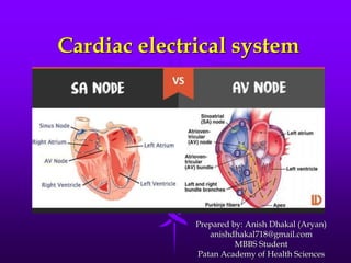
- 4. Sinoatrial (SA) Node :Pacemaker • A primary pacemaker of the heart, • The electrical impulses initiated by the SA node are conducted along the myocardial cells of the atria via internodal pathways • The impulses cause electrical stimulation and subsequent contraction of the atria
- 6. Self Excitation of Sinus Nodal Fibers
- 7. Conduction system • The S-A node generate and coordinate the transmission of electrical impulses to myocardial cells • The result is sequential atrioventricular contraction which provides for the most effective flow of blood , thereby optimizing cardiac out put
- 8. Continued….. • Cardiac conduction system: The electrical conduction system controls the heart rate • This system creates the electrical impulses and sends them throughout the heart. These impulses make the heart contract and pump blood.
- 9. Transmission of the cardiac impulse through Atria • Sinus nodal fibres directly connect with surrounding atrial muscle fibre • Several small bands of more conductive atrial fibers are present in atria Anterior Interatrial band (Bachmann’s Bundle) 3 Internodal pathways Anterior Internodal pathway Middle Internodal pathway Posterior Internodal pathways
- 10. Contd…. • Action potential travels outward into the atrial muscle • Action potential spread through the entire atrial muscle mass, eventually to AV node where A-V nodal delay occurs
- 12. Atrioventricular node • The AV node consist of specialized muscle cells similar to those of SA node • The AV node coordinate the incoming electrical impulses from atria Cause of slow conduction in AV node: • Diminished number of gap junctions • Absence of this delay causes abnormal pathway leads to Wolff-Parkinson-White syndrome
- 13. Bundle of His (A-V Bundle) • From the AV node, impulses travel through to the right and left bundle branches • These branches extend to the right and left sides of the septum and bottom of the
- 14. Bundle of His •These branch a lot to form the Purkinje fibers that transmit the impulses to the myocardium (muscle tissue) •The bundle of His, bundle branches and Purkinje fibers transmit quickly and cause both ventricles to contract at the same time
- 15. Contraction of Ventricles • As the ventricles contract, blood is forced out through the semilunar valves into the pulmonary trunk and the aorta. • After the ventricles complete their contraction phase, they relax and the SA node initiates another impulse to start another cardiac cycle.
Editor's Notes
- SA node - small, flattened, ellipsoid strip of specialized cardiac muscle, 3mm wide, 15 mm long and 1 mm thick. located in the superior posterolateral wall of right atrium immediately below and slightly lateral to the opening of superior vena-cava
- Velocity of conduction in most atrial muscle is 0.3 m/s Velocity of conduction in small bands of atrial fibres is 1 m/s
- Located in posterior wall of right atrium behind tricuspid valve. Another cause for slow conduction is smaller size of cells of AV node