Carpal Tunnel Syndrome Estimation through Median Nerve Segmentation in Ultrasound Videos
•
0 likes•108 views

EMBC2023
Recommended

Recommended
More Related Content
Similar to Carpal Tunnel Syndrome Estimation through Median Nerve Segmentation in Ultrasound Videos
Similar to Carpal Tunnel Syndrome Estimation through Median Nerve Segmentation in Ultrasound Videos (20)
More from sugiuralab
More from sugiuralab (20)
Recently uploaded
Recently uploaded (20)
Carpal Tunnel Syndrome Estimation through Median Nerve Segmentation in Ultrasound Videos
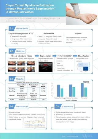
- 1. Yukina Sato1), Kana Matsuo1), Yohei Kawasaki1), Takafumi Koyama2), Eriku Yamada2), Koji Fujita2), and Yuta Sugiura1) 1) Keio University 2) Tokyo Medical and Dental University 01 Introduction 02 Methods Record ultrasound videos • Numbness of the fingers • Compression of the median nerve • The most common upper extremity neuropathy Carpal Tunnel Syndrome (CTS) • Detect CTS using deep learning-based analyses of ultrasound images • Automatic tracking of the median nerve in ultrasound videos Related work Clarifying whether using ultrasound video improves accuracy of CTS estimation Purpose 03 Results 04 Discussions • Improve accuracy • Take into account the time variation in median nerve tracking • Obtain features for each part of the same finger movement • Use a larger dataset 05 Conclusion • Segmented the median nerve in ultrasound videos • Performed CTS estimations • Estimations using features obtained from videos and time series data showed higher accuracy than those using features obtained from images. E-mail: info-lcl-group@keio.jp Carpal Tunnel Syndrome Estimation through Median Nerve Segmentation in Ultrasound Videos 0 0.1 0.2 0.3 0.4 0.5 0.6 0.7 0.8 0.9 1 Features from images Features from videos Time series Sensitivity Specificity Features from images Features from videos Time series Sensitivity 0.842 0.855 0.947 Specificity 0.581 0.677 0.645 Features Frame 1 second after the start of recording Median and maximum of all frames All frames Model XGBoost XGBoost KNN Comparison of the results of the classification Accuracy Comparison Segmentation Feature extraction Classification 134 cases taken by three skilled physicians Segment the nerve area by Mask R-CNN Obtain the features by image processing • Area • Perimeter • Aspect ratio • Centroid coordinates etc. Group 63-fold cross- validation