Immunofluorescence Antibody Validation Report for Anti-Tak1 Antibody (STJ95901)

Serine/threonine kinase which acts as an essential component of the MAP kinase signal transduction pathway. Plays an important role in the cascades of cellular responses evoked by changes in the environment. Mediates signal transduction of TRAF6, various cytokines including interleukin-1 (IL-1), transforming growth factor-beta (TGFB), TGFB-related factors like BMP2 and BMP4, toll-like receptors (TLR), tumor necrosis factor receptor CD40 and B-cell receptor (BCR). Ceramides are also able to activate MAP3K7/TAK1. Once activated, acts as an upstream activator of the MKK/JNK signal transduction cascade and the p38 MAPK signal transduction cascade through the phosphorylation and activation of several MAP kinase kinases like MAP2K1/MEK1, MAP2K3/MKK3, MAP2K6/MKK6 and MAP2K7/MKK7. Anti-Tak1 -http://www.stjohnslabs.com/tak1-antibody-p-94520 Join our Antibody Validation Project - http://www.stjohnslabs.com/services/antibody-validation

Recommended
Recommended
More Related Content
What's hot
What's hot (20)
Viewers also liked
Viewers also liked (17)
Similar to Immunofluorescence Antibody Validation Report for Anti-Tak1 Antibody (STJ95901)
Similar to Immunofluorescence Antibody Validation Report for Anti-Tak1 Antibody (STJ95901) (12)
More from St John's Laboratory Ltd
More from St John's Laboratory Ltd (20)
Recently uploaded
Recently uploaded (20)
Immunofluorescence Antibody Validation Report for Anti-Tak1 Antibody (STJ95901)
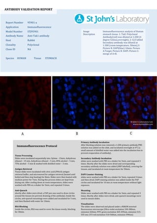
- 1. ANTIBODY VALIDATION REPORT Report Number 95901-a Application Immunofluorescence Model Number STJ95901 Antibody Name Anti-Tak1 antibody Host Rabbit Clonality Polyclonal Clone ID NA Species HUMAN Tissue STOMACH Image Description Immunofluorescence analysis of Human stomach tissue. 1: Tak1 Polyclonal Antibody(red) was diluted at 1:200 (4 degree Celsius,overnight). 2: Cy3 labled Secondary antibody was diluted at 1:300 (room temperature, 50min).3: Picture B: DAPI(blue) 10min. Picture A:Target. Picture B: DAPI. Picture C: merge of A+B. Primary Antibody Incubation After blocking solution was removed a 1:200 primary antibody/PBS solution was added on the slide, and incubated overnight at 4°C (a small amount of distilled water was added into the incubation box to prevent evaporation of antibody). Secondary Antibody Incubation slides were washed with PBS on a shaker for 5min, and repeated 3 times. Shortly after the slides were dried and corresponding secondary antibody solution was added (HRP labelled), covering the tissues, and incubated at room temperature for 50min. DAPI Counter-Staining slides were washed with PBS on a shaker for 5min, repeated 3 times and then dried. DAPI staining solution was added inside the PAP circles and incubated for 10 min at room temperature without light exposure. Mounting Slides were washed with PBS on a shaker for 5min, and repeated 3 times. Shortly after slides were dried, anti-quench mountings were used to mount slides. Visualization The slides were observed and placed under a NIKON inverted fluorescence microscope (Ultra violet excitation 330-380nm, emission 420nm; FITC green excitation 465-495nm, emission 515- 555 nm; CY3 red excitation 510-560nm, emission 590nm) Immunofluorescence Protocol Tissue Processing Slides were incubated sequentially into: Xylene - 15min, Anhydrous ethanol – 15 min, Anhydrous ethanol – 5 min, 85% alcohol – 5 min, 75% alcohol – 5 min & washed with distilled water – 5 min. Antigen Retrieval Tissue slides were incubated with citric acid (PH6.0) antigen retrieval buffer, and microwaved for antigen retrieval (heated until boiled and then stop heating) for 8min. Slides were then heated with medium power for 7min. During this process slides are kept from drying out. After cooling down at room temperature, slides were washed with PBS on a shaker for 5min, and repeated 3 times. Anti-Quench shortly after slides were dried, a PAP pen was used to draw circles around the tissues (to prevent draining of the antibody). Inside the circles, anti-quench mountings were added and incubated for 5 min, and then flushed with water for 10min. BSA Blocking Inside the circles, BSA was used to cover the tissue evenly, blocking for 30min. St John's Laboratory Ltd. www.stjohnslabs.com
- 2. ANTIBODY VALIDATION REPORT Report Number 95901-b Application Immunofluorescence Model Number STJ95901 Antibody Name Anti-Tak1 antibody Host Rabbit Clonality Polyclonal Clone ID NA Species HUMAN Tissue STOMACH Image Description Immunofluorescence analysis of Human stomach tissue. 1: Tak1 Polyclonal Antibody(red) was diluted at 1:200 (4 degree Celsius,overnight). 2: Cy3 labled Secondary antibody was diluted at 1:300 (room temperature, 50min).3: Picture B: DAPI(blue) 10min. Picture A:Target. Picture B: DAPI. Picture C: merge of A+B. Primary Antibody Incubation After blocking solution was removed a 1:200 primary antibody/PBS solution was added on the slide, and incubated overnight at 4°C (a small amount of distilled water was added into the incubation box to prevent evaporation of antibody). Secondary Antibody Incubation slides were washed with PBS on a shaker for 5min, and repeated 3 times. Shortly after the slides were dried and corresponding secondary antibody solution was added (HRP labelled), covering the tissues, and incubated at room temperature for 50min. DAPI Counter-Staining slides were washed with PBS on a shaker for 5min, repeated 3 times and then dried. DAPI staining solution was added inside the PAP circles and incubated for 10 min at room temperature without light exposure. Mounting Slides were washed with PBS on a shaker for 5min, and repeated 3 times. Shortly after slides were dried, anti-quench mountings were used to mount slides. Visualization The slides were observed and placed under a NIKON inverted fluorescence microscope (Ultra violet excitation 330-380nm, emission 420nm; FITC green excitation 465-495nm, emission 515- 555 nm; CY3 red excitation 510-560nm, emission 590nm) Immunofluorescence Protocol Tissue Processing Slides were incubated sequentially into: Xylene - 15min, Anhydrous ethanol – 15 min, Anhydrous ethanol – 5 min, 85% alcohol – 5 min, 75% alcohol – 5 min & washed with distilled water – 5 min. Antigen Retrieval Tissue slides were incubated with citric acid (PH6.0) antigen retrieval buffer, and microwaved for antigen retrieval (heated until boiled and then stop heating) for 8min. Slides were then heated with medium power for 7min. During this process slides are kept from drying out. After cooling down at room temperature, slides were washed with PBS on a shaker for 5min, and repeated 3 times. Anti-Quench shortly after slides were dried, a PAP pen was used to draw circles around the tissues (to prevent draining of the antibody). Inside the circles, anti-quench mountings were added and incubated for 5 min, and then flushed with water for 10min. BSA Blocking Inside the circles, BSA was used to cover the tissue evenly, blocking for 30min. St John's Laboratory Ltd. www.stjohnslabs.com
- 3. ANTIBODY VALIDATION REPORT Report Number 95901-c Application Immunofluorescence Model Number STJ95901 Antibody Name Anti-Tak1 antibody Host Rabbit Clonality Polyclonal Clone ID NA Species RAT Tissue KIDNEY Image Description Immunofluorescence analysis of Rat kidney tissue. 1: Tak1 Polyclonal Antibody(red) was diluted at 1:200 (4 degree Celsius,overnight). 2: Cy3 labled Secondary antibody was diluted at 1:300 (room temperature, 50min).3: Picture B: DAPI(blue) 10min. Picture A:Target. Picture B: DAPI. Picture C: merge of A+B. Primary Antibody Incubation After blocking solution was removed a 1:200 primary antibody/PBS solution was added on the slide, and incubated overnight at 4°C (a small amount of distilled water was added into the incubation box to prevent evaporation of antibody). Secondary Antibody Incubation slides were washed with PBS on a shaker for 5min, and repeated 3 times. Shortly after the slides were dried and corresponding secondary antibody solution was added (HRP labelled), covering the tissues, and incubated at room temperature for 50min. DAPI Counter-Staining slides were washed with PBS on a shaker for 5min, repeated 3 times and then dried. DAPI staining solution was added inside the PAP circles and incubated for 10 min at room temperature without light exposure. Mounting Slides were washed with PBS on a shaker for 5min, and repeated 3 times. Shortly after slides were dried, anti-quench mountings were used to mount slides. Visualization The slides were observed and placed under a NIKON inverted fluorescence microscope (Ultra violet excitation 330-380nm, emission 420nm; FITC green excitation 465-495nm, emission 515- 555 nm; CY3 red excitation 510-560nm, emission 590nm) Immunofluorescence Protocol Tissue Processing Slides were incubated sequentially into: Xylene - 15min, Anhydrous ethanol – 15 min, Anhydrous ethanol – 5 min, 85% alcohol – 5 min, 75% alcohol – 5 min & washed with distilled water – 5 min. Antigen Retrieval Tissue slides were incubated with citric acid (PH6.0) antigen retrieval buffer, and microwaved for antigen retrieval (heated until boiled and then stop heating) for 8min. Slides were then heated with medium power for 7min. During this process slides are kept from drying out. After cooling down at room temperature, slides were washed with PBS on a shaker for 5min, and repeated 3 times. Anti-Quench shortly after slides were dried, a PAP pen was used to draw circles around the tissues (to prevent draining of the antibody). Inside the circles, anti-quench mountings were added and incubated for 5 min, and then flushed with water for 10min. BSA Blocking Inside the circles, BSA was used to cover the tissue evenly, blocking for 30min. St John's Laboratory Ltd. www.stjohnslabs.com
- 4. ANTIBODY VALIDATION REPORT Report Number 95901-d Application Immunofluorescence Model Number STJ95901 Antibody Name Anti-Tak1 antibody Host Rabbit Clonality Polyclonal Clone ID NA Species RAT Tissue KIDNEY Image Description Immunofluorescence analysis of Rat kidney tissue. 1: Tak1 Polyclonal Antibody(red) was diluted at 1:200 (4 degree Celsius,overnight). 2: Cy3 labled Secondary antibody was diluted at 1:300 (room temperature, 50min).3: Picture B: DAPI(blue) 10min. Picture A:Target. Picture B: DAPI. Picture C: merge of A+B. Primary Antibody Incubation After blocking solution was removed a 1:200 primary antibody/PBS solution was added on the slide, and incubated overnight at 4°C (a small amount of distilled water was added into the incubation box to prevent evaporation of antibody). Secondary Antibody Incubation slides were washed with PBS on a shaker for 5min, and repeated 3 times. Shortly after the slides were dried and corresponding secondary antibody solution was added (HRP labelled), covering the tissues, and incubated at room temperature for 50min. DAPI Counter-Staining slides were washed with PBS on a shaker for 5min, repeated 3 times and then dried. DAPI staining solution was added inside the PAP circles and incubated for 10 min at room temperature without light exposure. Mounting Slides were washed with PBS on a shaker for 5min, and repeated 3 times. Shortly after slides were dried, anti-quench mountings were used to mount slides. Visualization The slides were observed and placed under a NIKON inverted fluorescence microscope (Ultra violet excitation 330-380nm, emission 420nm; FITC green excitation 465-495nm, emission 515- 555 nm; CY3 red excitation 510-560nm, emission 590nm) Immunofluorescence Protocol Tissue Processing Slides were incubated sequentially into: Xylene - 15min, Anhydrous ethanol – 15 min, Anhydrous ethanol – 5 min, 85% alcohol – 5 min, 75% alcohol – 5 min & washed with distilled water – 5 min. Antigen Retrieval Tissue slides were incubated with citric acid (PH6.0) antigen retrieval buffer, and microwaved for antigen retrieval (heated until boiled and then stop heating) for 8min. Slides were then heated with medium power for 7min. During this process slides are kept from drying out. After cooling down at room temperature, slides were washed with PBS on a shaker for 5min, and repeated 3 times. Anti-Quench shortly after slides were dried, a PAP pen was used to draw circles around the tissues (to prevent draining of the antibody). Inside the circles, anti-quench mountings were added and incubated for 5 min, and then flushed with water for 10min. BSA Blocking Inside the circles, BSA was used to cover the tissue evenly, blocking for 30min. St John's Laboratory Ltd. www.stjohnslabs.com
- 5. ANTIBODY VALIDATION REPORT Report Number 95901-e Application Immunofluorescence Model Number STJ95901 Antibody Name Anti-Tak1 antibody Host Rabbit Clonality Polyclonal Clone ID NA Species MOUSE Tissue KIDNEY Image Description Immunofluorescence analysis of Mouse kidney tissue. 1: Tak1 Polyclonal Antibody(red) was diluted at 1:200 (4 degree Celsius,overnight). 2: Cy3 labled Secondary antibody was diluted at 1:300 (room temperature, 50min).3: Picture B: DAPI(blue) 10min. Picture A:Target. Picture B: DAPI. Picture C: merge of A+B. Primary Antibody Incubation After blocking solution was removed a 1:200 primary antibody/PBS solution was added on the slide, and incubated overnight at 4°C (a small amount of distilled water was added into the incubation box to prevent evaporation of antibody). Secondary Antibody Incubation slides were washed with PBS on a shaker for 5min, and repeated 3 times. Shortly after the slides were dried and corresponding secondary antibody solution was added (HRP labelled), covering the tissues, and incubated at room temperature for 50min. DAPI Counter-Staining slides were washed with PBS on a shaker for 5min, repeated 3 times and then dried. DAPI staining solution was added inside the PAP circles and incubated for 10 min at room temperature without light exposure. Mounting Slides were washed with PBS on a shaker for 5min, and repeated 3 times. Shortly after slides were dried, anti-quench mountings were used to mount slides. Visualization The slides were observed and placed under a NIKON inverted fluorescence microscope (Ultra violet excitation 330-380nm, emission 420nm; FITC green excitation 465-495nm, emission 515- 555 nm; CY3 red excitation 510-560nm, emission 590nm) Immunofluorescence Protocol Tissue Processing Slides were incubated sequentially into: Xylene - 15min, Anhydrous ethanol – 15 min, Anhydrous ethanol – 5 min, 85% alcohol – 5 min, 75% alcohol – 5 min & washed with distilled water – 5 min. Antigen Retrieval Tissue slides were incubated with citric acid (PH6.0) antigen retrieval buffer, and microwaved for antigen retrieval (heated until boiled and then stop heating) for 8min. Slides were then heated with medium power for 7min. During this process slides are kept from drying out. After cooling down at room temperature, slides were washed with PBS on a shaker for 5min, and repeated 3 times. Anti-Quench shortly after slides were dried, a PAP pen was used to draw circles around the tissues (to prevent draining of the antibody). Inside the circles, anti-quench mountings were added and incubated for 5 min, and then flushed with water for 10min. BSA Blocking Inside the circles, BSA was used to cover the tissue evenly, blocking for 30min. St John's Laboratory Ltd. www.stjohnslabs.com
- 6. ANTIBODY VALIDATION REPORT Report Number 95901-f Application Immunofluorescence Model Number STJ95901 Antibody Name Anti-Tak1 antibody Host Rabbit Clonality Polyclonal Clone ID NA Species MOUSE Tissue KIDNEY Image Description Immunofluorescence analysis of Mouse kidney tissue. 1: Tak1 Polyclonal Antibody(red) was diluted at 1:200 (4 degree Celsius,overnight). 2: Cy3 labled Secondary antibody was diluted at 1:300 (room temperature, 50min).3: Picture B: DAPI(blue) 10min. Picture A:Target. Picture B: DAPI. Picture C: merge of A+B. Primary Antibody Incubation After blocking solution was removed a 1:200 primary antibody/PBS solution was added on the slide, and incubated overnight at 4°C (a small amount of distilled water was added into the incubation box to prevent evaporation of antibody). Secondary Antibody Incubation slides were washed with PBS on a shaker for 5min, and repeated 3 times. Shortly after the slides were dried and corresponding secondary antibody solution was added (HRP labelled), covering the tissues, and incubated at room temperature for 50min. DAPI Counter-Staining slides were washed with PBS on a shaker for 5min, repeated 3 times and then dried. DAPI staining solution was added inside the PAP circles and incubated for 10 min at room temperature without light exposure. Mounting Slides were washed with PBS on a shaker for 5min, and repeated 3 times. Shortly after slides were dried, anti-quench mountings were used to mount slides. Visualization The slides were observed and placed under a NIKON inverted fluorescence microscope (Ultra violet excitation 330-380nm, emission 420nm; FITC green excitation 465-495nm, emission 515- 555 nm; CY3 red excitation 510-560nm, emission 590nm) Immunofluorescence Protocol Tissue Processing Slides were incubated sequentially into: Xylene - 15min, Anhydrous ethanol – 15 min, Anhydrous ethanol – 5 min, 85% alcohol – 5 min, 75% alcohol – 5 min & washed with distilled water – 5 min. Antigen Retrieval Tissue slides were incubated with citric acid (PH6.0) antigen retrieval buffer, and microwaved for antigen retrieval (heated until boiled and then stop heating) for 8min. Slides were then heated with medium power for 7min. During this process slides are kept from drying out. After cooling down at room temperature, slides were washed with PBS on a shaker for 5min, and repeated 3 times. Anti-Quench shortly after slides were dried, a PAP pen was used to draw circles around the tissues (to prevent draining of the antibody). Inside the circles, anti-quench mountings were added and incubated for 5 min, and then flushed with water for 10min. BSA Blocking Inside the circles, BSA was used to cover the tissue evenly, blocking for 30min. St John's Laboratory Ltd. www.stjohnslabs.com