SlideShare a Scribd company logo
1 of 9
Download to read offline
ANTIBODY VALIDATION REPORT
Report Number 90129-a
Application Immunofluorescence
Model Number STJ90129
Antibody Name Anti-Histone H4 (Acetyl K16) antibody
Host Rabbit
Clonality Polyclonal
Clone ID NA
Species HUMAN Tissue KIDNEY
Image
Description
Immunofluorescence analysis of Human
kidney tissue. 1: Histone H4 (Acetyl
Lys16) Polyclonal Antibody(red) was
diluted at 1:200 (4 degree
Celsius,overnight). 2: Cy3 labled
Secondary antibody was diluted at
1:300 (room temperature, 50min).3:
Picture B: DAPI(blue) 10min. Picture
A:Target. Picture B: DAPI. Picture C:
merge of A+B.
Primary Antibody Incubation
After blocking solution was removed a 1:200 primary antibody/PBS
solution was added on the slide, and incubated overnight at 4°C (a
small amount of distilled water was added into the incubation box to
prevent evaporation of antibody).
Secondary Antibody Incubation
slides were washed with PBS on a shaker for 5min, and repeated 3
times. Shortly after the slides were dried and corresponding
secondary antibody solution was added (HRP labelled), covering the
tissues, and incubated at room temperature for 50min.
DAPI Counter-Staining
slides were washed with PBS on a shaker for 5min, repeated 3 times
and then dried. DAPI staining solution was added inside the PAP
circles and incubated for 10 min at room temperature without light
exposure.
Mounting
Slides were washed with PBS on a shaker for 5min, and repeated 3
times. Shortly after slides were dried, anti-quench mountings were
used to mount slides.
Visualization
The slides were observed and placed under a NIKON inverted
fluorescence microscope (Ultra violet excitation 330-380nm,
emission 420nm; FITC green excitation 465-495nm, emission 515-
555 nm; CY3 red excitation 510-560nm, emission 590nm)
Immunofluorescence Protocol
Tissue Processing
Slides were incubated sequentially into: Xylene - 15min, Anhydrous
ethanol – 15 min, Anhydrous ethanol – 5 min, 85% alcohol – 5 min,
75% alcohol – 5 min & washed with distilled water – 5 min.
Antigen Retrieval
Tissue slides were incubated with citric acid (PH6.0) antigen
retrieval buffer, and microwaved for antigen retrieval (heated until
boiled and then stop heating) for 8min. Slides were then heated with
medium power for 7min. During this process slides are kept from
drying out. After cooling down at room temperature, slides were
washed with PBS on a shaker for 5min, and repeated 3 times.
Anti-Quench
shortly after slides were dried, a PAP pen was used to draw circles
around the tissues (to prevent draining of the antibody). Inside the
circles, anti-quench mountings were added and incubated for 5 min,
and then flushed with water for 10min.
BSA Blocking
Inside the circles, BSA was used to cover the tissue evenly, blocking
for 30min.
ANTIBODY VALIDATION REPORT
Report Number 90129-b
Application Immunofluorescence
Model Number STJ90129
Antibody Name Anti-Histone H4 (Acetyl K16) antibody
Host Rabbit
Clonality Polyclonal
Clone ID NA
Species HUMAN Tissue KIDNEY
Image
Description
Immunofluorescence analysis of Human
kidney tissue. 1: Histone H4 (Acetyl
Lys16) Polyclonal Antibody(red) was
diluted at 1:200 (4 degree
Celsius,overnight). 2: Cy3 labled
Secondary antibody was diluted at
1:300 (room temperature, 50min).3:
Picture B: DAPI(blue) 10min. Picture
A:Target. Picture B: DAPI. Picture C:
merge of A+B.
Primary Antibody Incubation
After blocking solution was removed a 1:200 primary antibody/PBS
solution was added on the slide, and incubated overnight at 4°C (a
small amount of distilled water was added into the incubation box to
prevent evaporation of antibody).
Secondary Antibody Incubation
slides were washed with PBS on a shaker for 5min, and repeated 3
times. Shortly after the slides were dried and corresponding
secondary antibody solution was added (HRP labelled), covering the
tissues, and incubated at room temperature for 50min.
DAPI Counter-Staining
slides were washed with PBS on a shaker for 5min, repeated 3 times
and then dried. DAPI staining solution was added inside the PAP
circles and incubated for 10 min at room temperature without light
exposure.
Mounting
Slides were washed with PBS on a shaker for 5min, and repeated 3
times. Shortly after slides were dried, anti-quench mountings were
used to mount slides.
Visualization
The slides were observed and placed under a NIKON inverted
fluorescence microscope (Ultra violet excitation 330-380nm,
emission 420nm; FITC green excitation 465-495nm, emission 515-
555 nm; CY3 red excitation 510-560nm, emission 590nm)
Immunofluorescence Protocol
Tissue Processing
Slides were incubated sequentially into: Xylene - 15min, Anhydrous
ethanol – 15 min, Anhydrous ethanol – 5 min, 85% alcohol – 5 min,
75% alcohol – 5 min & washed with distilled water – 5 min.
Antigen Retrieval
Tissue slides were incubated with citric acid (PH6.0) antigen
retrieval buffer, and microwaved for antigen retrieval (heated until
boiled and then stop heating) for 8min. Slides were then heated with
medium power for 7min. During this process slides are kept from
drying out. After cooling down at room temperature, slides were
washed with PBS on a shaker for 5min, and repeated 3 times.
Anti-Quench
shortly after slides were dried, a PAP pen was used to draw circles
around the tissues (to prevent draining of the antibody). Inside the
circles, anti-quench mountings were added and incubated for 5 min,
and then flushed with water for 10min.
BSA Blocking
Inside the circles, BSA was used to cover the tissue evenly, blocking
for 30min.
ANTIBODY VALIDATION REPORT
Report Number 90129-c
Application Immunofluorescence
Model Number STJ90129
Antibody Name Anti-Histone H4 (Acetyl K16) antibody
Host Rabbit
Clonality Polyclonal
Clone ID NA
Species HUMAN Tissue KIDNEY
Image
Description
Immunofluorescence analysis of Human
kidney tissue. 1: Histone H4 (Acetyl
Lys16) Polyclonal Antibody(red) was
diluted at 1:200 (4 degree
Celsius,overnight). 2: Cy3 labled
Secondary antibody was diluted at
1:300 (room temperature, 50min).3:
Picture B: DAPI(blue) 10min. Picture
A:Target. Picture B: DAPI. Picture C:
merge of A+B.
Primary Antibody Incubation
After blocking solution was removed a 1:200 primary antibody/PBS
solution was added on the slide, and incubated overnight at 4°C (a
small amount of distilled water was added into the incubation box to
prevent evaporation of antibody).
Secondary Antibody Incubation
slides were washed with PBS on a shaker for 5min, and repeated 3
times. Shortly after the slides were dried and corresponding
secondary antibody solution was added (HRP labelled), covering the
tissues, and incubated at room temperature for 50min.
DAPI Counter-Staining
slides were washed with PBS on a shaker for 5min, repeated 3 times
and then dried. DAPI staining solution was added inside the PAP
circles and incubated for 10 min at room temperature without light
exposure.
Mounting
Slides were washed with PBS on a shaker for 5min, and repeated 3
times. Shortly after slides were dried, anti-quench mountings were
used to mount slides.
Visualization
The slides were observed and placed under a NIKON inverted
fluorescence microscope (Ultra violet excitation 330-380nm,
emission 420nm; FITC green excitation 465-495nm, emission 515-
555 nm; CY3 red excitation 510-560nm, emission 590nm)
Immunofluorescence Protocol
Tissue Processing
Slides were incubated sequentially into: Xylene - 15min, Anhydrous
ethanol – 15 min, Anhydrous ethanol – 5 min, 85% alcohol – 5 min,
75% alcohol – 5 min & washed with distilled water – 5 min.
Antigen Retrieval
Tissue slides were incubated with citric acid (PH6.0) antigen
retrieval buffer, and microwaved for antigen retrieval (heated until
boiled and then stop heating) for 8min. Slides were then heated with
medium power for 7min. During this process slides are kept from
drying out. After cooling down at room temperature, slides were
washed with PBS on a shaker for 5min, and repeated 3 times.
Anti-Quench
shortly after slides were dried, a PAP pen was used to draw circles
around the tissues (to prevent draining of the antibody). Inside the
circles, anti-quench mountings were added and incubated for 5 min,
and then flushed with water for 10min.
BSA Blocking
Inside the circles, BSA was used to cover the tissue evenly, blocking
for 30min.
ANTIBODY VALIDATION REPORT
Report Number 90129-d
Application Immunofluorescence
Model Number STJ90129
Antibody Name Anti-Histone H4 (Acetyl K16) antibody
Host Rabbit
Clonality Polyclonal
Clone ID NA
Species HUMAN Tissue LUNG CANCER
Image
Description
Immunofluorescence analysis of Human
lung cancer tissue. 1: Histone H4 (Acetyl
Lys16) Polyclonal Antibody(red) was
diluted at 1:200 (4 degree
Celsius,overnight). 2: Cy3 labled
Secondary antibody was diluted at
1:300 (room temperature, 50min).3:
Picture B: DAPI(blue) 10min. Picture
A:Target. Picture B: DAPI. Picture C:
merge of A+B.
Primary Antibody Incubation
After blocking solution was removed a 1:200 primary antibody/PBS
solution was added on the slide, and incubated overnight at 4°C (a
small amount of distilled water was added into the incubation box to
prevent evaporation of antibody).
Secondary Antibody Incubation
slides were washed with PBS on a shaker for 5min, and repeated 3
times. Shortly after the slides were dried and corresponding
secondary antibody solution was added (HRP labelled), covering the
tissues, and incubated at room temperature for 50min.
DAPI Counter-Staining
slides were washed with PBS on a shaker for 5min, repeated 3 times
and then dried. DAPI staining solution was added inside the PAP
circles and incubated for 10 min at room temperature without light
exposure.
Mounting
Slides were washed with PBS on a shaker for 5min, and repeated 3
times. Shortly after slides were dried, anti-quench mountings were
used to mount slides.
Visualization
The slides were observed and placed under a NIKON inverted
fluorescence microscope (Ultra violet excitation 330-380nm,
emission 420nm; FITC green excitation 465-495nm, emission 515-
555 nm; CY3 red excitation 510-560nm, emission 590nm)
Immunofluorescence Protocol
Tissue Processing
Slides were incubated sequentially into: Xylene - 15min, Anhydrous
ethanol – 15 min, Anhydrous ethanol – 5 min, 85% alcohol – 5 min,
75% alcohol – 5 min & washed with distilled water – 5 min.
Antigen Retrieval
Tissue slides were incubated with citric acid (PH6.0) antigen
retrieval buffer, and microwaved for antigen retrieval (heated until
boiled and then stop heating) for 8min. Slides were then heated with
medium power for 7min. During this process slides are kept from
drying out. After cooling down at room temperature, slides were
washed with PBS on a shaker for 5min, and repeated 3 times.
Anti-Quench
shortly after slides were dried, a PAP pen was used to draw circles
around the tissues (to prevent draining of the antibody). Inside the
circles, anti-quench mountings were added and incubated for 5 min,
and then flushed with water for 10min.
BSA Blocking
Inside the circles, BSA was used to cover the tissue evenly, blocking
for 30min.
ANTIBODY VALIDATION REPORT
Report Number 90129-e
Application Immunofluorescence
Model Number STJ90129
Antibody Name Anti-Histone H4 (Acetyl K16) antibody
Host Rabbit
Clonality Polyclonal
Clone ID NA
Species HUMAN Tissue LUNG CANCER
Image
Description
Immunofluorescence analysis of Human
lung cancer tissue. 1: Histone H4 (Acetyl
Lys16) Polyclonal Antibody(red) was
diluted at 1:200 (4 degree
Celsius,overnight). 2: Cy3 labled
Secondary antibody was diluted at
1:300 (room temperature, 50min).3:
Picture B: DAPI(blue) 10min. Picture
A:Target. Picture B: DAPI. Picture C:
merge of A+B.
Primary Antibody Incubation
After blocking solution was removed a 1:200 primary antibody/PBS
solution was added on the slide, and incubated overnight at 4°C (a
small amount of distilled water was added into the incubation box to
prevent evaporation of antibody).
Secondary Antibody Incubation
slides were washed with PBS on a shaker for 5min, and repeated 3
times. Shortly after the slides were dried and corresponding
secondary antibody solution was added (HRP labelled), covering the
tissues, and incubated at room temperature for 50min.
DAPI Counter-Staining
slides were washed with PBS on a shaker for 5min, repeated 3 times
and then dried. DAPI staining solution was added inside the PAP
circles and incubated for 10 min at room temperature without light
exposure.
Mounting
Slides were washed with PBS on a shaker for 5min, and repeated 3
times. Shortly after slides were dried, anti-quench mountings were
used to mount slides.
Visualization
The slides were observed and placed under a NIKON inverted
fluorescence microscope (Ultra violet excitation 330-380nm,
emission 420nm; FITC green excitation 465-495nm, emission 515-
555 nm; CY3 red excitation 510-560nm, emission 590nm)
Immunofluorescence Protocol
Tissue Processing
Slides were incubated sequentially into: Xylene - 15min, Anhydrous
ethanol – 15 min, Anhydrous ethanol – 5 min, 85% alcohol – 5 min,
75% alcohol – 5 min & washed with distilled water – 5 min.
Antigen Retrieval
Tissue slides were incubated with citric acid (PH6.0) antigen
retrieval buffer, and microwaved for antigen retrieval (heated until
boiled and then stop heating) for 8min. Slides were then heated with
medium power for 7min. During this process slides are kept from
drying out. After cooling down at room temperature, slides were
washed with PBS on a shaker for 5min, and repeated 3 times.
Anti-Quench
shortly after slides were dried, a PAP pen was used to draw circles
around the tissues (to prevent draining of the antibody). Inside the
circles, anti-quench mountings were added and incubated for 5 min,
and then flushed with water for 10min.
BSA Blocking
Inside the circles, BSA was used to cover the tissue evenly, blocking
for 30min.
ANTIBODY VALIDATION REPORT
Report Number 90129-f
Application Immunofluorescence
Model Number STJ90129
Antibody Name Anti-Histone H4 (Acetyl K16) antibody
Host Rabbit
Clonality Polyclonal
Clone ID NA
Species HUMAN Tissue LUNG CANCER
Image
Description
Immunofluorescence analysis of Human
lung cancer tissue. 1: Histone H4 (Acetyl
Lys16) Polyclonal Antibody(red) was
diluted at 1:200 (4 degree
Celsius,overnight). 2: Cy3 labled
Secondary antibody was diluted at
1:300 (room temperature, 50min).3:
Picture B: DAPI(blue) 10min. Picture
A:Target. Picture B: DAPI. Picture C:
merge of A+B.
Primary Antibody Incubation
After blocking solution was removed a 1:200 primary antibody/PBS
solution was added on the slide, and incubated overnight at 4°C (a
small amount of distilled water was added into the incubation box to
prevent evaporation of antibody).
Secondary Antibody Incubation
slides were washed with PBS on a shaker for 5min, and repeated 3
times. Shortly after the slides were dried and corresponding
secondary antibody solution was added (HRP labelled), covering the
tissues, and incubated at room temperature for 50min.
DAPI Counter-Staining
slides were washed with PBS on a shaker for 5min, repeated 3 times
and then dried. DAPI staining solution was added inside the PAP
circles and incubated for 10 min at room temperature without light
exposure.
Mounting
Slides were washed with PBS on a shaker for 5min, and repeated 3
times. Shortly after slides were dried, anti-quench mountings were
used to mount slides.
Visualization
The slides were observed and placed under a NIKON inverted
fluorescence microscope (Ultra violet excitation 330-380nm,
emission 420nm; FITC green excitation 465-495nm, emission 515-
555 nm; CY3 red excitation 510-560nm, emission 590nm)
Immunofluorescence Protocol
Tissue Processing
Slides were incubated sequentially into: Xylene - 15min, Anhydrous
ethanol – 15 min, Anhydrous ethanol – 5 min, 85% alcohol – 5 min,
75% alcohol – 5 min & washed with distilled water – 5 min.
Antigen Retrieval
Tissue slides were incubated with citric acid (PH6.0) antigen
retrieval buffer, and microwaved for antigen retrieval (heated until
boiled and then stop heating) for 8min. Slides were then heated with
medium power for 7min. During this process slides are kept from
drying out. After cooling down at room temperature, slides were
washed with PBS on a shaker for 5min, and repeated 3 times.
Anti-Quench
shortly after slides were dried, a PAP pen was used to draw circles
around the tissues (to prevent draining of the antibody). Inside the
circles, anti-quench mountings were added and incubated for 5 min,
and then flushed with water for 10min.
BSA Blocking
Inside the circles, BSA was used to cover the tissue evenly, blocking
for 30min.
ANTIBODY VALIDATION REPORT
Report Number 90129-g
Application Immunofluorescence
Model Number STJ90129
Antibody Name Anti-Histone H4 (Acetyl K16) antibody
Host Rabbit
Clonality Polyclonal
Clone ID NA
Species HUMAN Tissue LUNG
Image
Description
Immunofluorescence analysis of Human
lung tissue. 1: Histone H4 (Acetyl
Lys16) Polyclonal Antibody(red) was
diluted at 1:200 (4 degree
Celsius,overnight). 2: Cy3 labled
Secondary antibody was diluted at
1:300 (room temperature, 50min).3:
Picture B: DAPI(blue) 10min. Picture
A:Target. Picture B: DAPI. Picture C:
merge of A+B.
Primary Antibody Incubation
After blocking solution was removed a 1:200 primary antibody/PBS
solution was added on the slide, and incubated overnight at 4°C (a
small amount of distilled water was added into the incubation box to
prevent evaporation of antibody).
Secondary Antibody Incubation
slides were washed with PBS on a shaker for 5min, and repeated 3
times. Shortly after the slides were dried and corresponding
secondary antibody solution was added (HRP labelled), covering the
tissues, and incubated at room temperature for 50min.
DAPI Counter-Staining
slides were washed with PBS on a shaker for 5min, repeated 3 times
and then dried. DAPI staining solution was added inside the PAP
circles and incubated for 10 min at room temperature without light
exposure.
Mounting
Slides were washed with PBS on a shaker for 5min, and repeated 3
times. Shortly after slides were dried, anti-quench mountings were
used to mount slides.
Visualization
The slides were observed and placed under a NIKON inverted
fluorescence microscope (Ultra violet excitation 330-380nm,
emission 420nm; FITC green excitation 465-495nm, emission 515-
555 nm; CY3 red excitation 510-560nm, emission 590nm)
Immunofluorescence Protocol
Tissue Processing
Slides were incubated sequentially into: Xylene - 15min, Anhydrous
ethanol – 15 min, Anhydrous ethanol – 5 min, 85% alcohol – 5 min,
75% alcohol – 5 min & washed with distilled water – 5 min.
Antigen Retrieval
Tissue slides were incubated with citric acid (PH6.0) antigen
retrieval buffer, and microwaved for antigen retrieval (heated until
boiled and then stop heating) for 8min. Slides were then heated with
medium power for 7min. During this process slides are kept from
drying out. After cooling down at room temperature, slides were
washed with PBS on a shaker for 5min, and repeated 3 times.
Anti-Quench
shortly after slides were dried, a PAP pen was used to draw circles
around the tissues (to prevent draining of the antibody). Inside the
circles, anti-quench mountings were added and incubated for 5 min,
and then flushed with water for 10min.
BSA Blocking
Inside the circles, BSA was used to cover the tissue evenly, blocking
for 30min.
ANTIBODY VALIDATION REPORT
Report Number 90129-h
Application Immunofluorescence
Model Number STJ90129
Antibody Name Anti-Histone H4 (Acetyl K16) antibody
Host Rabbit
Clonality Polyclonal
Clone ID NA
Species HUMAN Tissue LUNG
Image
Description
Immunofluorescence analysis of Human
lung tissue. 1: Histone H4 (Acetyl
Lys16) Polyclonal Antibody(red) was
diluted at 1:200 (4 degree
Celsius,overnight). 2: Cy3 labled
Secondary antibody was diluted at
1:300 (room temperature, 50min).3:
Picture B: DAPI(blue) 10min. Picture
A:Target. Picture B: DAPI. Picture C:
merge of A+B.
Primary Antibody Incubation
After blocking solution was removed a 1:200 primary antibody/PBS
solution was added on the slide, and incubated overnight at 4°C (a
small amount of distilled water was added into the incubation box to
prevent evaporation of antibody).
Secondary Antibody Incubation
slides were washed with PBS on a shaker for 5min, and repeated 3
times. Shortly after the slides were dried and corresponding
secondary antibody solution was added (HRP labelled), covering the
tissues, and incubated at room temperature for 50min.
DAPI Counter-Staining
slides were washed with PBS on a shaker for 5min, repeated 3 times
and then dried. DAPI staining solution was added inside the PAP
circles and incubated for 10 min at room temperature without light
exposure.
Mounting
Slides were washed with PBS on a shaker for 5min, and repeated 3
times. Shortly after slides were dried, anti-quench mountings were
used to mount slides.
Visualization
The slides were observed and placed under a NIKON inverted
fluorescence microscope (Ultra violet excitation 330-380nm,
emission 420nm; FITC green excitation 465-495nm, emission 515-
555 nm; CY3 red excitation 510-560nm, emission 590nm)
Immunofluorescence Protocol
Tissue Processing
Slides were incubated sequentially into: Xylene - 15min, Anhydrous
ethanol – 15 min, Anhydrous ethanol – 5 min, 85% alcohol – 5 min,
75% alcohol – 5 min & washed with distilled water – 5 min.
Antigen Retrieval
Tissue slides were incubated with citric acid (PH6.0) antigen
retrieval buffer, and microwaved for antigen retrieval (heated until
boiled and then stop heating) for 8min. Slides were then heated with
medium power for 7min. During this process slides are kept from
drying out. After cooling down at room temperature, slides were
washed with PBS on a shaker for 5min, and repeated 3 times.
Anti-Quench
shortly after slides were dried, a PAP pen was used to draw circles
around the tissues (to prevent draining of the antibody). Inside the
circles, anti-quench mountings were added and incubated for 5 min,
and then flushed with water for 10min.
BSA Blocking
Inside the circles, BSA was used to cover the tissue evenly, blocking
for 30min.
ANTIBODY VALIDATION REPORT
Report Number 90129-i
Application Immunofluorescence
Model Number STJ90129
Antibody Name Anti-Histone H4 (Acetyl K16) antibody
Host Rabbit
Clonality Polyclonal
Clone ID NA
Species HUMAN Tissue LUNG
Image
Description
Immunofluorescence analysis of Human
lung tissue. 1: Histone H4 (Acetyl
Lys16) Polyclonal Antibody(red) was
diluted at 1:200 (4 degree
Celsius,overnight). 2: Cy3 labled
Secondary antibody was diluted at
1:300 (room temperature, 50min).3:
Picture B: DAPI(blue) 10min. Picture
A:Target. Picture B: DAPI. Picture C:
merge of A+B.
Primary Antibody Incubation
After blocking solution was removed a 1:200 primary antibody/PBS
solution was added on the slide, and incubated overnight at 4°C (a
small amount of distilled water was added into the incubation box to
prevent evaporation of antibody).
Secondary Antibody Incubation
slides were washed with PBS on a shaker for 5min, and repeated 3
times. Shortly after the slides were dried and corresponding
secondary antibody solution was added (HRP labelled), covering the
tissues, and incubated at room temperature for 50min.
DAPI Counter-Staining
slides were washed with PBS on a shaker for 5min, repeated 3 times
and then dried. DAPI staining solution was added inside the PAP
circles and incubated for 10 min at room temperature without light
exposure.
Mounting
Slides were washed with PBS on a shaker for 5min, and repeated 3
times. Shortly after slides were dried, anti-quench mountings were
used to mount slides.
Visualization
The slides were observed and placed under a NIKON inverted
fluorescence microscope (Ultra violet excitation 330-380nm,
emission 420nm; FITC green excitation 465-495nm, emission 515-
555 nm; CY3 red excitation 510-560nm, emission 590nm)
Immunofluorescence Protocol
Tissue Processing
Slides were incubated sequentially into: Xylene - 15min, Anhydrous
ethanol – 15 min, Anhydrous ethanol – 5 min, 85% alcohol – 5 min,
75% alcohol – 5 min & washed with distilled water – 5 min.
Antigen Retrieval
Tissue slides were incubated with citric acid (PH6.0) antigen
retrieval buffer, and microwaved for antigen retrieval (heated until
boiled and then stop heating) for 8min. Slides were then heated with
medium power for 7min. During this process slides are kept from
drying out. After cooling down at room temperature, slides were
washed with PBS on a shaker for 5min, and repeated 3 times.
Anti-Quench
shortly after slides were dried, a PAP pen was used to draw circles
around the tissues (to prevent draining of the antibody). Inside the
circles, anti-quench mountings were added and incubated for 5 min,
and then flushed with water for 10min.
BSA Blocking
Inside the circles, BSA was used to cover the tissue evenly, blocking
for 30min.

More Related Content

What's hot

What's hot (20)

Immunofluorescence Antibody Validation Report for Anti-Tubulin α Antibody (ST...
Immunofluorescence Antibody Validation Report for Anti-Tubulin α Antibody (ST...Immunofluorescence Antibody Validation Report for Anti-Tubulin α Antibody (ST...
Immunofluorescence Antibody Validation Report for Anti-Tubulin α Antibody (ST...
 
Immunofluorescence Antibody Validation Report for Anti-CD23 Antibody (STJ96951)
Immunofluorescence Antibody Validation Report for Anti-CD23 Antibody (STJ96951)Immunofluorescence Antibody Validation Report for Anti-CD23 Antibody (STJ96951)
Immunofluorescence Antibody Validation Report for Anti-CD23 Antibody (STJ96951)
 
Immunofluorescence Antibody Validation Report for Anti-Histone H2B Antibody (...
Immunofluorescence Antibody Validation Report for Anti-Histone H2B Antibody (...Immunofluorescence Antibody Validation Report for Anti-Histone H2B Antibody (...
Immunofluorescence Antibody Validation Report for Anti-Histone H2B Antibody (...
 
Immunofluorescence Antibody Validation Report for Anti-CK17 Antibody (STJ96983)
Immunofluorescence Antibody Validation Report for Anti-CK17 Antibody (STJ96983)Immunofluorescence Antibody Validation Report for Anti-CK17 Antibody (STJ96983)
Immunofluorescence Antibody Validation Report for Anti-CK17 Antibody (STJ96983)
 
Immunofluorescence Antibody Validation Report for Anti-Phospho-Akt (T308) Ant...
Immunofluorescence Antibody Validation Report for Anti-Phospho-Akt (T308) Ant...Immunofluorescence Antibody Validation Report for Anti-Phospho-Akt (T308) Ant...
Immunofluorescence Antibody Validation Report for Anti-Phospho-Akt (T308) Ant...
 
Immunofluorescence Antibody Validation Report for Anti-AR Antibody (STJ97233)
Immunofluorescence Antibody Validation Report for Anti-AR Antibody (STJ97233)Immunofluorescence Antibody Validation Report for Anti-AR Antibody (STJ97233)
Immunofluorescence Antibody Validation Report for Anti-AR Antibody (STJ97233)
 
Immunofluorescence Antibody Validation Report for Anti-HAO1 Antibody (STJ97395)
Immunofluorescence Antibody Validation Report for Anti-HAO1 Antibody (STJ97395)Immunofluorescence Antibody Validation Report for Anti-HAO1 Antibody (STJ97395)
Immunofluorescence Antibody Validation Report for Anti-HAO1 Antibody (STJ97395)
 
Immunofluorescence Antibody Validation Report for Anti-CDX2 Antibody (STJ96959)
Immunofluorescence Antibody Validation Report for Anti-CDX2 Antibody (STJ96959)Immunofluorescence Antibody Validation Report for Anti-CDX2 Antibody (STJ96959)
Immunofluorescence Antibody Validation Report for Anti-CDX2 Antibody (STJ96959)
 
Immunofluorescence Antibody Validation Report for Anti-Luciferase Antibody (S...
Immunofluorescence Antibody Validation Report for Anti-Luciferase Antibody (S...Immunofluorescence Antibody Validation Report for Anti-Luciferase Antibody (S...
Immunofluorescence Antibody Validation Report for Anti-Luciferase Antibody (S...
 
Immunofluorescence Antibody Validation Report for Anti-HP-1α Antibody (STJ98895)
Immunofluorescence Antibody Validation Report for Anti-HP-1α Antibody (STJ98895)Immunofluorescence Antibody Validation Report for Anti-HP-1α Antibody (STJ98895)
Immunofluorescence Antibody Validation Report for Anti-HP-1α Antibody (STJ98895)
 
Immunofluorescence Antibody Validation Report for Anti-Tak1 Antibody (STJ95901)
Immunofluorescence Antibody Validation Report for Anti-Tak1 Antibody (STJ95901)Immunofluorescence Antibody Validation Report for Anti-Tak1 Antibody (STJ95901)
Immunofluorescence Antibody Validation Report for Anti-Tak1 Antibody (STJ95901)
 
Immunofluorescence Antibody Validation Report for Anti-AP-1 Antibody (STJ91616)
Immunofluorescence Antibody Validation Report for Anti-AP-1 Antibody (STJ91616)Immunofluorescence Antibody Validation Report for Anti-AP-1 Antibody (STJ91616)
Immunofluorescence Antibody Validation Report for Anti-AP-1 Antibody (STJ91616)
 
Immunofluorescence Antibody Validation Report for Anti-EPAS-1 Antibody (STJ96...
Immunofluorescence Antibody Validation Report for Anti-EPAS-1 Antibody (STJ96...Immunofluorescence Antibody Validation Report for Anti-EPAS-1 Antibody (STJ96...
Immunofluorescence Antibody Validation Report for Anti-EPAS-1 Antibody (STJ96...
 
Immunofluorescence Antibody Validation Report for Anti-β-tubulin (HRP)Antibo...
Immunofluorescence Antibody Validation Report for Anti-β-tubulin  (HRP)Antibo...Immunofluorescence Antibody Validation Report for Anti-β-tubulin  (HRP)Antibo...
Immunofluorescence Antibody Validation Report for Anti-β-tubulin (HRP)Antibo...
 
Immunofluorescence Antibody Validation Report for Anti-PDGF-B Antibody (STJ94...
Immunofluorescence Antibody Validation Report for Anti-PDGF-B Antibody (STJ94...Immunofluorescence Antibody Validation Report for Anti-PDGF-B Antibody (STJ94...
Immunofluorescence Antibody Validation Report for Anti-PDGF-B Antibody (STJ94...
 
Immunofluorescence Antibody Validation Report for Anti-BMP-2 Antibody (STJ97346)
Immunofluorescence Antibody Validation Report for Anti-BMP-2 Antibody (STJ97346)Immunofluorescence Antibody Validation Report for Anti-BMP-2 Antibody (STJ97346)
Immunofluorescence Antibody Validation Report for Anti-BMP-2 Antibody (STJ97346)
 
Immunofluorescence Antibody Validation Report for Anti-PTEN Antibody (STJ98583)
Immunofluorescence Antibody Validation Report for Anti-PTEN Antibody (STJ98583)Immunofluorescence Antibody Validation Report for Anti-PTEN Antibody (STJ98583)
Immunofluorescence Antibody Validation Report for Anti-PTEN Antibody (STJ98583)
 
Immunofluorescence Antibody Validation Report for Anti-CYCS Antibody (STJ98953)
Immunofluorescence Antibody Validation Report for Anti-CYCS Antibody (STJ98953)Immunofluorescence Antibody Validation Report for Anti-CYCS Antibody (STJ98953)
Immunofluorescence Antibody Validation Report for Anti-CYCS Antibody (STJ98953)
 
Immunofluorescence Antibody Validation Report for Anti-IDE Antibody (STJ96985)
Immunofluorescence Antibody Validation Report for Anti-IDE Antibody (STJ96985)Immunofluorescence Antibody Validation Report for Anti-IDE Antibody (STJ96985)
Immunofluorescence Antibody Validation Report for Anti-IDE Antibody (STJ96985)
 
Immunofluorescence Antibody Validation Report for Anti-SNAI 1 Antibody (STJ95...
Immunofluorescence Antibody Validation Report for Anti-SNAI 1 Antibody (STJ95...Immunofluorescence Antibody Validation Report for Anti-SNAI 1 Antibody (STJ95...
Immunofluorescence Antibody Validation Report for Anti-SNAI 1 Antibody (STJ95...
 

Viewers also liked

Viewers also liked (20)

Immunohistochemistry Antibody Validation Report for Anti-Histone H4 (Acetyl K...
Immunohistochemistry Antibody Validation Report for Anti-Histone H4 (Acetyl K...Immunohistochemistry Antibody Validation Report for Anti-Histone H4 (Acetyl K...
Immunohistochemistry Antibody Validation Report for Anti-Histone H4 (Acetyl K...
 
Immunofluorescence Antibody Validation Report for Anti-Phospho-Chk2 (T68) Ant...
Immunofluorescence Antibody Validation Report for Anti-Phospho-Chk2 (T68) Ant...Immunofluorescence Antibody Validation Report for Anti-Phospho-Chk2 (T68) Ant...
Immunofluorescence Antibody Validation Report for Anti-Phospho-Chk2 (T68) Ant...
 
Immunofluorescence Antibody Validation Report for Anti-Cleaved-Notch 2 (D1733...
Immunofluorescence Antibody Validation Report for Anti-Cleaved-Notch 2 (D1733...Immunofluorescence Antibody Validation Report for Anti-Cleaved-Notch 2 (D1733...
Immunofluorescence Antibody Validation Report for Anti-Cleaved-Notch 2 (D1733...
 
Immunohistochemistry Antibody Validation Report for Anti-Phospho-AP-1 (S63) A...
Immunohistochemistry Antibody Validation Report for Anti-Phospho-AP-1 (S63) A...Immunohistochemistry Antibody Validation Report for Anti-Phospho-AP-1 (S63) A...
Immunohistochemistry Antibody Validation Report for Anti-Phospho-AP-1 (S63) A...
 
Immunohistochemistry Antibody Validation Report for Anti-Phospho-Chk2 (T68) A...
Immunohistochemistry Antibody Validation Report for Anti-Phospho-Chk2 (T68) A...Immunohistochemistry Antibody Validation Report for Anti-Phospho-Chk2 (T68) A...
Immunohistochemistry Antibody Validation Report for Anti-Phospho-Chk2 (T68) A...
 
Immunofluorescence Antibody Validation Report for Anti-Histone H3 (Acetyl K9)...
Immunofluorescence Antibody Validation Report for Anti-Histone H3 (Acetyl K9)...Immunofluorescence Antibody Validation Report for Anti-Histone H3 (Acetyl K9)...
Immunofluorescence Antibody Validation Report for Anti-Histone H3 (Acetyl K9)...
 
Immunofluorescence Antibody Validation Report for Anti-Caspase-7 Antibody (ST...
Immunofluorescence Antibody Validation Report for Anti-Caspase-7 Antibody (ST...Immunofluorescence Antibody Validation Report for Anti-Caspase-7 Antibody (ST...
Immunofluorescence Antibody Validation Report for Anti-Caspase-7 Antibody (ST...
 
Immunohistochemistry Antibody Validation Report for Anti-Cleaved-Caspase-3 p1...
Immunohistochemistry Antibody Validation Report for Anti-Cleaved-Caspase-3 p1...Immunohistochemistry Antibody Validation Report for Anti-Cleaved-Caspase-3 p1...
Immunohistochemistry Antibody Validation Report for Anti-Cleaved-Caspase-3 p1...
 
Immunofluorescence Antibody Validation Report for Anti-Cleaved-Caspase-3 p17 ...
Immunofluorescence Antibody Validation Report for Anti-Cleaved-Caspase-3 p17 ...Immunofluorescence Antibody Validation Report for Anti-Cleaved-Caspase-3 p17 ...
Immunofluorescence Antibody Validation Report for Anti-Cleaved-Caspase-3 p17 ...
 
Immunofluorescence Antibody Validation Report for Anti-Cleaved-Caspase-3 p12 ...
Immunofluorescence Antibody Validation Report for Anti-Cleaved-Caspase-3 p12 ...Immunofluorescence Antibody Validation Report for Anti-Cleaved-Caspase-3 p12 ...
Immunofluorescence Antibody Validation Report for Anti-Cleaved-Caspase-3 p12 ...
 
Immunohistochemistry Antibody Validation Report for Anti-Cleaved-Caspase-3 p1...
Immunohistochemistry Antibody Validation Report for Anti-Cleaved-Caspase-3 p1...Immunohistochemistry Antibody Validation Report for Anti-Cleaved-Caspase-3 p1...
Immunohistochemistry Antibody Validation Report for Anti-Cleaved-Caspase-3 p1...
 
Immunofluorescence Antibody Validation Report for Anti-Cleaved-Caspase-9 (D35...
Immunofluorescence Antibody Validation Report for Anti-Cleaved-Caspase-9 (D35...Immunofluorescence Antibody Validation Report for Anti-Cleaved-Caspase-9 (D35...
Immunofluorescence Antibody Validation Report for Anti-Cleaved-Caspase-9 (D35...
 
Immunohistochemistry Antibody Validation Report for Anti-Cleaved-Caspase-8 (D...
Immunohistochemistry Antibody Validation Report for Anti-Cleaved-Caspase-8 (D...Immunohistochemistry Antibody Validation Report for Anti-Cleaved-Caspase-8 (D...
Immunohistochemistry Antibody Validation Report for Anti-Cleaved-Caspase-8 (D...
 
Immunofluorescence Antibody Validation Report for Anti-Cleaved-Caspase-8 (D38...
Immunofluorescence Antibody Validation Report for Anti-Cleaved-Caspase-8 (D38...Immunofluorescence Antibody Validation Report for Anti-Cleaved-Caspase-8 (D38...
Immunofluorescence Antibody Validation Report for Anti-Cleaved-Caspase-8 (D38...
 
Immunofluorescence Antibody Validation Report for Anti-Cleaved-Caspase-1 (D21...
Immunofluorescence Antibody Validation Report for Anti-Cleaved-Caspase-1 (D21...Immunofluorescence Antibody Validation Report for Anti-Cleaved-Caspase-1 (D21...
Immunofluorescence Antibody Validation Report for Anti-Cleaved-Caspase-1 (D21...
 
Immunohistochemistry Antibody Validation Report for Anti-Cleaved-Caspase-1 (D...
Immunohistochemistry Antibody Validation Report for Anti-Cleaved-Caspase-1 (D...Immunohistochemistry Antibody Validation Report for Anti-Cleaved-Caspase-1 (D...
Immunohistochemistry Antibody Validation Report for Anti-Cleaved-Caspase-1 (D...
 
Immunofluorescence Antibody Validation Report for Anti-Cleaved-PARP-1 (D214) ...
Immunofluorescence Antibody Validation Report for Anti-Cleaved-PARP-1 (D214) ...Immunofluorescence Antibody Validation Report for Anti-Cleaved-PARP-1 (D214) ...
Immunofluorescence Antibody Validation Report for Anti-Cleaved-PARP-1 (D214) ...
 
Immunohistochemistry Antibody Validation Report for Anti-Phospho-Catenin-β (S...
Immunohistochemistry Antibody Validation Report for Anti-Phospho-Catenin-β (S...Immunohistochemistry Antibody Validation Report for Anti-Phospho-Catenin-β (S...
Immunohistochemistry Antibody Validation Report for Anti-Phospho-Catenin-β (S...
 
Immunofluorescence Antibody Validation Report for Anti-Cleaved-Notch 1 (V1754...
Immunofluorescence Antibody Validation Report for Anti-Cleaved-Notch 1 (V1754...Immunofluorescence Antibody Validation Report for Anti-Cleaved-Notch 1 (V1754...
Immunofluorescence Antibody Validation Report for Anti-Cleaved-Notch 1 (V1754...
 
Immunohistochemistry Antibody Validation Report for Anti-Phospho-Akt (S473) A...
Immunohistochemistry Antibody Validation Report for Anti-Phospho-Akt (S473) A...Immunohistochemistry Antibody Validation Report for Anti-Phospho-Akt (S473) A...
Immunohistochemistry Antibody Validation Report for Anti-Phospho-Akt (S473) A...
 

Similar to Immunofluorescence Antibody Validation Report for Anti-Histone H4 (Acetyl K16) Antibody (STJ90129)

Similar to Immunofluorescence Antibody Validation Report for Anti-Histone H4 (Acetyl K16) Antibody (STJ90129) (14)

Immunofluorescence Antibody Validation Report for Anti-Histone H3 Antibody (S...
Immunofluorescence Antibody Validation Report for Anti-Histone H3 Antibody (S...Immunofluorescence Antibody Validation Report for Anti-Histone H3 Antibody (S...
Immunofluorescence Antibody Validation Report for Anti-Histone H3 Antibody (S...
 
Immunofluorescence Antibody Validation Report for Anti-VE-Cadherin Antibody (...
Immunofluorescence Antibody Validation Report for Anti-VE-Cadherin Antibody (...Immunofluorescence Antibody Validation Report for Anti-VE-Cadherin Antibody (...
Immunofluorescence Antibody Validation Report for Anti-VE-Cadherin Antibody (...
 
Immunofluorescence Antibody Validation Report for Anti-α skeletal muscle acti...
Immunofluorescence Antibody Validation Report for Anti-α skeletal muscle acti...Immunofluorescence Antibody Validation Report for Anti-α skeletal muscle acti...
Immunofluorescence Antibody Validation Report for Anti-α skeletal muscle acti...
 
Immunofluorescence Antibody Validation Report for Anti-SIRT1 Antibody (STJ95667)
Immunofluorescence Antibody Validation Report for Anti-SIRT1 Antibody (STJ95667)Immunofluorescence Antibody Validation Report for Anti-SIRT1 Antibody (STJ95667)
Immunofluorescence Antibody Validation Report for Anti-SIRT1 Antibody (STJ95667)
 
Immunofluorescence Antibody Validation Report for Anti-FAS Antibody (STJ93041)
Immunofluorescence Antibody Validation Report for Anti-FAS Antibody (STJ93041)Immunofluorescence Antibody Validation Report for Anti-FAS Antibody (STJ93041)
Immunofluorescence Antibody Validation Report for Anti-FAS Antibody (STJ93041)
 
Immunofluorescence Antibody Validation Report for Anti-Collagen IV Antibody (...
Immunofluorescence Antibody Validation Report for Anti-Collagen IV Antibody (...Immunofluorescence Antibody Validation Report for Anti-Collagen IV Antibody (...
Immunofluorescence Antibody Validation Report for Anti-Collagen IV Antibody (...
 
Immunofluorescence Antibody Validation Report for Anti-α-tubulin Antibody (ST...
Immunofluorescence Antibody Validation Report for Anti-α-tubulin Antibody (ST...Immunofluorescence Antibody Validation Report for Anti-α-tubulin Antibody (ST...
Immunofluorescence Antibody Validation Report for Anti-α-tubulin Antibody (ST...
 
Immunofluorescence Antibody Validation Report for Anti-Lamin B1 Antibody (STJ...
Immunofluorescence Antibody Validation Report for Anti-Lamin B1 Antibody (STJ...Immunofluorescence Antibody Validation Report for Anti-Lamin B1 Antibody (STJ...
Immunofluorescence Antibody Validation Report for Anti-Lamin B1 Antibody (STJ...
 
Immunofluorescence Antibody Validation Report for Anti-IL8 Antibody (STJ96518)
Immunofluorescence Antibody Validation Report for Anti-IL8 Antibody (STJ96518)Immunofluorescence Antibody Validation Report for Anti-IL8 Antibody (STJ96518)
Immunofluorescence Antibody Validation Report for Anti-IL8 Antibody (STJ96518)
 
Immunofluorescence Antibody Validation Report for Anti-COX IV Antibody (STJ96...
Immunofluorescence Antibody Validation Report for Anti-COX IV Antibody (STJ96...Immunofluorescence Antibody Validation Report for Anti-COX IV Antibody (STJ96...
Immunofluorescence Antibody Validation Report for Anti-COX IV Antibody (STJ96...
 
Immunofluorescence Antibody Validation Report for Anti-Phospho-JNK1/2/3 (Y185...
Immunofluorescence Antibody Validation Report for Anti-Phospho-JNK1/2/3 (Y185...Immunofluorescence Antibody Validation Report for Anti-Phospho-JNK1/2/3 (Y185...
Immunofluorescence Antibody Validation Report for Anti-Phospho-JNK1/2/3 (Y185...
 
Immunofluorescence Antibody Validation Report for Anti-HER2 Antibody (STJ96947)
Immunofluorescence Antibody Validation Report for Anti-HER2 Antibody (STJ96947)Immunofluorescence Antibody Validation Report for Anti-HER2 Antibody (STJ96947)
Immunofluorescence Antibody Validation Report for Anti-HER2 Antibody (STJ96947)
 
Immunofluorescence Antibody Validation Report for Anti-OCT1 Antibody (STJ96991)
Immunofluorescence Antibody Validation Report for Anti-OCT1 Antibody (STJ96991)Immunofluorescence Antibody Validation Report for Anti-OCT1 Antibody (STJ96991)
Immunofluorescence Antibody Validation Report for Anti-OCT1 Antibody (STJ96991)
 
Immunofluorescence Antibody Validation Report for Anti-CD4 Antibody (STJ96972)
Immunofluorescence Antibody Validation Report for Anti-CD4 Antibody (STJ96972)Immunofluorescence Antibody Validation Report for Anti-CD4 Antibody (STJ96972)
Immunofluorescence Antibody Validation Report for Anti-CD4 Antibody (STJ96972)
 

More from St John's Laboratory Ltd

Western Blot Customer Review Anti Glucocorticoid Receptor antibody (STJ97101)
Western Blot Customer Review Anti Glucocorticoid Receptor antibody (STJ97101)Western Blot Customer Review Anti Glucocorticoid Receptor antibody (STJ97101)
Western Blot Customer Review Anti Glucocorticoid Receptor antibody (STJ97101)
St John's Laboratory Ltd
 
Immunohistochemistry Antibody Validation Report for Anti-Caspase-8 Antibody (...
Immunohistochemistry Antibody Validation Report for Anti-Caspase-8 Antibody (...Immunohistochemistry Antibody Validation Report for Anti-Caspase-8 Antibody (...
Immunohistochemistry Antibody Validation Report for Anti-Caspase-8 Antibody (...
St John's Laboratory Ltd
 

More from St John's Laboratory Ltd (20)

Immunohistochemistry Customer Review Anti-Smo antibody (STJ95710)
Immunohistochemistry Customer Review Anti-Smo antibody (STJ95710)Immunohistochemistry Customer Review Anti-Smo antibody (STJ95710)
Immunohistochemistry Customer Review Anti-Smo antibody (STJ95710)
 
Western Blot Customer Review Anti Glucocorticoid Receptor antibody (STJ97101)
Western Blot Customer Review Anti Glucocorticoid Receptor antibody (STJ97101)Western Blot Customer Review Anti Glucocorticoid Receptor antibody (STJ97101)
Western Blot Customer Review Anti Glucocorticoid Receptor antibody (STJ97101)
 
Western Blot Customer Review Anti-Phospho-Cofilin (S3) Antibody (STJ90230)
Western Blot Customer Review Anti-Phospho-Cofilin (S3) Antibody (STJ90230)Western Blot Customer Review Anti-Phospho-Cofilin (S3) Antibody (STJ90230)
Western Blot Customer Review Anti-Phospho-Cofilin (S3) Antibody (STJ90230)
 
Scientist of the Month - May Byron Baron
Scientist of the Month - May Byron BaronScientist of the Month - May Byron Baron
Scientist of the Month - May Byron Baron
 
Western Blot Customer Review Anti-WAVE2 Antibody (STJ96263)
Western Blot Customer Review Anti-WAVE2 Antibody (STJ96263)Western Blot Customer Review Anti-WAVE2 Antibody (STJ96263)
Western Blot Customer Review Anti-WAVE2 Antibody (STJ96263)
 
Western Blot Customer Review Anti-Endophilin I Antibody (STJ92924)
Western Blot Customer Review Anti-Endophilin I Antibody (STJ92924)Western Blot Customer Review Anti-Endophilin I Antibody (STJ92924)
Western Blot Customer Review Anti-Endophilin I Antibody (STJ92924)
 
Western Blot Customer Review Anti-β-tubulin Antibody (STJ97037)
Western Blot Customer Review Anti-β-tubulin Antibody (STJ97037)Western Blot Customer Review Anti-β-tubulin Antibody (STJ97037)
Western Blot Customer Review Anti-β-tubulin Antibody (STJ97037)
 
Immunohistochemistry Antibody Validation Report for Anti-Caspase-8 Antibody (...
Immunohistochemistry Antibody Validation Report for Anti-Caspase-8 Antibody (...Immunohistochemistry Antibody Validation Report for Anti-Caspase-8 Antibody (...
Immunohistochemistry Antibody Validation Report for Anti-Caspase-8 Antibody (...
 
Immunohistochemistry Antibody Validation Report for Anti-Gamma Tubulin Antibo...
Immunohistochemistry Antibody Validation Report for Anti-Gamma Tubulin Antibo...Immunohistochemistry Antibody Validation Report for Anti-Gamma Tubulin Antibo...
Immunohistochemistry Antibody Validation Report for Anti-Gamma Tubulin Antibo...
 
Immunohistochemistry Antibody Validation Report for Anti-Epsilon Tubulin Anti...
Immunohistochemistry Antibody Validation Report for Anti-Epsilon Tubulin Anti...Immunohistochemistry Antibody Validation Report for Anti-Epsilon Tubulin Anti...
Immunohistochemistry Antibody Validation Report for Anti-Epsilon Tubulin Anti...
 
Immunohistochemistry Antibody Validation Report for Anti-Luciferase Antibody ...
Immunohistochemistry Antibody Validation Report for Anti-Luciferase Antibody ...Immunohistochemistry Antibody Validation Report for Anti-Luciferase Antibody ...
Immunohistochemistry Antibody Validation Report for Anti-Luciferase Antibody ...
 
Immunohistochemistry Antibody Validation Report for Anti-LC3A Antibody (STJ97...
Immunohistochemistry Antibody Validation Report for Anti-LC3A Antibody (STJ97...Immunohistochemistry Antibody Validation Report for Anti-LC3A Antibody (STJ97...
Immunohistochemistry Antibody Validation Report for Anti-LC3A Antibody (STJ97...
 
Immunohistochemistry Antibody Validation Report for Anti-CHOP Antibody (STJ97...
Immunohistochemistry Antibody Validation Report for Anti-CHOP Antibody (STJ97...Immunohistochemistry Antibody Validation Report for Anti-CHOP Antibody (STJ97...
Immunohistochemistry Antibody Validation Report for Anti-CHOP Antibody (STJ97...
 
Immunohistochemistry Antibody Validation Report for Anti-phospho-MLKL (S358) ...
Immunohistochemistry Antibody Validation Report for Anti-phospho-MLKL (S358) ...Immunohistochemistry Antibody Validation Report for Anti-phospho-MLKL (S358) ...
Immunohistochemistry Antibody Validation Report for Anti-phospho-MLKL (S358) ...
 
Immunohistochemistry Antibody Validation Report for Anti-ERK1 Antibody (STJ97...
Immunohistochemistry Antibody Validation Report for Anti-ERK1 Antibody (STJ97...Immunohistochemistry Antibody Validation Report for Anti-ERK1 Antibody (STJ97...
Immunohistochemistry Antibody Validation Report for Anti-ERK1 Antibody (STJ97...
 
Immunohistochemistry Antibody Validation Report for Anti-PDGFRα Antibody (STJ...
Immunohistochemistry Antibody Validation Report for Anti-PDGFRα Antibody (STJ...Immunohistochemistry Antibody Validation Report for Anti-PDGFRα Antibody (STJ...
Immunohistochemistry Antibody Validation Report for Anti-PDGFRα Antibody (STJ...
 
Immunohistochemistry Antibody Validation Report for Anti-Caspase-1 Antibody (...
Immunohistochemistry Antibody Validation Report for Anti-Caspase-1 Antibody (...Immunohistochemistry Antibody Validation Report for Anti-Caspase-1 Antibody (...
Immunohistochemistry Antibody Validation Report for Anti-Caspase-1 Antibody (...
 
Immunohistochemistry Antibody Validation Report for Anti-Amyloid-β Antibody (...
Immunohistochemistry Antibody Validation Report for Anti-Amyloid-β Antibody (...Immunohistochemistry Antibody Validation Report for Anti-Amyloid-β Antibody (...
Immunohistochemistry Antibody Validation Report for Anti-Amyloid-β Antibody (...
 
Immunohistochemistry Antibody Validation Report for Anti-Bcl-2 Antibody (STJ9...
Immunohistochemistry Antibody Validation Report for Anti-Bcl-2 Antibody (STJ9...Immunohistochemistry Antibody Validation Report for Anti-Bcl-2 Antibody (STJ9...
Immunohistochemistry Antibody Validation Report for Anti-Bcl-2 Antibody (STJ9...
 
Immunohistochemistry Antibody Validation Report for Anti-α-SMA Antibody (STJ9...
Immunohistochemistry Antibody Validation Report for Anti-α-SMA Antibody (STJ9...Immunohistochemistry Antibody Validation Report for Anti-α-SMA Antibody (STJ9...
Immunohistochemistry Antibody Validation Report for Anti-α-SMA Antibody (STJ9...
 

Recently uploaded

Bacterial Identification and Classifications
Bacterial Identification and ClassificationsBacterial Identification and Classifications
Bacterial Identification and Classifications
Areesha Ahmad
 
Module for Grade 9 for Asynchronous/Distance learning
Module for Grade 9 for Asynchronous/Distance learningModule for Grade 9 for Asynchronous/Distance learning
Module for Grade 9 for Asynchronous/Distance learning
levieagacer
 
Asymmetry in the atmosphere of the ultra-hot Jupiter WASP-76 b
Asymmetry in the atmosphere of the ultra-hot Jupiter WASP-76 bAsymmetry in the atmosphere of the ultra-hot Jupiter WASP-76 b
Asymmetry in the atmosphere of the ultra-hot Jupiter WASP-76 b
Sérgio Sacani
 

Recently uploaded (20)

9999266834 Call Girls In Noida Sector 22 (Delhi) Call Girl Service
9999266834 Call Girls In Noida Sector 22 (Delhi) Call Girl Service9999266834 Call Girls In Noida Sector 22 (Delhi) Call Girl Service
9999266834 Call Girls In Noida Sector 22 (Delhi) Call Girl Service
 
Bacterial Identification and Classifications
Bacterial Identification and ClassificationsBacterial Identification and Classifications
Bacterial Identification and Classifications
 
Zoology 5th semester notes( Sumit_yadav).pdf
Zoology 5th semester notes( Sumit_yadav).pdfZoology 5th semester notes( Sumit_yadav).pdf
Zoology 5th semester notes( Sumit_yadav).pdf
 
chemical bonding Essentials of Physical Chemistry2.pdf
chemical bonding Essentials of Physical Chemistry2.pdfchemical bonding Essentials of Physical Chemistry2.pdf
chemical bonding Essentials of Physical Chemistry2.pdf
 
PSYCHOSOCIAL NEEDS. in nursing II sem pptx
PSYCHOSOCIAL NEEDS. in nursing II sem pptxPSYCHOSOCIAL NEEDS. in nursing II sem pptx
PSYCHOSOCIAL NEEDS. in nursing II sem pptx
 
Clean In Place(CIP).pptx .
Clean In Place(CIP).pptx                 .Clean In Place(CIP).pptx                 .
Clean In Place(CIP).pptx .
 
Pulmonary drug delivery system M.pharm -2nd sem P'ceutics
Pulmonary drug delivery system M.pharm -2nd sem P'ceuticsPulmonary drug delivery system M.pharm -2nd sem P'ceutics
Pulmonary drug delivery system M.pharm -2nd sem P'ceutics
 
Connaught Place, Delhi Call girls :8448380779 Model Escorts | 100% verified
Connaught Place, Delhi Call girls :8448380779 Model Escorts | 100% verifiedConnaught Place, Delhi Call girls :8448380779 Model Escorts | 100% verified
Connaught Place, Delhi Call girls :8448380779 Model Escorts | 100% verified
 
Introduction to Viruses
Introduction to VirusesIntroduction to Viruses
Introduction to Viruses
 
Molecular markers- RFLP, RAPD, AFLP, SNP etc.
Molecular markers- RFLP, RAPD, AFLP, SNP etc.Molecular markers- RFLP, RAPD, AFLP, SNP etc.
Molecular markers- RFLP, RAPD, AFLP, SNP etc.
 
module for grade 9 for distance learning
module for grade 9 for distance learningmodule for grade 9 for distance learning
module for grade 9 for distance learning
 
High Profile 🔝 8250077686 📞 Call Girls Service in GTB Nagar🍑
High Profile 🔝 8250077686 📞 Call Girls Service in GTB Nagar🍑High Profile 🔝 8250077686 📞 Call Girls Service in GTB Nagar🍑
High Profile 🔝 8250077686 📞 Call Girls Service in GTB Nagar🍑
 
High Class Escorts in Hyderabad ₹7.5k Pick Up & Drop With Cash Payment 969456...
High Class Escorts in Hyderabad ₹7.5k Pick Up & Drop With Cash Payment 969456...High Class Escorts in Hyderabad ₹7.5k Pick Up & Drop With Cash Payment 969456...
High Class Escorts in Hyderabad ₹7.5k Pick Up & Drop With Cash Payment 969456...
 
Human & Veterinary Respiratory Physilogy_DR.E.Muralinath_Associate Professor....
Human & Veterinary Respiratory Physilogy_DR.E.Muralinath_Associate Professor....Human & Veterinary Respiratory Physilogy_DR.E.Muralinath_Associate Professor....
Human & Veterinary Respiratory Physilogy_DR.E.Muralinath_Associate Professor....
 
9654467111 Call Girls In Raj Nagar Delhi Short 1500 Night 6000
9654467111 Call Girls In Raj Nagar Delhi Short 1500 Night 60009654467111 Call Girls In Raj Nagar Delhi Short 1500 Night 6000
9654467111 Call Girls In Raj Nagar Delhi Short 1500 Night 6000
 
Module for Grade 9 for Asynchronous/Distance learning
Module for Grade 9 for Asynchronous/Distance learningModule for Grade 9 for Asynchronous/Distance learning
Module for Grade 9 for Asynchronous/Distance learning
 
Locating and isolating a gene, FISH, GISH, Chromosome walking and jumping, te...
Locating and isolating a gene, FISH, GISH, Chromosome walking and jumping, te...Locating and isolating a gene, FISH, GISH, Chromosome walking and jumping, te...
Locating and isolating a gene, FISH, GISH, Chromosome walking and jumping, te...
 
Site Acceptance Test .
Site Acceptance Test                    .Site Acceptance Test                    .
Site Acceptance Test .
 
Asymmetry in the atmosphere of the ultra-hot Jupiter WASP-76 b
Asymmetry in the atmosphere of the ultra-hot Jupiter WASP-76 bAsymmetry in the atmosphere of the ultra-hot Jupiter WASP-76 b
Asymmetry in the atmosphere of the ultra-hot Jupiter WASP-76 b
 
Grade 7 - Lesson 1 - Microscope and Its Functions
Grade 7 - Lesson 1 - Microscope and Its FunctionsGrade 7 - Lesson 1 - Microscope and Its Functions
Grade 7 - Lesson 1 - Microscope and Its Functions
 

Immunofluorescence Antibody Validation Report for Anti-Histone H4 (Acetyl K16) Antibody (STJ90129)

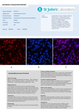
  • 1. ANTIBODY VALIDATION REPORT Report Number 90129-a Application Immunofluorescence Model Number STJ90129 Antibody Name Anti-Histone H4 (Acetyl K16) antibody Host Rabbit Clonality Polyclonal Clone ID NA Species HUMAN Tissue KIDNEY Image Description Immunofluorescence analysis of Human kidney tissue. 1: Histone H4 (Acetyl Lys16) Polyclonal Antibody(red) was diluted at 1:200 (4 degree Celsius,overnight). 2: Cy3 labled Secondary antibody was diluted at 1:300 (room temperature, 50min).3: Picture B: DAPI(blue) 10min. Picture A:Target. Picture B: DAPI. Picture C: merge of A+B. Primary Antibody Incubation After blocking solution was removed a 1:200 primary antibody/PBS solution was added on the slide, and incubated overnight at 4°C (a small amount of distilled water was added into the incubation box to prevent evaporation of antibody). Secondary Antibody Incubation slides were washed with PBS on a shaker for 5min, and repeated 3 times. Shortly after the slides were dried and corresponding secondary antibody solution was added (HRP labelled), covering the tissues, and incubated at room temperature for 50min. DAPI Counter-Staining slides were washed with PBS on a shaker for 5min, repeated 3 times and then dried. DAPI staining solution was added inside the PAP circles and incubated for 10 min at room temperature without light exposure. Mounting Slides were washed with PBS on a shaker for 5min, and repeated 3 times. Shortly after slides were dried, anti-quench mountings were used to mount slides. Visualization The slides were observed and placed under a NIKON inverted fluorescence microscope (Ultra violet excitation 330-380nm, emission 420nm; FITC green excitation 465-495nm, emission 515- 555 nm; CY3 red excitation 510-560nm, emission 590nm) Immunofluorescence Protocol Tissue Processing Slides were incubated sequentially into: Xylene - 15min, Anhydrous ethanol – 15 min, Anhydrous ethanol – 5 min, 85% alcohol – 5 min, 75% alcohol – 5 min & washed with distilled water – 5 min. Antigen Retrieval Tissue slides were incubated with citric acid (PH6.0) antigen retrieval buffer, and microwaved for antigen retrieval (heated until boiled and then stop heating) for 8min. Slides were then heated with medium power for 7min. During this process slides are kept from drying out. After cooling down at room temperature, slides were washed with PBS on a shaker for 5min, and repeated 3 times. Anti-Quench shortly after slides were dried, a PAP pen was used to draw circles around the tissues (to prevent draining of the antibody). Inside the circles, anti-quench mountings were added and incubated for 5 min, and then flushed with water for 10min. BSA Blocking Inside the circles, BSA was used to cover the tissue evenly, blocking for 30min.
  • 2. ANTIBODY VALIDATION REPORT Report Number 90129-b Application Immunofluorescence Model Number STJ90129 Antibody Name Anti-Histone H4 (Acetyl K16) antibody Host Rabbit Clonality Polyclonal Clone ID NA Species HUMAN Tissue KIDNEY Image Description Immunofluorescence analysis of Human kidney tissue. 1: Histone H4 (Acetyl Lys16) Polyclonal Antibody(red) was diluted at 1:200 (4 degree Celsius,overnight). 2: Cy3 labled Secondary antibody was diluted at 1:300 (room temperature, 50min).3: Picture B: DAPI(blue) 10min. Picture A:Target. Picture B: DAPI. Picture C: merge of A+B. Primary Antibody Incubation After blocking solution was removed a 1:200 primary antibody/PBS solution was added on the slide, and incubated overnight at 4°C (a small amount of distilled water was added into the incubation box to prevent evaporation of antibody). Secondary Antibody Incubation slides were washed with PBS on a shaker for 5min, and repeated 3 times. Shortly after the slides were dried and corresponding secondary antibody solution was added (HRP labelled), covering the tissues, and incubated at room temperature for 50min. DAPI Counter-Staining slides were washed with PBS on a shaker for 5min, repeated 3 times and then dried. DAPI staining solution was added inside the PAP circles and incubated for 10 min at room temperature without light exposure. Mounting Slides were washed with PBS on a shaker for 5min, and repeated 3 times. Shortly after slides were dried, anti-quench mountings were used to mount slides. Visualization The slides were observed and placed under a NIKON inverted fluorescence microscope (Ultra violet excitation 330-380nm, emission 420nm; FITC green excitation 465-495nm, emission 515- 555 nm; CY3 red excitation 510-560nm, emission 590nm) Immunofluorescence Protocol Tissue Processing Slides were incubated sequentially into: Xylene - 15min, Anhydrous ethanol – 15 min, Anhydrous ethanol – 5 min, 85% alcohol – 5 min, 75% alcohol – 5 min & washed with distilled water – 5 min. Antigen Retrieval Tissue slides were incubated with citric acid (PH6.0) antigen retrieval buffer, and microwaved for antigen retrieval (heated until boiled and then stop heating) for 8min. Slides were then heated with medium power for 7min. During this process slides are kept from drying out. After cooling down at room temperature, slides were washed with PBS on a shaker for 5min, and repeated 3 times. Anti-Quench shortly after slides were dried, a PAP pen was used to draw circles around the tissues (to prevent draining of the antibody). Inside the circles, anti-quench mountings were added and incubated for 5 min, and then flushed with water for 10min. BSA Blocking Inside the circles, BSA was used to cover the tissue evenly, blocking for 30min.
  • 3. ANTIBODY VALIDATION REPORT Report Number 90129-c Application Immunofluorescence Model Number STJ90129 Antibody Name Anti-Histone H4 (Acetyl K16) antibody Host Rabbit Clonality Polyclonal Clone ID NA Species HUMAN Tissue KIDNEY Image Description Immunofluorescence analysis of Human kidney tissue. 1: Histone H4 (Acetyl Lys16) Polyclonal Antibody(red) was diluted at 1:200 (4 degree Celsius,overnight). 2: Cy3 labled Secondary antibody was diluted at 1:300 (room temperature, 50min).3: Picture B: DAPI(blue) 10min. Picture A:Target. Picture B: DAPI. Picture C: merge of A+B. Primary Antibody Incubation After blocking solution was removed a 1:200 primary antibody/PBS solution was added on the slide, and incubated overnight at 4°C (a small amount of distilled water was added into the incubation box to prevent evaporation of antibody). Secondary Antibody Incubation slides were washed with PBS on a shaker for 5min, and repeated 3 times. Shortly after the slides were dried and corresponding secondary antibody solution was added (HRP labelled), covering the tissues, and incubated at room temperature for 50min. DAPI Counter-Staining slides were washed with PBS on a shaker for 5min, repeated 3 times and then dried. DAPI staining solution was added inside the PAP circles and incubated for 10 min at room temperature without light exposure. Mounting Slides were washed with PBS on a shaker for 5min, and repeated 3 times. Shortly after slides were dried, anti-quench mountings were used to mount slides. Visualization The slides were observed and placed under a NIKON inverted fluorescence microscope (Ultra violet excitation 330-380nm, emission 420nm; FITC green excitation 465-495nm, emission 515- 555 nm; CY3 red excitation 510-560nm, emission 590nm) Immunofluorescence Protocol Tissue Processing Slides were incubated sequentially into: Xylene - 15min, Anhydrous ethanol – 15 min, Anhydrous ethanol – 5 min, 85% alcohol – 5 min, 75% alcohol – 5 min & washed with distilled water – 5 min. Antigen Retrieval Tissue slides were incubated with citric acid (PH6.0) antigen retrieval buffer, and microwaved for antigen retrieval (heated until boiled and then stop heating) for 8min. Slides were then heated with medium power for 7min. During this process slides are kept from drying out. After cooling down at room temperature, slides were washed with PBS on a shaker for 5min, and repeated 3 times. Anti-Quench shortly after slides were dried, a PAP pen was used to draw circles around the tissues (to prevent draining of the antibody). Inside the circles, anti-quench mountings were added and incubated for 5 min, and then flushed with water for 10min. BSA Blocking Inside the circles, BSA was used to cover the tissue evenly, blocking for 30min.
  • 4. ANTIBODY VALIDATION REPORT Report Number 90129-d Application Immunofluorescence Model Number STJ90129 Antibody Name Anti-Histone H4 (Acetyl K16) antibody Host Rabbit Clonality Polyclonal Clone ID NA Species HUMAN Tissue LUNG CANCER Image Description Immunofluorescence analysis of Human lung cancer tissue. 1: Histone H4 (Acetyl Lys16) Polyclonal Antibody(red) was diluted at 1:200 (4 degree Celsius,overnight). 2: Cy3 labled Secondary antibody was diluted at 1:300 (room temperature, 50min).3: Picture B: DAPI(blue) 10min. Picture A:Target. Picture B: DAPI. Picture C: merge of A+B. Primary Antibody Incubation After blocking solution was removed a 1:200 primary antibody/PBS solution was added on the slide, and incubated overnight at 4°C (a small amount of distilled water was added into the incubation box to prevent evaporation of antibody). Secondary Antibody Incubation slides were washed with PBS on a shaker for 5min, and repeated 3 times. Shortly after the slides were dried and corresponding secondary antibody solution was added (HRP labelled), covering the tissues, and incubated at room temperature for 50min. DAPI Counter-Staining slides were washed with PBS on a shaker for 5min, repeated 3 times and then dried. DAPI staining solution was added inside the PAP circles and incubated for 10 min at room temperature without light exposure. Mounting Slides were washed with PBS on a shaker for 5min, and repeated 3 times. Shortly after slides were dried, anti-quench mountings were used to mount slides. Visualization The slides were observed and placed under a NIKON inverted fluorescence microscope (Ultra violet excitation 330-380nm, emission 420nm; FITC green excitation 465-495nm, emission 515- 555 nm; CY3 red excitation 510-560nm, emission 590nm) Immunofluorescence Protocol Tissue Processing Slides were incubated sequentially into: Xylene - 15min, Anhydrous ethanol – 15 min, Anhydrous ethanol – 5 min, 85% alcohol – 5 min, 75% alcohol – 5 min & washed with distilled water – 5 min. Antigen Retrieval Tissue slides were incubated with citric acid (PH6.0) antigen retrieval buffer, and microwaved for antigen retrieval (heated until boiled and then stop heating) for 8min. Slides were then heated with medium power for 7min. During this process slides are kept from drying out. After cooling down at room temperature, slides were washed with PBS on a shaker for 5min, and repeated 3 times. Anti-Quench shortly after slides were dried, a PAP pen was used to draw circles around the tissues (to prevent draining of the antibody). Inside the circles, anti-quench mountings were added and incubated for 5 min, and then flushed with water for 10min. BSA Blocking Inside the circles, BSA was used to cover the tissue evenly, blocking for 30min.
  • 5. ANTIBODY VALIDATION REPORT Report Number 90129-e Application Immunofluorescence Model Number STJ90129 Antibody Name Anti-Histone H4 (Acetyl K16) antibody Host Rabbit Clonality Polyclonal Clone ID NA Species HUMAN Tissue LUNG CANCER Image Description Immunofluorescence analysis of Human lung cancer tissue. 1: Histone H4 (Acetyl Lys16) Polyclonal Antibody(red) was diluted at 1:200 (4 degree Celsius,overnight). 2: Cy3 labled Secondary antibody was diluted at 1:300 (room temperature, 50min).3: Picture B: DAPI(blue) 10min. Picture A:Target. Picture B: DAPI. Picture C: merge of A+B. Primary Antibody Incubation After blocking solution was removed a 1:200 primary antibody/PBS solution was added on the slide, and incubated overnight at 4°C (a small amount of distilled water was added into the incubation box to prevent evaporation of antibody). Secondary Antibody Incubation slides were washed with PBS on a shaker for 5min, and repeated 3 times. Shortly after the slides were dried and corresponding secondary antibody solution was added (HRP labelled), covering the tissues, and incubated at room temperature for 50min. DAPI Counter-Staining slides were washed with PBS on a shaker for 5min, repeated 3 times and then dried. DAPI staining solution was added inside the PAP circles and incubated for 10 min at room temperature without light exposure. Mounting Slides were washed with PBS on a shaker for 5min, and repeated 3 times. Shortly after slides were dried, anti-quench mountings were used to mount slides. Visualization The slides were observed and placed under a NIKON inverted fluorescence microscope (Ultra violet excitation 330-380nm, emission 420nm; FITC green excitation 465-495nm, emission 515- 555 nm; CY3 red excitation 510-560nm, emission 590nm) Immunofluorescence Protocol Tissue Processing Slides were incubated sequentially into: Xylene - 15min, Anhydrous ethanol – 15 min, Anhydrous ethanol – 5 min, 85% alcohol – 5 min, 75% alcohol – 5 min & washed with distilled water – 5 min. Antigen Retrieval Tissue slides were incubated with citric acid (PH6.0) antigen retrieval buffer, and microwaved for antigen retrieval (heated until boiled and then stop heating) for 8min. Slides were then heated with medium power for 7min. During this process slides are kept from drying out. After cooling down at room temperature, slides were washed with PBS on a shaker for 5min, and repeated 3 times. Anti-Quench shortly after slides were dried, a PAP pen was used to draw circles around the tissues (to prevent draining of the antibody). Inside the circles, anti-quench mountings were added and incubated for 5 min, and then flushed with water for 10min. BSA Blocking Inside the circles, BSA was used to cover the tissue evenly, blocking for 30min.
  • 6. ANTIBODY VALIDATION REPORT Report Number 90129-f Application Immunofluorescence Model Number STJ90129 Antibody Name Anti-Histone H4 (Acetyl K16) antibody Host Rabbit Clonality Polyclonal Clone ID NA Species HUMAN Tissue LUNG CANCER Image Description Immunofluorescence analysis of Human lung cancer tissue. 1: Histone H4 (Acetyl Lys16) Polyclonal Antibody(red) was diluted at 1:200 (4 degree Celsius,overnight). 2: Cy3 labled Secondary antibody was diluted at 1:300 (room temperature, 50min).3: Picture B: DAPI(blue) 10min. Picture A:Target. Picture B: DAPI. Picture C: merge of A+B. Primary Antibody Incubation After blocking solution was removed a 1:200 primary antibody/PBS solution was added on the slide, and incubated overnight at 4°C (a small amount of distilled water was added into the incubation box to prevent evaporation of antibody). Secondary Antibody Incubation slides were washed with PBS on a shaker for 5min, and repeated 3 times. Shortly after the slides were dried and corresponding secondary antibody solution was added (HRP labelled), covering the tissues, and incubated at room temperature for 50min. DAPI Counter-Staining slides were washed with PBS on a shaker for 5min, repeated 3 times and then dried. DAPI staining solution was added inside the PAP circles and incubated for 10 min at room temperature without light exposure. Mounting Slides were washed with PBS on a shaker for 5min, and repeated 3 times. Shortly after slides were dried, anti-quench mountings were used to mount slides. Visualization The slides were observed and placed under a NIKON inverted fluorescence microscope (Ultra violet excitation 330-380nm, emission 420nm; FITC green excitation 465-495nm, emission 515- 555 nm; CY3 red excitation 510-560nm, emission 590nm) Immunofluorescence Protocol Tissue Processing Slides were incubated sequentially into: Xylene - 15min, Anhydrous ethanol – 15 min, Anhydrous ethanol – 5 min, 85% alcohol – 5 min, 75% alcohol – 5 min & washed with distilled water – 5 min. Antigen Retrieval Tissue slides were incubated with citric acid (PH6.0) antigen retrieval buffer, and microwaved for antigen retrieval (heated until boiled and then stop heating) for 8min. Slides were then heated with medium power for 7min. During this process slides are kept from drying out. After cooling down at room temperature, slides were washed with PBS on a shaker for 5min, and repeated 3 times. Anti-Quench shortly after slides were dried, a PAP pen was used to draw circles around the tissues (to prevent draining of the antibody). Inside the circles, anti-quench mountings were added and incubated for 5 min, and then flushed with water for 10min. BSA Blocking Inside the circles, BSA was used to cover the tissue evenly, blocking for 30min.
  • 7. ANTIBODY VALIDATION REPORT Report Number 90129-g Application Immunofluorescence Model Number STJ90129 Antibody Name Anti-Histone H4 (Acetyl K16) antibody Host Rabbit Clonality Polyclonal Clone ID NA Species HUMAN Tissue LUNG Image Description Immunofluorescence analysis of Human lung tissue. 1: Histone H4 (Acetyl Lys16) Polyclonal Antibody(red) was diluted at 1:200 (4 degree Celsius,overnight). 2: Cy3 labled Secondary antibody was diluted at 1:300 (room temperature, 50min).3: Picture B: DAPI(blue) 10min. Picture A:Target. Picture B: DAPI. Picture C: merge of A+B. Primary Antibody Incubation After blocking solution was removed a 1:200 primary antibody/PBS solution was added on the slide, and incubated overnight at 4°C (a small amount of distilled water was added into the incubation box to prevent evaporation of antibody). Secondary Antibody Incubation slides were washed with PBS on a shaker for 5min, and repeated 3 times. Shortly after the slides were dried and corresponding secondary antibody solution was added (HRP labelled), covering the tissues, and incubated at room temperature for 50min. DAPI Counter-Staining slides were washed with PBS on a shaker for 5min, repeated 3 times and then dried. DAPI staining solution was added inside the PAP circles and incubated for 10 min at room temperature without light exposure. Mounting Slides were washed with PBS on a shaker for 5min, and repeated 3 times. Shortly after slides were dried, anti-quench mountings were used to mount slides. Visualization The slides were observed and placed under a NIKON inverted fluorescence microscope (Ultra violet excitation 330-380nm, emission 420nm; FITC green excitation 465-495nm, emission 515- 555 nm; CY3 red excitation 510-560nm, emission 590nm) Immunofluorescence Protocol Tissue Processing Slides were incubated sequentially into: Xylene - 15min, Anhydrous ethanol – 15 min, Anhydrous ethanol – 5 min, 85% alcohol – 5 min, 75% alcohol – 5 min & washed with distilled water – 5 min. Antigen Retrieval Tissue slides were incubated with citric acid (PH6.0) antigen retrieval buffer, and microwaved for antigen retrieval (heated until boiled and then stop heating) for 8min. Slides were then heated with medium power for 7min. During this process slides are kept from drying out. After cooling down at room temperature, slides were washed with PBS on a shaker for 5min, and repeated 3 times. Anti-Quench shortly after slides were dried, a PAP pen was used to draw circles around the tissues (to prevent draining of the antibody). Inside the circles, anti-quench mountings were added and incubated for 5 min, and then flushed with water for 10min. BSA Blocking Inside the circles, BSA was used to cover the tissue evenly, blocking for 30min.
  • 8. ANTIBODY VALIDATION REPORT Report Number 90129-h Application Immunofluorescence Model Number STJ90129 Antibody Name Anti-Histone H4 (Acetyl K16) antibody Host Rabbit Clonality Polyclonal Clone ID NA Species HUMAN Tissue LUNG Image Description Immunofluorescence analysis of Human lung tissue. 1: Histone H4 (Acetyl Lys16) Polyclonal Antibody(red) was diluted at 1:200 (4 degree Celsius,overnight). 2: Cy3 labled Secondary antibody was diluted at 1:300 (room temperature, 50min).3: Picture B: DAPI(blue) 10min. Picture A:Target. Picture B: DAPI. Picture C: merge of A+B. Primary Antibody Incubation After blocking solution was removed a 1:200 primary antibody/PBS solution was added on the slide, and incubated overnight at 4°C (a small amount of distilled water was added into the incubation box to prevent evaporation of antibody). Secondary Antibody Incubation slides were washed with PBS on a shaker for 5min, and repeated 3 times. Shortly after the slides were dried and corresponding secondary antibody solution was added (HRP labelled), covering the tissues, and incubated at room temperature for 50min. DAPI Counter-Staining slides were washed with PBS on a shaker for 5min, repeated 3 times and then dried. DAPI staining solution was added inside the PAP circles and incubated for 10 min at room temperature without light exposure. Mounting Slides were washed with PBS on a shaker for 5min, and repeated 3 times. Shortly after slides were dried, anti-quench mountings were used to mount slides. Visualization The slides were observed and placed under a NIKON inverted fluorescence microscope (Ultra violet excitation 330-380nm, emission 420nm; FITC green excitation 465-495nm, emission 515- 555 nm; CY3 red excitation 510-560nm, emission 590nm) Immunofluorescence Protocol Tissue Processing Slides were incubated sequentially into: Xylene - 15min, Anhydrous ethanol – 15 min, Anhydrous ethanol – 5 min, 85% alcohol – 5 min, 75% alcohol – 5 min & washed with distilled water – 5 min. Antigen Retrieval Tissue slides were incubated with citric acid (PH6.0) antigen retrieval buffer, and microwaved for antigen retrieval (heated until boiled and then stop heating) for 8min. Slides were then heated with medium power for 7min. During this process slides are kept from drying out. After cooling down at room temperature, slides were washed with PBS on a shaker for 5min, and repeated 3 times. Anti-Quench shortly after slides were dried, a PAP pen was used to draw circles around the tissues (to prevent draining of the antibody). Inside the circles, anti-quench mountings were added and incubated for 5 min, and then flushed with water for 10min. BSA Blocking Inside the circles, BSA was used to cover the tissue evenly, blocking for 30min.
  • 9. ANTIBODY VALIDATION REPORT Report Number 90129-i Application Immunofluorescence Model Number STJ90129 Antibody Name Anti-Histone H4 (Acetyl K16) antibody Host Rabbit Clonality Polyclonal Clone ID NA Species HUMAN Tissue LUNG Image Description Immunofluorescence analysis of Human lung tissue. 1: Histone H4 (Acetyl Lys16) Polyclonal Antibody(red) was diluted at 1:200 (4 degree Celsius,overnight). 2: Cy3 labled Secondary antibody was diluted at 1:300 (room temperature, 50min).3: Picture B: DAPI(blue) 10min. Picture A:Target. Picture B: DAPI. Picture C: merge of A+B. Primary Antibody Incubation After blocking solution was removed a 1:200 primary antibody/PBS solution was added on the slide, and incubated overnight at 4°C (a small amount of distilled water was added into the incubation box to prevent evaporation of antibody). Secondary Antibody Incubation slides were washed with PBS on a shaker for 5min, and repeated 3 times. Shortly after the slides were dried and corresponding secondary antibody solution was added (HRP labelled), covering the tissues, and incubated at room temperature for 50min. DAPI Counter-Staining slides were washed with PBS on a shaker for 5min, repeated 3 times and then dried. DAPI staining solution was added inside the PAP circles and incubated for 10 min at room temperature without light exposure. Mounting Slides were washed with PBS on a shaker for 5min, and repeated 3 times. Shortly after slides were dried, anti-quench mountings were used to mount slides. Visualization The slides were observed and placed under a NIKON inverted fluorescence microscope (Ultra violet excitation 330-380nm, emission 420nm; FITC green excitation 465-495nm, emission 515- 555 nm; CY3 red excitation 510-560nm, emission 590nm) Immunofluorescence Protocol Tissue Processing Slides were incubated sequentially into: Xylene - 15min, Anhydrous ethanol – 15 min, Anhydrous ethanol – 5 min, 85% alcohol – 5 min, 75% alcohol – 5 min & washed with distilled water – 5 min. Antigen Retrieval Tissue slides were incubated with citric acid (PH6.0) antigen retrieval buffer, and microwaved for antigen retrieval (heated until boiled and then stop heating) for 8min. Slides were then heated with medium power for 7min. During this process slides are kept from drying out. After cooling down at room temperature, slides were washed with PBS on a shaker for 5min, and repeated 3 times. Anti-Quench shortly after slides were dried, a PAP pen was used to draw circles around the tissues (to prevent draining of the antibody). Inside the circles, anti-quench mountings were added and incubated for 5 min, and then flushed with water for 10min. BSA Blocking Inside the circles, BSA was used to cover the tissue evenly, blocking for 30min.