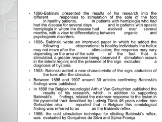
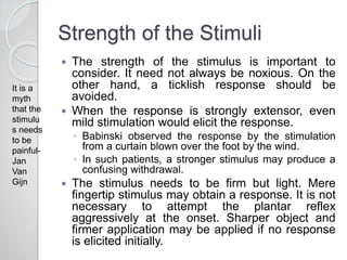
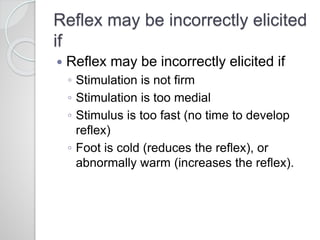
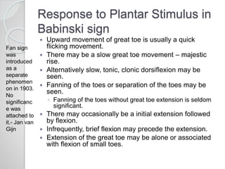
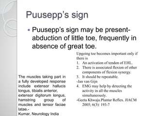
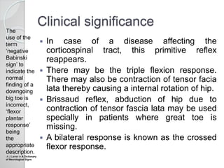
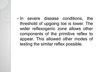
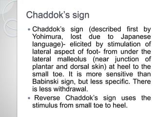
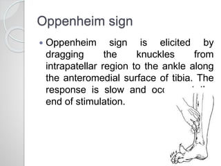
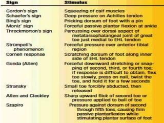
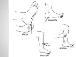
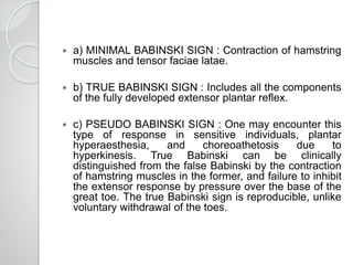
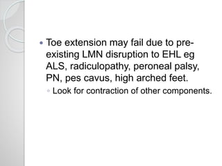
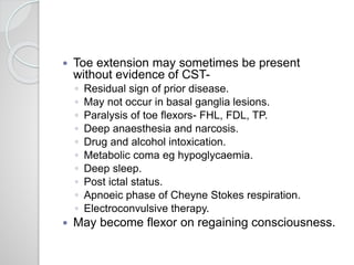
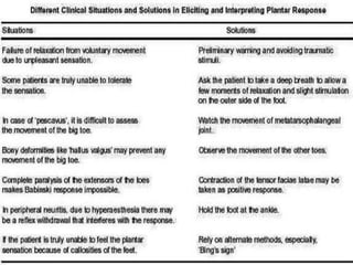
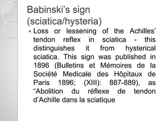
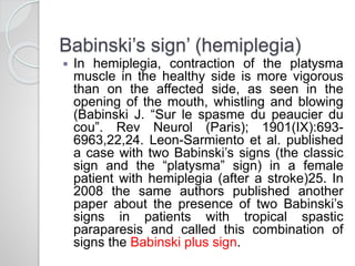
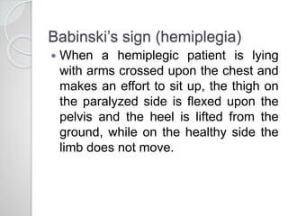
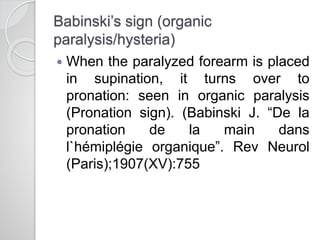
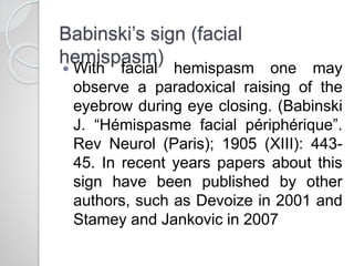
2. Joseph Jules Francois Felix
Babinski
Born to a Polish couple in Paris, on
November 17, 1857, Joseph François Felix
Babinski, he became head of neurology
clinics under Charcot, after his thesis on
pathological anatomy of multiple sclerosis
was ranked second. He left the Salpêtrière
Hospital and became the head of neurology
service at La Pitié Hospital in 1895 where he
continued till his retirement. 9 years
thereafter he died on October 29, 1932 at the
age of 74 having established the first
neurological service at France and leaving
behind a great contribution to neurology.
3. 1896-Babinski presented the results of his research into the
different responses to stimulation of the sole of the foot
in healthy patients, in patients with hemiplegia who had
had the disease for several days and in patients with spastic
hemiplegia in whom the disease had evolved over several
months, with a view to differentiating between organic and
psychogenic disorders.
1898- Babinski wrote an improved paper in which he added the
following observations: in healthy individuals the hallux
may not move after the stimulation; the response may vary
depending on the area of the sole of the foot that is
stimulated, a greater response being observed if stimulation occurs
in the lateral region; and the presence of the sign excludes a
diagnosis of hysteria.
1903- Babinski added a new characteristic of the sign: abduction of
the toes after the stimulus.
Between 1898 and 1907 around 30 articles confirming Babinski’s
findings were published.
In 1898 the Belgian neurologist Arthur Van Gehuchten published the
results of his research, which, in addition to supporting
Babinski’s findings, related the extensor response to the lesion in
the pyramidal tract described by Ludwig Türck 46 years earlier. Van
Gehuchten also reported that in Belgium this semiological
finding was referred to as as the Babinski reflex.
1966- the cold stimulation technique for eliciting Babinski’s reflex,
was evaluated by Gonçalves da Silva and Spina-França
4. How to Elicit
The patient should be explained the
procedure and should be relaxed. The
knees should be extended, whether in
supine position (preferred) or sitting
position (support the foot in hand or on
knee of examiner)- this helps expose
plantar surface.
Flexed knee may abolish the upgoing
toe.
The stimulation should be done far
laterally (S1 root or sural nerve sensory
distribution).
◦ More medially, a positive response may be
missed.
5. The stimulus starts at the heel and the
stimulator is moved up at a steady pace
turning at the metatarsophalangeal joint
and then continuing along metatarsal
pad from the little toe, but not extending
to the base of great toe.
Frequently, the response may be elicited
till one reaches the mid portion of the
foot.
The reflex is reinforced by rotating the
patient’s head to opposite side.
6. Instrument of Stimulation
Stimulate the plantar surface of the
foot with a blunt point eg handle of
reflex hammer, tongue blade and
thumbnail. The instrument of use has
been a subject of debate.
◦ Henry Miller preferred use of Bentley key.
◦ Babinski favoured goose quill.
7. Strength of the Stimuli
The strength of the stimulus is important to
consider. It need not always be noxious. On the
other hand, a ticklish response should be
avoided.
When the response is strongly extensor, even
mild stimulation would elicit the response.
◦ Babinski observed the response by the stimulation
from a curtain blown over the foot by the wind.
◦ In such patients, a stronger stimulus may produce a
confusing withdrawal.
The stimulus needs to be firm but light. Mere
fingertip stimulus may obtain a response. It is not
necessary to attempt the plantar reflex
aggressively at the onset. Sharper object and
firmer application may be applied if no response
is elicited initially.
It is a
myth
that the
stimulu
s needs
to be
painful-
Jan
Van
Gijn
8. Reflex may be incorrectly elicited
if
Reflex may be incorrectly elicited if
◦ Stimulation is not firm
◦ Stimulation is too medial
◦ Stimulus is too fast (no time to develop
reflex)
◦ Foot is cold (reduces the reflex), or
abnormally warm (increases the reflex).
9. Response to Plantar Stimulus in
Babinski sign
Upward movement of great toe is usually a quick
flicking movement.
There may be a slow great toe movement – majestic
rise.
Alternatively slow, tonic, clonic dorsiflexion may be
seen.
Fanning of the toes or separation of the toes may be
seen.
◦ Fanning of the toes without great toe extension is seldom
significant.
There may occasionally be a initial extension followed
by flexion.
Infrequently, brief flexion may precede the extension.
Extension of the great toe may be alone or associated
with flexion of small toes.
Fan sign
was
introduced
as a
separate
phenomen
on in 1903.
No
significanc
e was
attached to
it.- Jan van
Gijn
10. Puusepp’s sign
Puusepp’s sign may be present-
abduction of little toe, frequently in
absence of great toe.
Upgoing toe becomes important only if
there is
1. An activation of tendon of EHL.
2. There is associated flexion of other
components of flexion synergy.
3. It should be repeatable.
-Jan van Gijn
4. EMG may help by detecting the
activity in all the muscles
simultaneously.
-Geeta Khwaja.Plantar Reflex. JIACM
2005; 6(3): 193-7
The muscles taking part in
a fully developed response
include extensor hallucis
longus, tibialis anterior,
extensor digitorum longus,
hamstring group of
muscles and tensor faciae
latae.-
Kumar, Neurology India
11. Known since Before Babinski to be
Extensor normally in infants-cause- lack
of myelination of CST.
Small Cowper
Madonna- Raphael
Madonna with
Child and 2
Angels- Botticelli
Nejm 1999 Jan
12. Babinski sign is a primitive flexion response that
emerges at 18 weeks in utero.
CNS is organized so as to avoid or withdraw from
noxious stimuli.
The flexion response includes flexion at hip and knee
and dorsiflexion at ankle and toes-flexion synergy.
◦ Even though the muscles involved are of the extensor
group, the overall action is shortening of the limb.
◦ Therefore its termed flexion response.
This persists normally from 1 year to 3 year of age. The
myelination of corticospinal tract completes by 1 year of
age.
◦ Thereafter, this primitive reflex is suppressed, which is
necessary for normal ambulation.
◦ The flexion is a part of local skin reflex, similar to
abdominal reflex.
The brisk
flexion
synergy is
brought
under
control of
CST which
inhibit the
spinal
motor
neuron.
- Kumar in
Neurology
India
13. The motor neurons of the leg muscles
are laminated into separate columns
within the anterior horns of the cord,
each of which supply proximal or distal
flexor or extensor muscles.
Both structural as well as functional
lesions of the pyramidal tract fibres
projecting onto the lumbosacral
anterior horn cells and interneurons
supplying the leg muscles subserving
the ‘flexion reflex synergy’ can release
the Babinski sign.
- Kumar in NI
15. The reflexogenic area is the first sacral
dermatome, with the receptor nerve endings
being located in the skin on the sole of the
foot.
The afferent fibres travel in the tibial nerve
which is a branch of the sciatic nerve, to relay
in the L4-5 to S1-2 cord segments.
The efferent fibres from the spinal cord travel
back in the sciatic nerve.
Fibres supplying the toe flexors travel in the
tibial nerve while those supplying the toe
extensors travel in the peroneal nerve to
reach the foot.
Supraspinal influences from the cortex also
influence and modify the spinal reflex or
response.
16. The Plantar Reflex being a
polysynaptic reflex, the interneurons in
the reflex arc connect with motor
neurons at several segmental levels,
leading to a co-ordinated motor
response or movement of the foot and
lower limb following cutaneous
stimulation of the sole of the foot.
18. Clinical significance
In case of a disease affecting the
corticospinal tract, this primitive reflex
reappears.
There may be the triple flexion response.
There may also be contraction of tensor facia
lata thereby causing a internal rotation of hip.
Brissaud reflex, abduction of hip due to
contraction of tensor fascia lata may be used
specially in patients where great toe is
missing.
A bilateral response is known as the crossed
flexor response.
The
use of the
term
‘negative
Babinski
sign’ to
indicate the
normal
finding of a
downgoing
big toe is
incorrect,
‘flexor
plantar
response’
being
the
appropriate
description.
-A J Larner in A Dictionary
of Neurological Signs
19. In severe disease conditions, the
threshold of upgoing toe is lower. The
wider reflexogenic zone allows other
components of the primitive reflex to
appear. This allowed other modes of
testing the similar reflex possible.
20. Chaddok’s sign
Chaddok’s sign (described first by
Yohimura, lost due to Japanese
language)- elicited by stimulation of
lateral aspect of foot- from under the
lateral malleolus (near junction of
plantar and dorsal skin) at heel to the
small toe. It is more sensitive than
Babinski sign, but less specific. There
is less withdrawal.
Reverse Chaddok’s sign uses the
stimulus from small toe to heel.
21. Oppenheim sign
Oppenheim sign is elicited by
dragging the knuckles from
intrapatellar region to the ankle along
the anteromedial surface of tibia. The
response is slow and occurs at the
end of stimulation.
25. a) MINIMAL BABINSKI SIGN : Contraction of hamstring
muscles and tensor faciae latae.
b) TRUE BABINSKI SIGN : Includes all the components
of the fully developed extensor plantar reflex.
c) PSEUDO BABINSKI SIGN : One may encounter this
type of response in sensitive individuals, plantar
hyperaesthesia, and choreoathetosis due to
hyperkinesis. True Babinski can be clinically
distinguished from the false Babinski by the contraction
of hamstring muscles in the former, and failure to inhibit
the extensor response by pressure over the base of the
great toe. The true Babinski sign is reproducible, unlike
voluntary withdrawal of the toes.
26. d) EXAGGERATED BABINSKI SIGN : It can either be in
the form of 'flexor spasm' or 'extensor spasm',
depending upon the muscles i.e. whether flexors or
extensors, have excess of tone. Flexor spasms occur in
spinal cord disease, bilateral upper motor neuron lesion
at a supraspinal level, multiple sclerosis and subacute
combined degeneration of the cord, while 'extensor
spasm' occurs in patients with corticospinal tract lesion
when the posterior column function is normal.
e) INVERSION OF PLANTAR REFLEX : If the short
flexors of the toe are paralysed or flexor tendons are
severed accidentally, an extensor response may be
obtained.
f) TONIC BABINSKI REFLEX : Characterised by slow
prolonged contraction of extensors of toe, seen in
frontal lobe lesions and extrapyramidal involvement.
27. g) CROSSED EXTENSOR
RESPONSE/BILATERAL BABINSKI
SIGN : Unilateral stimulation produces
bilateral Babinski in patients with bilateral
cerebral disease and spinal cord
disease.
h) SPONTANEOUS BABINSKI : In
infants and children following
manipulation of the foot, and in patients
with extensive pyramidal tract diseases,
passive extension of the knee or passive
flexion of the hip and the knee, may
produce a positive Babinski sign.
28. PROBLEMS WITH
BABINSKI SIGN
Reflex may be incorrectly elicited if
Stimulation is not firm
Stimulation is too medial
Stimulus is too fast (no time to develop reflex)
Foot is cold (reduces the reflex), or abnormally warm (increases the
reflex).
29. Differentiating extensor sign from withdrawal-
Hip and knee flexion are not reliable since the
Babinski sign is actually a withdrawal
response.
◦ Dorsiflexion of ankle is rare with voluntary
withdrawal.
◦ Withdrawal occurs with intense and
uncomfortable stimuli.
◦ In ticklish person, holding the ankle may suffice.
◦ Some favour auto Babinski, but this may not be
useful.
◦ Pressure on base of great toe eliminates
withdrawal response, but not extensor response.
◦ Presence of Brissaud reflex favours reflex
30. Toe movement may be circumspect-
Movement may be absent or decreased as in
on repeated stimulation, or with withdrawal,
or may be mute or silent.
◦ Initial toe movement is most important.
◦ Associated components of reflex
◦ Asymmetry of plantar response may be
significant (including sluggish flexion).
◦ May become extensor later in the day, or once
tired.
◦ Preknowledge of history and rest of the
examination may help.
◦ Use of Chaddok or Oppenheim test or Gordon
test and reinforcement.
It is a myth
that 1st
movement
counts.
Other
movements
of flexion
synergy and
even prior
flexion of
great toe may
occur-
Jan van Gijn
Its a myth that
Babinski
mimics may be
used if it is
doubtul
because the
resulting
phenomenon is
the same.
- Jan van Gijn
31. Toe extension may fail due to pre-
existing LMN disruption to EHL eg
ALS, radiculopathy, peroneal palsy,
PN, pes cavus, high arched feet.
◦ Look for contraction of other components.
32. Toe extension may sometimes be present
without evidence of CST-
◦ Residual sign of prior disease.
◦ May not occur in basal ganglia lesions.
◦ Paralysis of toe flexors- FHL, FDL, TP.
◦ Deep anaesthesia and narcosis.
◦ Drug and alcohol intoxication.
◦ Metabolic coma eg hypoglycaemia.
◦ Deep sleep.
◦ Post ictal status.
◦ Apnoeic phase of Cheyne Stokes respiration.
◦ Electroconvulsive therapy.
May become flexor on regaining consciousness.
35. Babinski’s sign
(sciatica/hysteria)
Loss or lessening of the Achilles’
tendon reflex in sciatica - this
distinguishes it from hysterical
sciatica. This sign was published in
1896 (Bulletins et Mémoires de la
Société Medicale des Hôpitaux de
Paris 1896; (XIII): 887-889), as
“Abolition du réflexe de tendon
d’Achille dans la sciatique
36. Babinski’s sign’ (hemiplegia)
In hemiplegia, contraction of the platysma
muscle in the healthy side is more vigorous
than on the affected side, as seen in the
opening of the mouth, whistling and blowing
(Babinski J. “Sur le spasme du peaucier du
cou”. Rev Neurol (Paris); 1901(IX):693-
6963,22,24. Leon-Sarmiento et al. published
a case with two Babinski’s signs (the classic
sign and the “platysma” sign) in a female
patient with hemiplegia (after a stroke)25. In
2008 the same authors published another
paper about the presence of two Babinski’s
signs in patients with tropical spastic
paraparesis and called this combination of
signs the Babinski plus sign.
37. Babinski’s sign (hemiplegia)
When a hemiplegic patient is lying
with arms crossed upon the chest and
makes an effort to sit up, the thigh on
the paralyzed side is flexed upon the
pelvis and the heel is lifted from the
ground, while on the healthy side the
limb does not move.
38. Babinski’s sign (organic
paralysis/hysteria)
When the paralyzed forearm is placed
in supination, it turns over to
pronation: seen in organic paralysis
(Pronation sign). (Babinski J. “De la
pronation de la main dans
l`hémiplégie organique”. Rev Neurol
(Paris);1907(XV):755
39. Babinski’s sign (facial
hemispasm)
With facial hemispasm one may
observe a paradoxical raising of the
eyebrow during eye closing. (Babinski
J. “Hémispasme facial périphérique”.
Rev Neurol (Paris); 1905 (XIII): 443-
45. In recent years papers about this
sign have been published by other
authors, such as Devoize in 2001 and
Stamey and Jankovic in 2007
41. Pseudo Babinski sign
This sign may be encountered in
patients with choreoathetosis where
the upgoing toe is a manifestation of
hyperkinesia.
42. Inversion of the plantar reflex
If the short flexors of the toe are
paralysed, or the flexor tendons have
been severed, an extensor plantar
response may be obtained even in the
absence of UMN lesions and is
termed inversion of the plantar reflex
of peripheral origin.
43.
44.
45. The Hoffmann's sign is sometimes
described as the upper limb equivalent
of the Babinski's sign because both
indicate upper motor neuron
dysfunction. Mechanistically, they
differ significantly; the finger flexor
reflex is a simple monosynaptic spinal
reflex involving the flexor digitorum
profundus that is normally fully
inhibited by upper motor neurons
47. DeJong. 7th Ed. Chap 40. Pathological reflexes.
The History, Physical, and Laboratory Examinations. 3rd edition.Walker HK, Hall
WD, Hurst JW, 2 - Journal, Indian Academy of Clinical Medicine Vol. 6 July-
September, 2005 Geeta Akhwaja
Jan van Gijn. The Babinski sign. PRACTICAL NEUROLOGY.2002.
The Babinski Sign for Diseases of the Corticospinal System. Steven D.
Waldman MD, JD. Physical Diagnosis of Pain, 157, 245-245a
A J Larner in A Dictionary of Neurological Signs
FRANCIS A. NEELON, ELISABETH N. HARVEY. The Babinski Sign. NEJM.
1999 JAN. 196.
SP Kumar, D Ramasubramanian. The Babinski sign-a reappraisal.
Neurology India. 2000;48:314-8.
Fabio T. Kakitani1, Daniel Collares, Adam Y. Kurozawa1, Plínio M.G. de
Lima, Hélio A.G. Teive. How many Babinski’s signs are there? Arq
Neuropsiquiatr 2010;68(4):662-665
Austin J summer. The Babinski Sign. Journal of the Neurological Sciences.
2014;343 :2