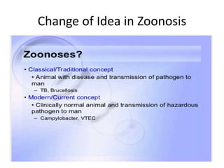
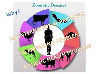
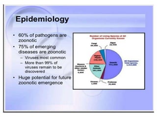
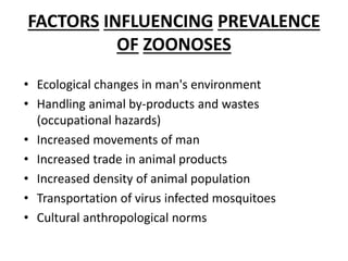
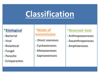
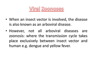
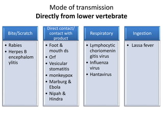
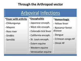
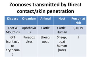
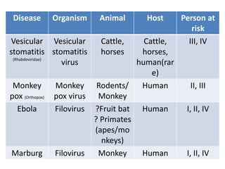
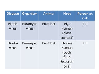
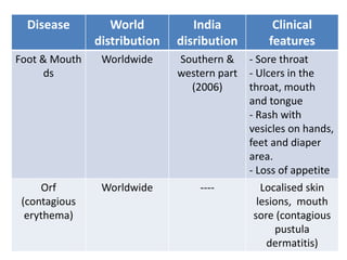
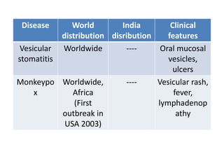
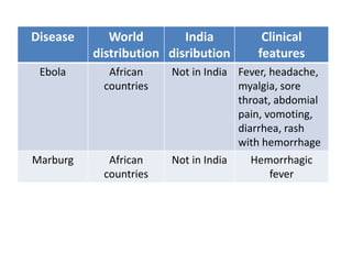
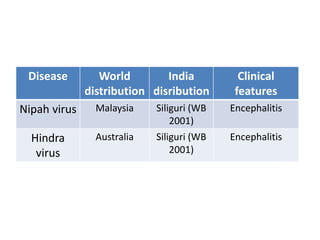
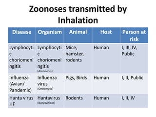
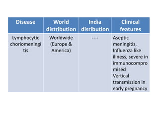
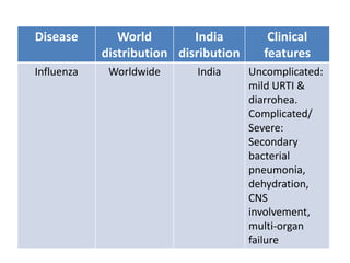
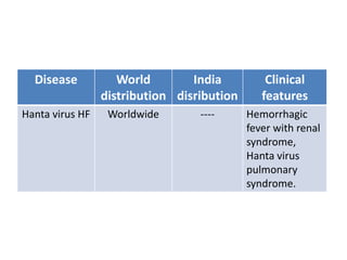
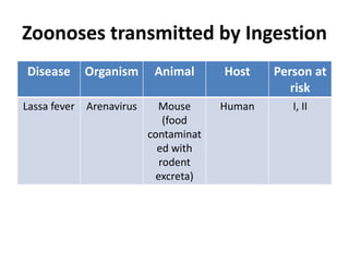
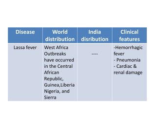
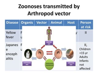
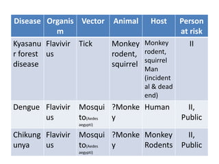
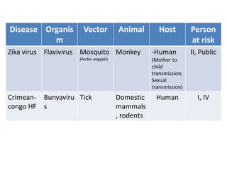
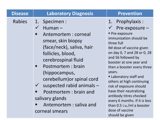
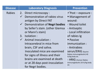
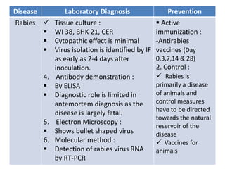
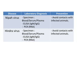
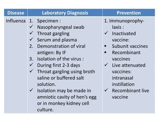
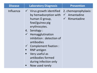
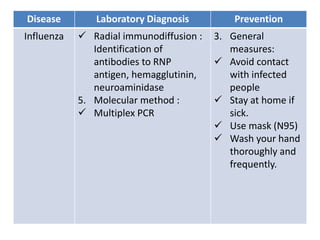
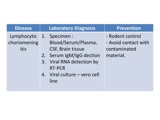
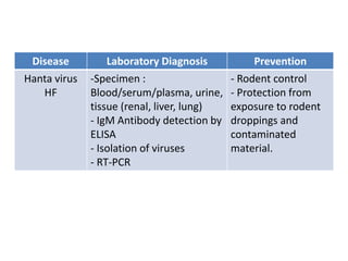
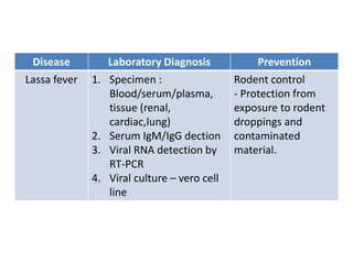
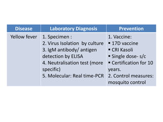
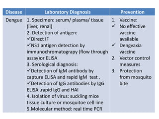
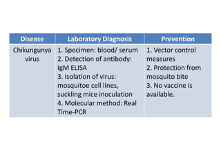
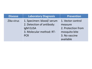
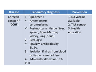
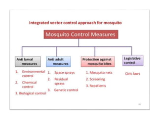
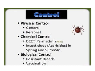
ZOONOSES
Dr. manoj vedpathak
• Scope
 Definition
 Epidemiology
 Factors influencing prevalence
 Stages of zoonotic diseases
 Modes of transmission
 Persons at risk
 Classification
 Viral zoonoses – classification, general clinical features,
diseases in brief
 Laboratory diagnosis
 Treatment
 Prevention
•The word 'Zoonosis' (Pleural: Zoonoses) was
introduced by Rudolf Virchow in 1880 to include
collectively the diseases shared in nature by man and
animals.
• Later WHO in 1959 defined that Zoonoses are "those
diseases and infections which are naturally transmitted
between vertebrate animals and man".
(http://www.who.int/topics/zoonoses/en/)
• Zoonoses include only those infections where there is
either a proof or a strong circumstantial evidence for
transmission between animals and man.
• Zoonotic Diseases (also known as zoonoses) are
caused by infections that are shared between animals
and people. (https://www.cdc.gov/onehealth/basics/zoonotic-diseases.html)
Change of Idea in Zoonosis
FACTORS INFLUENCING PREVALENCE
OF ZOONOSES
• Ecological changes in man's environment
• Handling animal by-products and wastes
(occupational hazards)
• Increased movements of man
• Increased trade in animal products
• Increased density of animal population
• Transportation of virus infected mosquitoes
• Cultural anthropological norms
Who are at Risk?
I. Farmers, livestock and animal processing workers
II. Outdoor recreational activities or vocational activities
in wild or underdeveloped regions
III. Persons in contact with pets, other animals in urban
areas
IV. Health care and laboratory personnel (human and
veterinary)
V. Fishers and others working in aquatic envirnment
VI. Immunocompromised hosts;
Public, anyone, any place
Classification
*Etiological
- Bacterial
- Viral
- Rickettsial
- Fungal
- Parasitic
- Ectoparasites
*Mode of
transmission
- Direct zoonoses
- Cyclozoonoses
- Metazoonoses
- Saprozoonoses
*Reservoir host
- Anthropozoonoses
- Zooanthroponoses
- Amphixenoses
• When an insect vector is involved, the disease
is also known as an arboviral disease.
• However, not all arboviral diseases are
zoonosis: where the transmission cycle takes
place exclusively between insect vector and
human e.g. dengue and yellow fever.
Mode of transmission
Directly from lower vertebrate
Bite/Scratch
• Rabies
• Herpes B
encephalom
ylitis
Direct contact/
contact with
product
• Foot &
mouth ds
• Orf
• Vesicular
stomatitis
• monkeypox
• Marburg &
Ebola
• Nipah &
Hindra
Respiratory
• Lymphocytic
choriomenin
gitis virus
• Influenza
virus
• Hantavirus
Ingestion
• Lassa fever
Through the Arthropod vector
Arboviral Infections
*Fever with arthrits
- Chikungunya
- Mayaro
- Ross river
- Sindbis
- Semiliki
*Encephalitis
- Japanese enceph.
- West nile enceph.
- Colorado tick fever
- California enceph.
- St. Louis enceph.
- Estern equine
- Western equine
- Venezuelan equine
*Hemorrhagic
- Yellow fever
- Kyasanur forest
disease
- Dengue
- Crimean congo HF
- Omsk HF
• According to Zoonotic Diseases of Public Health
Importance (manual) published by Zoonosis Division
of National Centre for Disease Control, Delhi.(20th
July 2016)
• Following viral zoonotic diseases seen in India are:
 Rabies
 Arboviral Infections
 Zika Virus Disease
 Crimean-Congo Haemorrhagic Fever
 Ebola Virus
General Clinical features of Viral
Diseases
• Fever
• Headache
• Bodyache
• Myalgia
• Nausea
Zoonoses transmitted by animal
bite
Disease Organism Animal Host Person at
risk
Rabies
(Rhabdoviridae)
Rabies
virus
Dogs, fox,
bats,
cattles
Human
(Accident
al)
II, IV,
Public
Herpes B
encephal
omylitis
(Herpesviridae)
Herpes
simiae
Monkey Human
(rare) II, III, IV
Disease World
distribution
India
disribution
Clinical
features
Rabies Worlswide,
rural areas of
Africa and
Asia, (Except
Australia, New
Zealand, UK,
Ireland,
Scandinavia,
Japan and
Taiwan
Except
andaman and
Nicobar
Islands,
Lakshadweep
headache,
malaise, sore
throat and
mild fever,
pain and
tingling,
Aerophobia,
Hydrophobia
Herpes B
encephalomyli
tis
---- ----
Acute
ascending
myelitis
Zoonoses transmitted by Direct
contact/skin penetration
Disease Organism Animal Host Person at
risk
Foot &
Mouth ds
Aphthovir
us
Cattle Cattle,
Human
I, III, IV
Orf
(contagio
us
erythema
)
Parapox
virus
Sheep,
goat
Sheep,
goat
human
(rare)
I
Disease Organism Animal Host Person at
risk
Vesicular
stomatitis
(Rhabdoviridae)
Vesicular
stomatitis
virus
Cattle,
horses
Cattle,
horses,
human(rar
e)
III, IV
Monkey
pox (Orthopox)
Monkey
pox virus
Rodents/
Monkey
Human II, III
Ebola Filovirus ?Fruit bat
? Primates
(apes/mo
nkeys)
Human I, II, IV
Marburg Filovirus Monkey Human I, II, IV
Disease Organism Animal Host Person at
risk
Nipah
virus
Paramyxo
virus
Fruit bat Pigs
Human
(close
contact)
I, II
Hindra
virus
Paramyxo
virus
Fruit bat Horses
Human
(body
fluid
&secreti
ons)
I, II
Disease World
distribution
India
disribution
Clinical
features
Foot & Mouth
ds
Worldwide Southern &
western part
(2006)
- Sore throat
- Ulcers in the
throat, mouth
and tongue
- Rash with
vesicles on hands,
feet and diaper
area.
- Loss of appetite
Orf
(contagious
erythema)
Worldwide ---- Localised skin
lesions, mouth
sore (contagious
pustula
dermatitis)
Disease World
distribution
India
disribution
Clinical
features
Vesicular
stomatitis
Worldwide ---- Oral mucosal
vesicles,
ulcers
Monkeypo
x
Worldwide,
Africa
(First
outbreak in
USA 2003)
---- Vesicular rash,
fever,
lymphadenop
athy
Disease World
distribution
India
disribution
Clinical
features
Ebola African
countries
Not in India Fever, headache,
myalgia, sore
throat, abdomial
pain, vomoting,
diarrhea, rash
with hemorrhage
Marburg African
countries
Not in India Hemorrhagic
fever
Disease World
distribution
India
disribution
Clinical
features
Nipah virus Malaysia Siliguri (WB
2001)
Encephalitis
Hindra
virus
Australia Siliguri (WB
2001)
Encephalitis
Zoonoses transmitted by
Inhalation
Disease Organism Animal Host Person at
risk
Lymphocyti
c
choriomeni
ngitis
Lymphocyti
c
choriomeni
ngitis
(Arenavirus)
Mice,
hamster,
rodents
Human I, III, IV,
Public
Influenza
(Avian/
Pandemic)
Influenza
virus
(Orthomyxo)
Pigs, Birds Human I, II, Public
Hanta virus
HF
Hantavirus
(Bunyaviridae)
Rodents Human I, II, IV
Disease World
distribution
India
disribution
Clinical
features
Lymphocytic
choriomeningi
tis
Worldwide
(Europe &
America)
---- Aseptic
meningitis,
Influenza like
illness, severe in
immunocompro
mised
Vertical
transmission in
early pregnancy
Disease World
distribution
India
disribution
Clinical
features
Influenza Worldwide India Uncomplicated:
mild URTI &
diarrohea.
Complicated/
Severe:
Secondary
bacterial
pneumonia,
dehydration,
CNS
involvement,
multi-organ
failure
Disease World
distribution
India
disribution
Clinical
features
Hanta virus HF Worldwide ---- Hemorrhagic
fever with renal
syndrome,
Hanta virus
pulmonary
syndrome.
Zoonoses transmitted by Ingestion
Disease Organism Animal Host Person at
risk
Lassa fever Arenavirus Mouse
(food
contaminat
ed with
rodent
excreta)
Human I, II
Disease World
distribution
India
disribution
Clinical
features
Lassa fever West Africa
Outbreaks
have occurred
in the Central
African
Republic,
Guinea,Liberia
Nigeria, and
Sierra
----
-Hemorrhagic
fever
- Pneumonia
- Cardiac &
renal damage
Zoonoses transmitted by
Arthropod vector
Disease Organis
m
Vector Animal Host Person
at risk
Yellow
fever
Flavivir
us
Mosqui
to (Aedes
aegypti)
Primate
s
Monkey
Human
II
Japanes
e
enceph
alitis
Flavivir
us
Mosqui
to
( Culex
tritaeniorhynch
us)
Pigs Animal
Birds
Human
(dead
end)
II,
Children
<15 yr
Adults
Infants
not
affected
Disease Organis
m
Vector Animal Host Person
at risk
Kyasanu
r forest
disease
Flavivir
us
Tick Monkey
rodent,
squirrel
Monkey
rodent,
squirrel
Man
(incident
al & dead
end)
II
Dengue Flavivir
us
Mosqui
to(Aedes
aegypti)
?Monke
y
Human II,
Public
Chikung
unya
Flavivir
us
Mosqui
to(Aedes
aegypti)
?Monke
y
Monkey
Rodents
II,
Public
Disease Organis
m
Vector Animal Host Person
at risk
Zika virus Flavivirus Mosquito
(Aedes aegypti)
Monkey -Human
(Mother to
child
transmission;
Sexual
transmission)
II, Public
Crimean-
congo HF
Bunyaviru
s
Tick Domestic
mammals
, rodents
Human I, IV
Disease World
distributio
n
India
disributi
on
Clinical features
Yellow fever Worlwide
Africa
Not in
India
Fever, headache,
myalgia, nausea,
vomiting, hemorrhagic
Japanese
encephalitis
Japan, India,
south
east Asia
Assam, UP,
Bihar,
Haryana,
Goa,
Maharasht
ra, South
India
signs of
increased intracranial
pressure,
difficulty of speech,
ocular palsies,
hemiplegia,
quadriplegia,
tremors, altered
sensorium,
convulsions, coma
Disease World
distributi
on
India
disribution
Clinical features
Kyasanur
forest
disease
India Karnataka Fever, headache
conjunctivitis,
myalgia, prostration,
gastrointestinal
disturbances.
Dengue India, far
east,
Caribbean
islands
Whole of India Saddle back fever,
headache, retro
bulbar pain, break
bone fever,
conjuctival injection,
lymphadenopathy,
maculopapular rash
Disease World
distributi
on
India
disribution
Clinical features
Chikungu
nya
Sub-
Saharan
Africa,
India, many
areas of
Asia
Tamil Nadu,
Kerala,
Karnataka,
Maharashtra,
Madhya Pradesh,
Gujarat,
Rajasthan,
Pondicherry,
Goa,
Orissa, West
Bengal,
UP and
Andaman.
Fever, chills,
vomiting, epistaxis,
petechiae,
arthropathy, pain,
swelling, stiffness of
the
metacarpophalange
a
l, wrist, elbow,
shoulder, knee,
ankle and
metatarsal
joints
Disease World
distribution
India
disribution
Clinical
features
Zika virus Brazil, South &
Central
America,
Caribbean,
Europe,
Australia.
Not present -Asymptomatic:
Symptomatic
(5:1)
- Minor: fever,
rash
- Congenital
transmission-
microcephaly
- GBS (very few
cases)
Disease World
distribution
India
disribution
Clinical
features
Crimean-congo
HF
Africa, Europe
Middle East
and Central
Asia, Pakistan
(50-60 cases
annually)
Gujrat (2011) Sore eyes,
photophobia,
sore throat,
diarrhoea,
abdominal pain,
hepatomegaly,
mood swings
Hemorrhagic
manifestations
(hematemisis,
petechiae,
malena etc.)
Disease Organism Vector Animal Person at
risk
Encephalitis
(Eastern equine,
western equine,
venezuelan
equine)
Alphavirus Mosquito
(Aades, culex)
Birds,
horses
II, III, IV,
Public
California Bunyavirus Mosquito
(Aades)
Mammals,
wild
rodents
I, II, Public
St. Louis
Encephalitis
Flavivirus Mosquito
(Culex)
Birds I, Public
West nile
encephalitis
Flavivirus Mosquito
(Aades, culex)
Birds II, Public
Disease Organism Vector Animal Person at
risk
Murray
valley
encephalitis
Flavivirus Mosquito
(Culex)
Birds II
Rift valley
fever
Bunyavirus Mosquito
(Aades)
Sheep,
goat, cattle
I, II, IV,
Public
Colorado
tick fever
Orbovirus Tick Rodents I, II
Sandfly
fever
Bunyavirus Sandfly Small
mammals
I, II
• Egg innoculation
 Yolk sac – JE virus, St. Louis virus,
West nile virus.
 Amniotic sac – Influenza virus
 Allantoic sac – larger cavity hence
used to derive viral vaccines.
Influenza vaccine
Yellow fever (17D) vaccine
Rabies (Flury strain) vaccine.
Laboratory Diagnosis & Prevention
Disease Laboratory Diagnosis Prevention
Rabies 1. Specimen :
 Human –
 Antemortem : corneal
smear, skin biopsy
(face/neck), saliva, hair
follicles, blood,
cerebrospinal fluid
 Postmortem : brain
(hippocampus,
cerebellum)or spinal cord
 suspected rabid animals –
 Postmortem : brain and
salivary glands
 Antemortem : saliva and
corneal smears
1. Prophylaxis :
 Pre-exposure –
 Pre-exposure
immunization should be
three full
IM dose of vaccine given
on day 0, 7 and 28 or 0, 28
and 56 followed by
booster at one year and
then a booster every three
years.
 Laboratory staff and
others at high continuing
risk of exposure should
have their neutralizing
antibody titres checked
every 6 months. If it is less
than 0.5 i.u./ml a booster
dose of vaccine
should be given
Disease Laboratory Diagnosis Prevention
Rabies 2. Direct microscopy :
 Demonstration of rabies virus
antigen by Direct FAT
 Demonstration of Negri bodies
by Seller’s stain. (other Giemsa
or Mann’s stain)
3. Isolation :
 Animal inoculation :
Intracerebral in mice from
brain, CSF and saliva.
Inoculated mice are examined
for signs of illness and their
brains are examined at death
or at 28 days post-inoculation
for Negri bodies.
Post – exposure :
 Management of
wound:
- Wound toilet
- Antiseptic
- Local infiltration
of rabies Ig
 Passive
immunization :
- Antirabies
serun/ERIG (Equine
Rabies immunoglobulin) 40
IU/kg body wt.
- HRIG (Human Rabies
immunoglobulin) 20 IU/kg body
wt.
Disease Laboratory Diagnosis Prevention
Rabies  Tissue culture :
 WI 38, BHK 21, CER
 Cytopathic effect is minimal
 Virus isolation is identified by IF
as early as 2-4 days after
inoculation.
4. Antibody demonstration :
 By ELISA
 Diagnostic role is limited in
antemortem diagnosis as the
disease is largely fatal.
5. Electron Microscopy :
 Shows bullet shaped virus
6. Molecular method :
 Detection of rabies virus RNA
by RT-PCR
 Active
immunization :
-Antirabies
vaccines (Day
0,3,7,14 & 28)
2. Control :
 Rabies is
primarily a disease
of animals and
control measures
have to be directed
towards the natural
reservoir of the
disease
 Vaccines for
animals
Name of the vaccine Fixed virus
strain
Substrate Available
1. Neural tissue vaccine
BPL inactivated sheep
brain vaccine
(Semple type)
PV – 11 Sheep brain Production
stopped since
December 2004
Cell Culture vaccines
i) Human Diploid Cell
Vaccine
(HDCV)
ii) Purified Chick Embryo
Cell
Vaccine (PCEC)
iii) Purified Vero Cell
Rabies
Vaccine (PVRV)
Pitman Moore
(PM)
LEP-Flury
Pitman Moore
(PM)
MRC-5
Primary SPF
chick
embryo cells
Vero Cells
Imported
Produced locally
in pvt. Sector
Imported +
produced locally
in public sector
Purified Duck Embryo
Vaccine
Pitman Moore
(PM)
Duck Embryo Imported
Disease Laboratory Diagnosis Prevention
Ebola - Specimen :
Blood/serum/plasma
- ELISA for Ag & Ab
detection
- RT-PCR
- Electron microscopy
- Vero cell line for virus
isolation
- Rodent control
- Avoid contacts
with rodents & their
products
Marburg - Specimen :
Blood/serum/plasma
- ELISA for Ag & Ab
detection
- RT-PCR
- Electron microscopy
- Vero cell line for virus
isolation
- Rodent control
- Avoid contacts
with rodents & their
products
Disease Laboratory Diagnosis Prevention
Nipah virus - Specimen :
Blood/Serum/Plasma
- ELISA (IgM/IgG)
- PCR (RNA)
- Avoid contacts with
infected animals.
Hindra virus - Specimen :
Blood/Serum/Plasma
- ELISA (IgM/IgG)
- PCR (RNA)
- Avoid contacts with
infected animals.
Disease Laboratory Diagnosis Prevention
Influenza 1. Specimen :
 Nasopharyngeal swab
 Throat gargling
 Serum and plasma
2. Demonstration of viral
antigen: By IF
3. Isolation of the virus :
 During first 2-3 days
 Throat gargling using broth
saline or buffered salt
solution.
 Isolation may be made in
amniotic cavity of hen’s egg
or in monkey kidney cell
culture.
1. Immunoprophy-
laxis :
 Inactivated
vaccine:
 Subunit vaccines
 Recombinant
vaccines
 Live attenuated
vaccines:
intranasal
instillation
 Recombinant live
vaccine
Disease Laboratory Diagnosis Prevention
Influenza  Virus growth identified
by hemadsorption with
human O group,
fowl/guinea pig
erythrocytes.
4. Serology :
 Hemagglutination
inhibition : detection of
antibodies
 Complement fixation :
 RNP antigen
 Very useful as
antibodies formed
during infection only
 Now used rarely
2. chemoprophylaxis:
 Amantadine
 Rimantadine
Disease Laboratory Diagnosis Prevention
Influenza  Radial immunodiffusion :
Identification of
antibodies to RNP
antigen, hemagglutinin,
neuroaminidase
5. Molecular method :
 Multiplex PCR
3. General
measures:
 Avoid contact
with infected
people
 Stay at home if
sick.
 Use mask (N95)
 Wash your hand
thoroughly and
frequently.
Disease Laboratory Diagnosis Prevention
Lymphocytic
choriomening
itis
1. Specimen :
Blood/Serum/Plasma,
CSF, Brain tissue
2. Serum IgM/IgG dection
3. Viral RNA detection by
RT-PCR
4. Viral culture – vero cell
line
- Rodent control
- Avoid contact with
contaminated
material.
Disease Laboratory Diagnosis Prevention
Hanta virus
HF
-Specimen :
Blood/serum/plasma, urine,
tissue (renal, liver, lung)
- IgM Antibody detection by
ELISA
- Isolation of viruses
- RT-PCR
- Rodent control
- Protection from
exposure to rodent
droppings and
contaminated
material.
Disease Laboratory Diagnosis Prevention
Lassa fever 1. Specimen :
Blood/serum/plasma,
tissue (renal,
cardiac,lung)
2. Serum IgM/IgG dection
3. Viral RNA detection by
RT-PCR
4. Viral culture – vero cell
line
Rodent control
- Protection from
exposure to rodent
droppings and
contaminated
material.
Disease Laboratory Diagnosis Prevention
Yellow fever 1. Specimen :
2. Virus Isolation by culture
3. IgM antibody/ antigen
detection by ELISA
4. Neutralisation test (more
specific)
5. Molecular: Real time-PCR
1. Vaccine:
 17D vaccine
 CRI Kasoli
 Single dose- s/c
 Certification for 10
years.
2. Control measures:
mosquito control
Disease Laboratory Diagnosis Prevention
Japanese
encephalitis
1. Specimen :
 Serum
 CSF
 Brain tissue
(postmortem)
2. Detection of antigen:
from autopsied brain
tissue by fluorescent
antibody test.
3. Isolation of virus:
mosquito cell line from
CSF or brain tissure
4. Detection of antibody:
HAI , IgM capture ELISA
5. Molecular: PCR
Control measures:
1. Interruption of
transmission:
Mosquito control
2. Vaccine:
 Live attenuated
:SA14-14-2,
 Inactivate vaccine:
nakayama, beijing
vaccine
3. Health education
Disease Laboratory Diagnosis Prevention
Kyasanur
forest disease
1. Specimen: serum / blood
2. Detection of
antibody:IgM ELISA
3. Isolation of virus from
blood
4. Molecular : nested PCR
and Real Time -PCR for viral
RNA
1. Protection of
man:
 Vaccine:
 Killed KFD vaccine-
2 doses at interval of
2 months , boster at
6 to 9 month then
every 5 year
 Prevention of tick
bite.
2. Protective
measures against
ticks: lindane
effective for 6 weeks
Disease Laboratory Diagnosis Prevention
Dengue 1. Specimen: serum/ plasma/ tissue
(liver, renal)
2. Detection of antigen:
Direct IF
NS1 antigen detection by
immunochromatograpy (flow through
assay)or ELISA
3. Serological diagnosis:
Detection of IgM antibody by
capture ELISA and rapid IgM test .
Detection of IgG antibodies by IgG
ELISA ,rapid IgG and HAI
4. Isolation of virus: suckling mice
tissue culture or mosquitoe cell line
5.Molecular method: real time PCR
1. Vaccine:
 No effective
vaccine
available
 Dengvaxia
vaccine
2. Vector control
measures
3. Protection
from mosquito
bite
Disease Laboratory Diagnosis Prevention
Chikungunya
virus
1. Specimen: blood/ serum
2. Detection of antibody:
IgM ELISA
3. Isolation of virus:
mosquitoe cell lines,
suckling mice inoculation
4. Molecular method: Real
Time-PCR
1. Vector control
measures
2. Protection from
mosquito bite
3. No vaccine is
available.
Disease Laboratory Diagnosis Prevention
Zika virus 1. Specimen: blood/ serum
2. Detection of antibody:
IgM ELISA
3. Molecular method: RT-
PCR
1. Vector control
measure
2. Protection from
mosquito bite
3. No vaccine
available
Disease Laboratory Diagnosis Prevention
Crimean-
congo HF
(CCHF)
1. Specimen :
 Antemorterm:
serum/plasma
 Postmorterm : tissue (liver,
spleen, Bone Marrow,
kidney, lung ,brain)
2. Serology:
 IgG/IgM antibodies by
ELISA.
3. Isolation if virus from blood
or tissue : vero cell line
4. Molecular detection : RT-
PCR
1. No vaccine
available
2. Tick control
3. Health
education
References
• www.who.int/topics/zoonoses/en/
• www.ncdc.gov.in
• www.cdc.gov.in
• Ghasemzadeh I, Namazi SH,Review of bacterial and viral zoonotic
infections transmitted by dogs, J Med Life;2015; 8(Spec Iss 4): 1–5
• Shanson DC, Microbiology in clinical practice, Indian ed; Wrights & Sons
pvt ltd; London, England, 1983, pp 400-11
• Text book of Microbiology, Ananthnarayan, Panikar R; 9
th
edition,2014
• Sastry AS, Sandhya BK; Essentials of Medical Microbiology; Jaypee
brothers Medical Publishers Pvt Ltd, 1
st
edition,2016
• Park’s textbook of Preventive and social medicine; Park K; Bhanot
Publication, Jabalpur, India;24
th
Ed, 2017
• Gandham Pavani, Review Article ZOONOTIC DISEASES WITH SPECIAL
REFERENCE TO INDIA, International Journal of Basic and Applied Medical
Sciences ISSN:2014 Vol. 4 (1) January-April, pp. 73-87.
• J Kay Richmond, Deborah J Baglole, Lassa fever: epidemiology, clinical
features, and social consequences, BMJ 2003;327:1271–5
Zoonoses   final

Zoonoses final

  • 1.
  • 2.
    • Scope  Definition Epidemiology  Factors influencing prevalence  Stages of zoonotic diseases  Modes of transmission  Persons at risk  Classification  Viral zoonoses – classification, general clinical features, diseases in brief  Laboratory diagnosis  Treatment  Prevention
  • 4.
    •The word 'Zoonosis'(Pleural: Zoonoses) was introduced by Rudolf Virchow in 1880 to include collectively the diseases shared in nature by man and animals. • Later WHO in 1959 defined that Zoonoses are "those diseases and infections which are naturally transmitted between vertebrate animals and man". (http://www.who.int/topics/zoonoses/en/) • Zoonoses include only those infections where there is either a proof or a strong circumstantial evidence for transmission between animals and man. • Zoonotic Diseases (also known as zoonoses) are caused by infections that are shared between animals and people. (https://www.cdc.gov/onehealth/basics/zoonotic-diseases.html)
  • 5.
    Change of Ideain Zoonosis
  • 8.
    FACTORS INFLUENCING PREVALENCE OFZOONOSES • Ecological changes in man's environment • Handling animal by-products and wastes (occupational hazards) • Increased movements of man • Increased trade in animal products • Increased density of animal population • Transportation of virus infected mosquitoes • Cultural anthropological norms
  • 12.
    Who are atRisk? I. Farmers, livestock and animal processing workers II. Outdoor recreational activities or vocational activities in wild or underdeveloped regions III. Persons in contact with pets, other animals in urban areas IV. Health care and laboratory personnel (human and veterinary) V. Fishers and others working in aquatic envirnment VI. Immunocompromised hosts; Public, anyone, any place
  • 13.
    Classification *Etiological - Bacterial - Viral -Rickettsial - Fungal - Parasitic - Ectoparasites *Mode of transmission - Direct zoonoses - Cyclozoonoses - Metazoonoses - Saprozoonoses *Reservoir host - Anthropozoonoses - Zooanthroponoses - Amphixenoses
  • 14.
    • When aninsect vector is involved, the disease is also known as an arboviral disease. • However, not all arboviral diseases are zoonosis: where the transmission cycle takes place exclusively between insect vector and human e.g. dengue and yellow fever.
  • 15.
    Mode of transmission Directlyfrom lower vertebrate Bite/Scratch • Rabies • Herpes B encephalom ylitis Direct contact/ contact with product • Foot & mouth ds • Orf • Vesicular stomatitis • monkeypox • Marburg & Ebola • Nipah & Hindra Respiratory • Lymphocytic choriomenin gitis virus • Influenza virus • Hantavirus Ingestion • Lassa fever
  • 16.
    Through the Arthropodvector Arboviral Infections *Fever with arthrits - Chikungunya - Mayaro - Ross river - Sindbis - Semiliki *Encephalitis - Japanese enceph. - West nile enceph. - Colorado tick fever - California enceph. - St. Louis enceph. - Estern equine - Western equine - Venezuelan equine *Hemorrhagic - Yellow fever - Kyasanur forest disease - Dengue - Crimean congo HF - Omsk HF
  • 17.
    • According toZoonotic Diseases of Public Health Importance (manual) published by Zoonosis Division of National Centre for Disease Control, Delhi.(20th July 2016) • Following viral zoonotic diseases seen in India are:  Rabies  Arboviral Infections  Zika Virus Disease  Crimean-Congo Haemorrhagic Fever  Ebola Virus
  • 18.
    General Clinical featuresof Viral Diseases • Fever • Headache • Bodyache • Myalgia • Nausea
  • 19.
    Zoonoses transmitted byanimal bite Disease Organism Animal Host Person at risk Rabies (Rhabdoviridae) Rabies virus Dogs, fox, bats, cattles Human (Accident al) II, IV, Public Herpes B encephal omylitis (Herpesviridae) Herpes simiae Monkey Human (rare) II, III, IV
  • 20.
    Disease World distribution India disribution Clinical features Rabies Worlswide, ruralareas of Africa and Asia, (Except Australia, New Zealand, UK, Ireland, Scandinavia, Japan and Taiwan Except andaman and Nicobar Islands, Lakshadweep headache, malaise, sore throat and mild fever, pain and tingling, Aerophobia, Hydrophobia Herpes B encephalomyli tis ---- ---- Acute ascending myelitis
  • 21.
    Zoonoses transmitted byDirect contact/skin penetration Disease Organism Animal Host Person at risk Foot & Mouth ds Aphthovir us Cattle Cattle, Human I, III, IV Orf (contagio us erythema ) Parapox virus Sheep, goat Sheep, goat human (rare) I
  • 22.
    Disease Organism AnimalHost Person at risk Vesicular stomatitis (Rhabdoviridae) Vesicular stomatitis virus Cattle, horses Cattle, horses, human(rar e) III, IV Monkey pox (Orthopox) Monkey pox virus Rodents/ Monkey Human II, III Ebola Filovirus ?Fruit bat ? Primates (apes/mo nkeys) Human I, II, IV Marburg Filovirus Monkey Human I, II, IV
  • 23.
    Disease Organism AnimalHost Person at risk Nipah virus Paramyxo virus Fruit bat Pigs Human (close contact) I, II Hindra virus Paramyxo virus Fruit bat Horses Human (body fluid &secreti ons) I, II
  • 24.
    Disease World distribution India disribution Clinical features Foot &Mouth ds Worldwide Southern & western part (2006) - Sore throat - Ulcers in the throat, mouth and tongue - Rash with vesicles on hands, feet and diaper area. - Loss of appetite Orf (contagious erythema) Worldwide ---- Localised skin lesions, mouth sore (contagious pustula dermatitis)
  • 25.
    Disease World distribution India disribution Clinical features Vesicular stomatitis Worldwide ----Oral mucosal vesicles, ulcers Monkeypo x Worldwide, Africa (First outbreak in USA 2003) ---- Vesicular rash, fever, lymphadenop athy
  • 26.
    Disease World distribution India disribution Clinical features Ebola African countries Notin India Fever, headache, myalgia, sore throat, abdomial pain, vomoting, diarrhea, rash with hemorrhage Marburg African countries Not in India Hemorrhagic fever
  • 27.
    Disease World distribution India disribution Clinical features Nipah virusMalaysia Siliguri (WB 2001) Encephalitis Hindra virus Australia Siliguri (WB 2001) Encephalitis
  • 28.
    Zoonoses transmitted by Inhalation DiseaseOrganism Animal Host Person at risk Lymphocyti c choriomeni ngitis Lymphocyti c choriomeni ngitis (Arenavirus) Mice, hamster, rodents Human I, III, IV, Public Influenza (Avian/ Pandemic) Influenza virus (Orthomyxo) Pigs, Birds Human I, II, Public Hanta virus HF Hantavirus (Bunyaviridae) Rodents Human I, II, IV
  • 29.
    Disease World distribution India disribution Clinical features Lymphocytic choriomeningi tis Worldwide (Europe & America) ----Aseptic meningitis, Influenza like illness, severe in immunocompro mised Vertical transmission in early pregnancy
  • 30.
    Disease World distribution India disribution Clinical features Influenza WorldwideIndia Uncomplicated: mild URTI & diarrohea. Complicated/ Severe: Secondary bacterial pneumonia, dehydration, CNS involvement, multi-organ failure
  • 31.
    Disease World distribution India disribution Clinical features Hanta virusHF Worldwide ---- Hemorrhagic fever with renal syndrome, Hanta virus pulmonary syndrome.
  • 32.
    Zoonoses transmitted byIngestion Disease Organism Animal Host Person at risk Lassa fever Arenavirus Mouse (food contaminat ed with rodent excreta) Human I, II
  • 33.
    Disease World distribution India disribution Clinical features Lassa feverWest Africa Outbreaks have occurred in the Central African Republic, Guinea,Liberia Nigeria, and Sierra ---- -Hemorrhagic fever - Pneumonia - Cardiac & renal damage
  • 34.
    Zoonoses transmitted by Arthropodvector Disease Organis m Vector Animal Host Person at risk Yellow fever Flavivir us Mosqui to (Aedes aegypti) Primate s Monkey Human II Japanes e enceph alitis Flavivir us Mosqui to ( Culex tritaeniorhynch us) Pigs Animal Birds Human (dead end) II, Children <15 yr Adults Infants not affected
  • 35.
    Disease Organis m Vector AnimalHost Person at risk Kyasanu r forest disease Flavivir us Tick Monkey rodent, squirrel Monkey rodent, squirrel Man (incident al & dead end) II Dengue Flavivir us Mosqui to(Aedes aegypti) ?Monke y Human II, Public Chikung unya Flavivir us Mosqui to(Aedes aegypti) ?Monke y Monkey Rodents II, Public
  • 36.
    Disease Organis m Vector AnimalHost Person at risk Zika virus Flavivirus Mosquito (Aedes aegypti) Monkey -Human (Mother to child transmission; Sexual transmission) II, Public Crimean- congo HF Bunyaviru s Tick Domestic mammals , rodents Human I, IV
  • 37.
    Disease World distributio n India disributi on Clinical features Yellowfever Worlwide Africa Not in India Fever, headache, myalgia, nausea, vomiting, hemorrhagic Japanese encephalitis Japan, India, south east Asia Assam, UP, Bihar, Haryana, Goa, Maharasht ra, South India signs of increased intracranial pressure, difficulty of speech, ocular palsies, hemiplegia, quadriplegia, tremors, altered sensorium, convulsions, coma
  • 38.
    Disease World distributi on India disribution Clinical features Kyasanur forest disease IndiaKarnataka Fever, headache conjunctivitis, myalgia, prostration, gastrointestinal disturbances. Dengue India, far east, Caribbean islands Whole of India Saddle back fever, headache, retro bulbar pain, break bone fever, conjuctival injection, lymphadenopathy, maculopapular rash
  • 39.
    Disease World distributi on India disribution Clinical features Chikungu nya Sub- Saharan Africa, India,many areas of Asia Tamil Nadu, Kerala, Karnataka, Maharashtra, Madhya Pradesh, Gujarat, Rajasthan, Pondicherry, Goa, Orissa, West Bengal, UP and Andaman. Fever, chills, vomiting, epistaxis, petechiae, arthropathy, pain, swelling, stiffness of the metacarpophalange a l, wrist, elbow, shoulder, knee, ankle and metatarsal joints
  • 40.
    Disease World distribution India disribution Clinical features Zika virusBrazil, South & Central America, Caribbean, Europe, Australia. Not present -Asymptomatic: Symptomatic (5:1) - Minor: fever, rash - Congenital transmission- microcephaly - GBS (very few cases)
  • 41.
    Disease World distribution India disribution Clinical features Crimean-congo HF Africa, Europe MiddleEast and Central Asia, Pakistan (50-60 cases annually) Gujrat (2011) Sore eyes, photophobia, sore throat, diarrhoea, abdominal pain, hepatomegaly, mood swings Hemorrhagic manifestations (hematemisis, petechiae, malena etc.)
  • 42.
    Disease Organism VectorAnimal Person at risk Encephalitis (Eastern equine, western equine, venezuelan equine) Alphavirus Mosquito (Aades, culex) Birds, horses II, III, IV, Public California Bunyavirus Mosquito (Aades) Mammals, wild rodents I, II, Public St. Louis Encephalitis Flavivirus Mosquito (Culex) Birds I, Public West nile encephalitis Flavivirus Mosquito (Aades, culex) Birds II, Public
  • 43.
    Disease Organism VectorAnimal Person at risk Murray valley encephalitis Flavivirus Mosquito (Culex) Birds II Rift valley fever Bunyavirus Mosquito (Aades) Sheep, goat, cattle I, II, IV, Public Colorado tick fever Orbovirus Tick Rodents I, II Sandfly fever Bunyavirus Sandfly Small mammals I, II
  • 44.
    • Egg innoculation Yolk sac – JE virus, St. Louis virus, West nile virus.  Amniotic sac – Influenza virus  Allantoic sac – larger cavity hence used to derive viral vaccines. Influenza vaccine Yellow fever (17D) vaccine Rabies (Flury strain) vaccine.
  • 45.
  • 46.
    Disease Laboratory DiagnosisPrevention Rabies 1. Specimen :  Human –  Antemortem : corneal smear, skin biopsy (face/neck), saliva, hair follicles, blood, cerebrospinal fluid  Postmortem : brain (hippocampus, cerebellum)or spinal cord  suspected rabid animals –  Postmortem : brain and salivary glands  Antemortem : saliva and corneal smears 1. Prophylaxis :  Pre-exposure –  Pre-exposure immunization should be three full IM dose of vaccine given on day 0, 7 and 28 or 0, 28 and 56 followed by booster at one year and then a booster every three years.  Laboratory staff and others at high continuing risk of exposure should have their neutralizing antibody titres checked every 6 months. If it is less than 0.5 i.u./ml a booster dose of vaccine should be given
  • 47.
    Disease Laboratory DiagnosisPrevention Rabies 2. Direct microscopy :  Demonstration of rabies virus antigen by Direct FAT  Demonstration of Negri bodies by Seller’s stain. (other Giemsa or Mann’s stain) 3. Isolation :  Animal inoculation : Intracerebral in mice from brain, CSF and saliva. Inoculated mice are examined for signs of illness and their brains are examined at death or at 28 days post-inoculation for Negri bodies. Post – exposure :  Management of wound: - Wound toilet - Antiseptic - Local infiltration of rabies Ig  Passive immunization : - Antirabies serun/ERIG (Equine Rabies immunoglobulin) 40 IU/kg body wt. - HRIG (Human Rabies immunoglobulin) 20 IU/kg body wt.
  • 49.
    Disease Laboratory DiagnosisPrevention Rabies  Tissue culture :  WI 38, BHK 21, CER  Cytopathic effect is minimal  Virus isolation is identified by IF as early as 2-4 days after inoculation. 4. Antibody demonstration :  By ELISA  Diagnostic role is limited in antemortem diagnosis as the disease is largely fatal. 5. Electron Microscopy :  Shows bullet shaped virus 6. Molecular method :  Detection of rabies virus RNA by RT-PCR  Active immunization : -Antirabies vaccines (Day 0,3,7,14 & 28) 2. Control :  Rabies is primarily a disease of animals and control measures have to be directed towards the natural reservoir of the disease  Vaccines for animals
  • 51.
    Name of thevaccine Fixed virus strain Substrate Available 1. Neural tissue vaccine BPL inactivated sheep brain vaccine (Semple type) PV – 11 Sheep brain Production stopped since December 2004 Cell Culture vaccines i) Human Diploid Cell Vaccine (HDCV) ii) Purified Chick Embryo Cell Vaccine (PCEC) iii) Purified Vero Cell Rabies Vaccine (PVRV) Pitman Moore (PM) LEP-Flury Pitman Moore (PM) MRC-5 Primary SPF chick embryo cells Vero Cells Imported Produced locally in pvt. Sector Imported + produced locally in public sector Purified Duck Embryo Vaccine Pitman Moore (PM) Duck Embryo Imported
  • 52.
    Disease Laboratory DiagnosisPrevention Ebola - Specimen : Blood/serum/plasma - ELISA for Ag & Ab detection - RT-PCR - Electron microscopy - Vero cell line for virus isolation - Rodent control - Avoid contacts with rodents & their products Marburg - Specimen : Blood/serum/plasma - ELISA for Ag & Ab detection - RT-PCR - Electron microscopy - Vero cell line for virus isolation - Rodent control - Avoid contacts with rodents & their products
  • 53.
    Disease Laboratory DiagnosisPrevention Nipah virus - Specimen : Blood/Serum/Plasma - ELISA (IgM/IgG) - PCR (RNA) - Avoid contacts with infected animals. Hindra virus - Specimen : Blood/Serum/Plasma - ELISA (IgM/IgG) - PCR (RNA) - Avoid contacts with infected animals.
  • 54.
    Disease Laboratory DiagnosisPrevention Influenza 1. Specimen :  Nasopharyngeal swab  Throat gargling  Serum and plasma 2. Demonstration of viral antigen: By IF 3. Isolation of the virus :  During first 2-3 days  Throat gargling using broth saline or buffered salt solution.  Isolation may be made in amniotic cavity of hen’s egg or in monkey kidney cell culture. 1. Immunoprophy- laxis :  Inactivated vaccine:  Subunit vaccines  Recombinant vaccines  Live attenuated vaccines: intranasal instillation  Recombinant live vaccine
  • 55.
    Disease Laboratory DiagnosisPrevention Influenza  Virus growth identified by hemadsorption with human O group, fowl/guinea pig erythrocytes. 4. Serology :  Hemagglutination inhibition : detection of antibodies  Complement fixation :  RNP antigen  Very useful as antibodies formed during infection only  Now used rarely 2. chemoprophylaxis:  Amantadine  Rimantadine
  • 56.
    Disease Laboratory DiagnosisPrevention Influenza  Radial immunodiffusion : Identification of antibodies to RNP antigen, hemagglutinin, neuroaminidase 5. Molecular method :  Multiplex PCR 3. General measures:  Avoid contact with infected people  Stay at home if sick.  Use mask (N95)  Wash your hand thoroughly and frequently.
  • 57.
    Disease Laboratory DiagnosisPrevention Lymphocytic choriomening itis 1. Specimen : Blood/Serum/Plasma, CSF, Brain tissue 2. Serum IgM/IgG dection 3. Viral RNA detection by RT-PCR 4. Viral culture – vero cell line - Rodent control - Avoid contact with contaminated material.
  • 58.
    Disease Laboratory DiagnosisPrevention Hanta virus HF -Specimen : Blood/serum/plasma, urine, tissue (renal, liver, lung) - IgM Antibody detection by ELISA - Isolation of viruses - RT-PCR - Rodent control - Protection from exposure to rodent droppings and contaminated material.
  • 59.
    Disease Laboratory DiagnosisPrevention Lassa fever 1. Specimen : Blood/serum/plasma, tissue (renal, cardiac,lung) 2. Serum IgM/IgG dection 3. Viral RNA detection by RT-PCR 4. Viral culture – vero cell line Rodent control - Protection from exposure to rodent droppings and contaminated material.
  • 60.
    Disease Laboratory DiagnosisPrevention Yellow fever 1. Specimen : 2. Virus Isolation by culture 3. IgM antibody/ antigen detection by ELISA 4. Neutralisation test (more specific) 5. Molecular: Real time-PCR 1. Vaccine:  17D vaccine  CRI Kasoli  Single dose- s/c  Certification for 10 years. 2. Control measures: mosquito control
  • 62.
    Disease Laboratory DiagnosisPrevention Japanese encephalitis 1. Specimen :  Serum  CSF  Brain tissue (postmortem) 2. Detection of antigen: from autopsied brain tissue by fluorescent antibody test. 3. Isolation of virus: mosquito cell line from CSF or brain tissure 4. Detection of antibody: HAI , IgM capture ELISA 5. Molecular: PCR Control measures: 1. Interruption of transmission: Mosquito control 2. Vaccine:  Live attenuated :SA14-14-2,  Inactivate vaccine: nakayama, beijing vaccine 3. Health education
  • 64.
    Disease Laboratory DiagnosisPrevention Kyasanur forest disease 1. Specimen: serum / blood 2. Detection of antibody:IgM ELISA 3. Isolation of virus from blood 4. Molecular : nested PCR and Real Time -PCR for viral RNA 1. Protection of man:  Vaccine:  Killed KFD vaccine- 2 doses at interval of 2 months , boster at 6 to 9 month then every 5 year  Prevention of tick bite. 2. Protective measures against ticks: lindane effective for 6 weeks
  • 65.
    Disease Laboratory DiagnosisPrevention Dengue 1. Specimen: serum/ plasma/ tissue (liver, renal) 2. Detection of antigen: Direct IF NS1 antigen detection by immunochromatograpy (flow through assay)or ELISA 3. Serological diagnosis: Detection of IgM antibody by capture ELISA and rapid IgM test . Detection of IgG antibodies by IgG ELISA ,rapid IgG and HAI 4. Isolation of virus: suckling mice tissue culture or mosquitoe cell line 5.Molecular method: real time PCR 1. Vaccine:  No effective vaccine available  Dengvaxia vaccine 2. Vector control measures 3. Protection from mosquito bite
  • 67.
    Disease Laboratory DiagnosisPrevention Chikungunya virus 1. Specimen: blood/ serum 2. Detection of antibody: IgM ELISA 3. Isolation of virus: mosquitoe cell lines, suckling mice inoculation 4. Molecular method: Real Time-PCR 1. Vector control measures 2. Protection from mosquito bite 3. No vaccine is available.
  • 69.
    Disease Laboratory DiagnosisPrevention Zika virus 1. Specimen: blood/ serum 2. Detection of antibody: IgM ELISA 3. Molecular method: RT- PCR 1. Vector control measure 2. Protection from mosquito bite 3. No vaccine available
  • 70.
    Disease Laboratory DiagnosisPrevention Crimean- congo HF (CCHF) 1. Specimen :  Antemorterm: serum/plasma  Postmorterm : tissue (liver, spleen, Bone Marrow, kidney, lung ,brain) 2. Serology:  IgG/IgM antibodies by ELISA. 3. Isolation if virus from blood or tissue : vero cell line 4. Molecular detection : RT- PCR 1. No vaccine available 2. Tick control 3. Health education
  • 73.
    References • www.who.int/topics/zoonoses/en/ • www.ncdc.gov.in •www.cdc.gov.in • Ghasemzadeh I, Namazi SH,Review of bacterial and viral zoonotic infections transmitted by dogs, J Med Life;2015; 8(Spec Iss 4): 1–5 • Shanson DC, Microbiology in clinical practice, Indian ed; Wrights & Sons pvt ltd; London, England, 1983, pp 400-11 • Text book of Microbiology, Ananthnarayan, Panikar R; 9 th edition,2014 • Sastry AS, Sandhya BK; Essentials of Medical Microbiology; Jaypee brothers Medical Publishers Pvt Ltd, 1 st edition,2016 • Park’s textbook of Preventive and social medicine; Park K; Bhanot Publication, Jabalpur, India;24 th Ed, 2017 • Gandham Pavani, Review Article ZOONOTIC DISEASES WITH SPECIAL REFERENCE TO INDIA, International Journal of Basic and Applied Medical Sciences ISSN:2014 Vol. 4 (1) January-April, pp. 73-87. • J Kay Richmond, Deborah J Baglole, Lassa fever: epidemiology, clinical features, and social consequences, BMJ 2003;327:1271–5

Editor's Notes

  • #18 Direct zoonoses- These are transmitted from an infected vertebrate host to a susceptible host (man) by direct contact, by contact with a fomite or by a mechanical vector. The agent itself undergoes little or no propagative or developmental changes during transmission, e.g. rabies, anthrax, brucellosis, leptospirosis, toxoplasmosis. Cyclozoonoses- These require more than one vertebrate host species, but no invertebrate host for the completion of the life cycle of the agent, e.g. echinococcosis, taeniasis. Metazoonoses- These are transmitted biologically by invertebrate vectors, in which the agent multiplies and/or develops and there is always an extrinsic incubation (prepatent) period before transmission to another vertebrate host e.g., plague, arbovirus infections, schistosomiasis, leishmaniasis. Saprozoonoses- These require a vertebrate host and a non-animal developmental site like soil, plant material, pigeon dropping etc. for the development of the infectious agent e.g. aspergillosis, coccidioidomycosis, cryptococosis, histoplasmosis, zygomycosis. Anthropozoonosis: Lower vertebrate to human Zooanthroponosis: Human to Lower vertebrate Amphixenoses: Maintained & transmitted Lower vertebrate Human