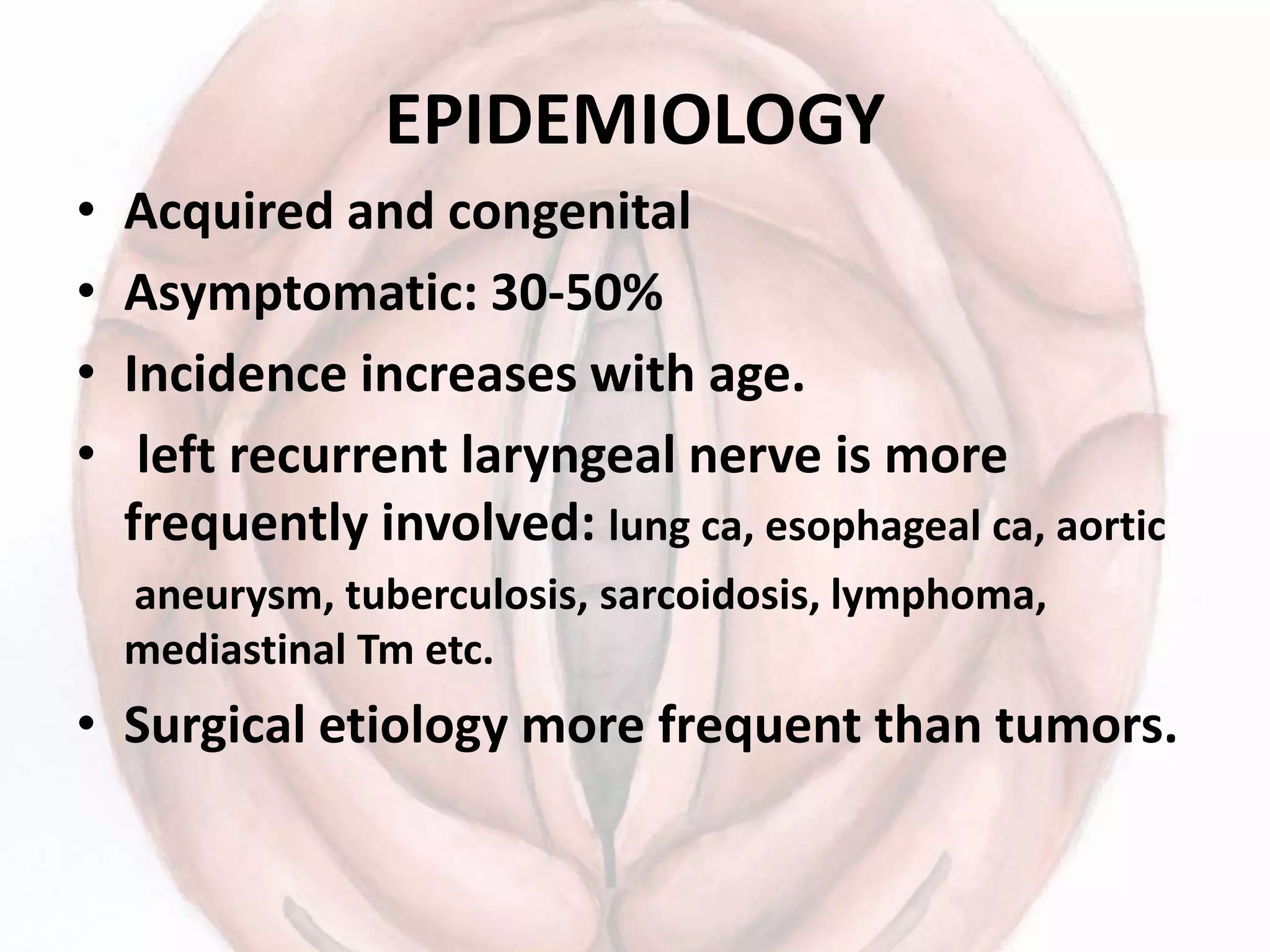
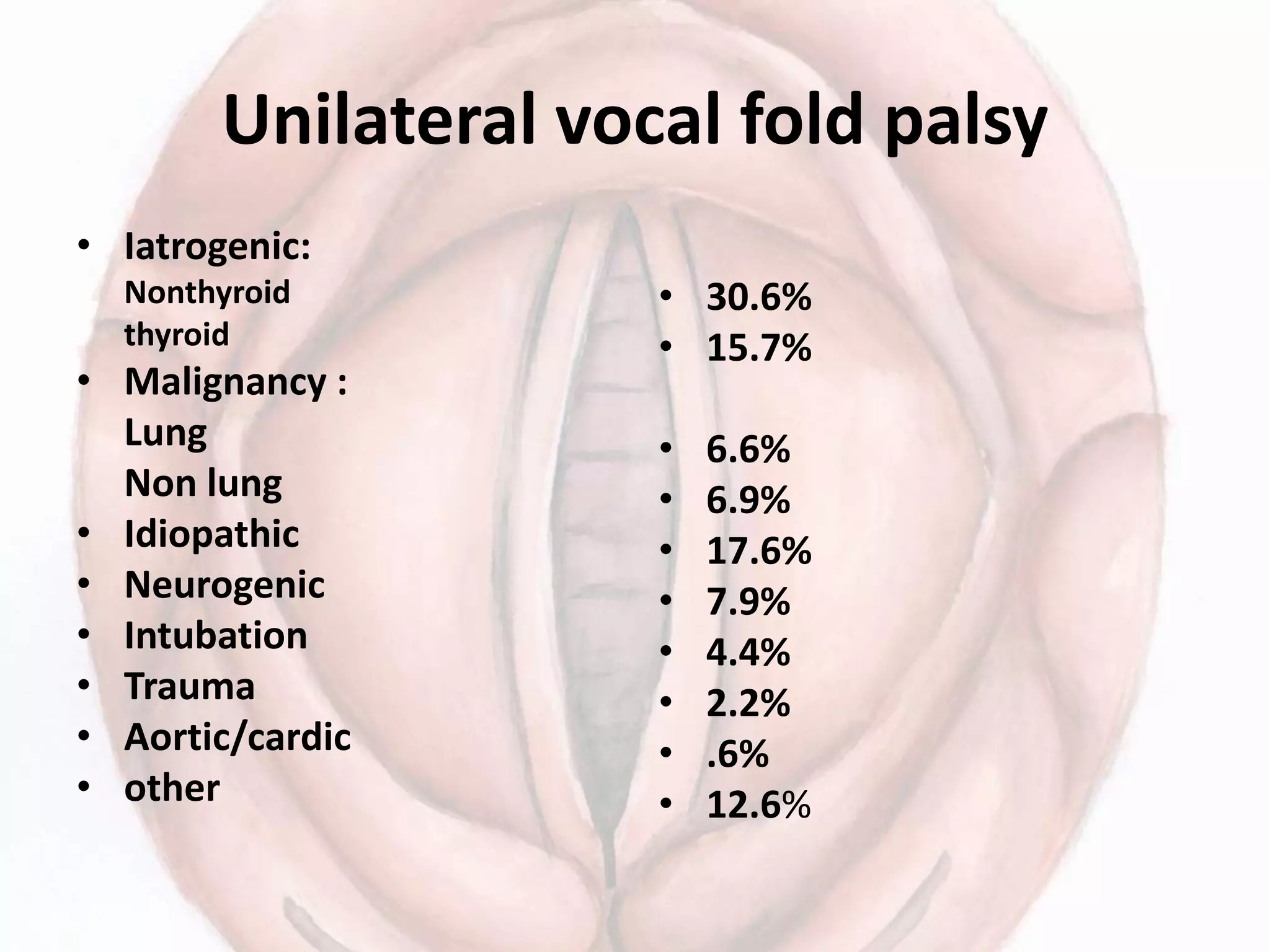
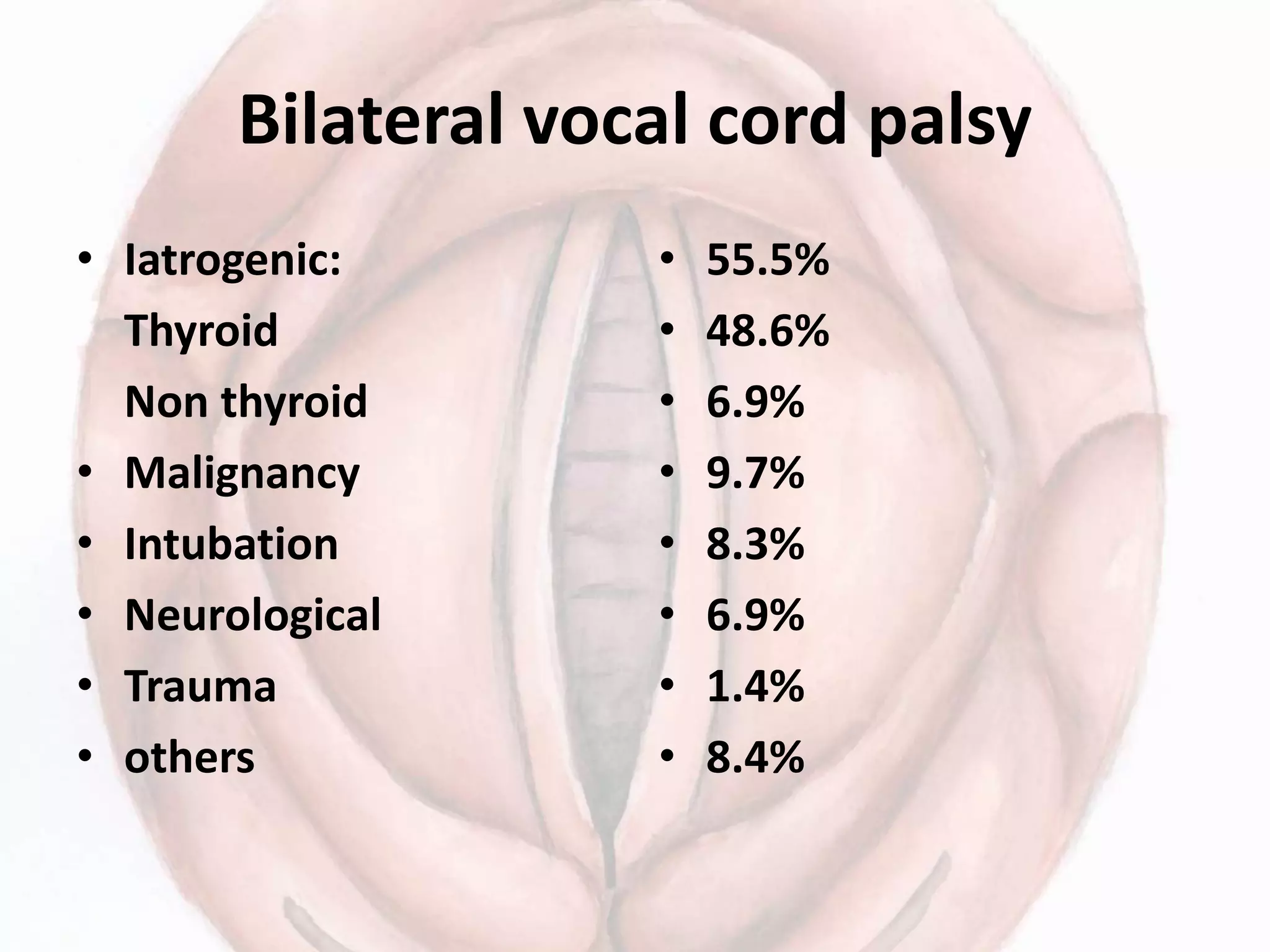
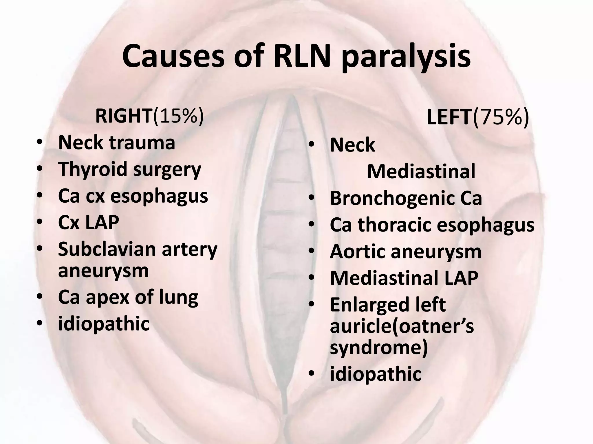
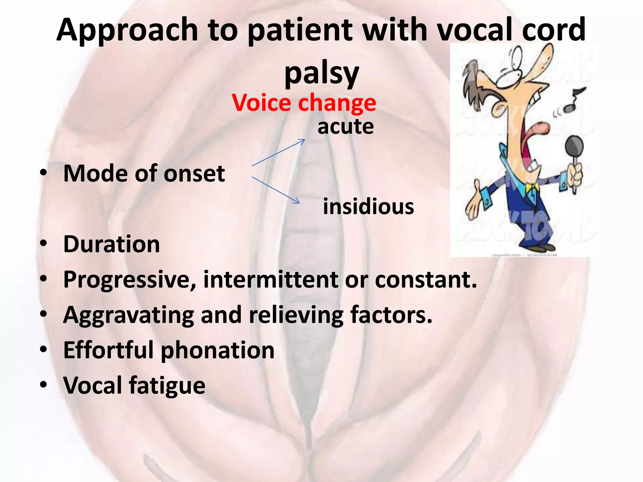
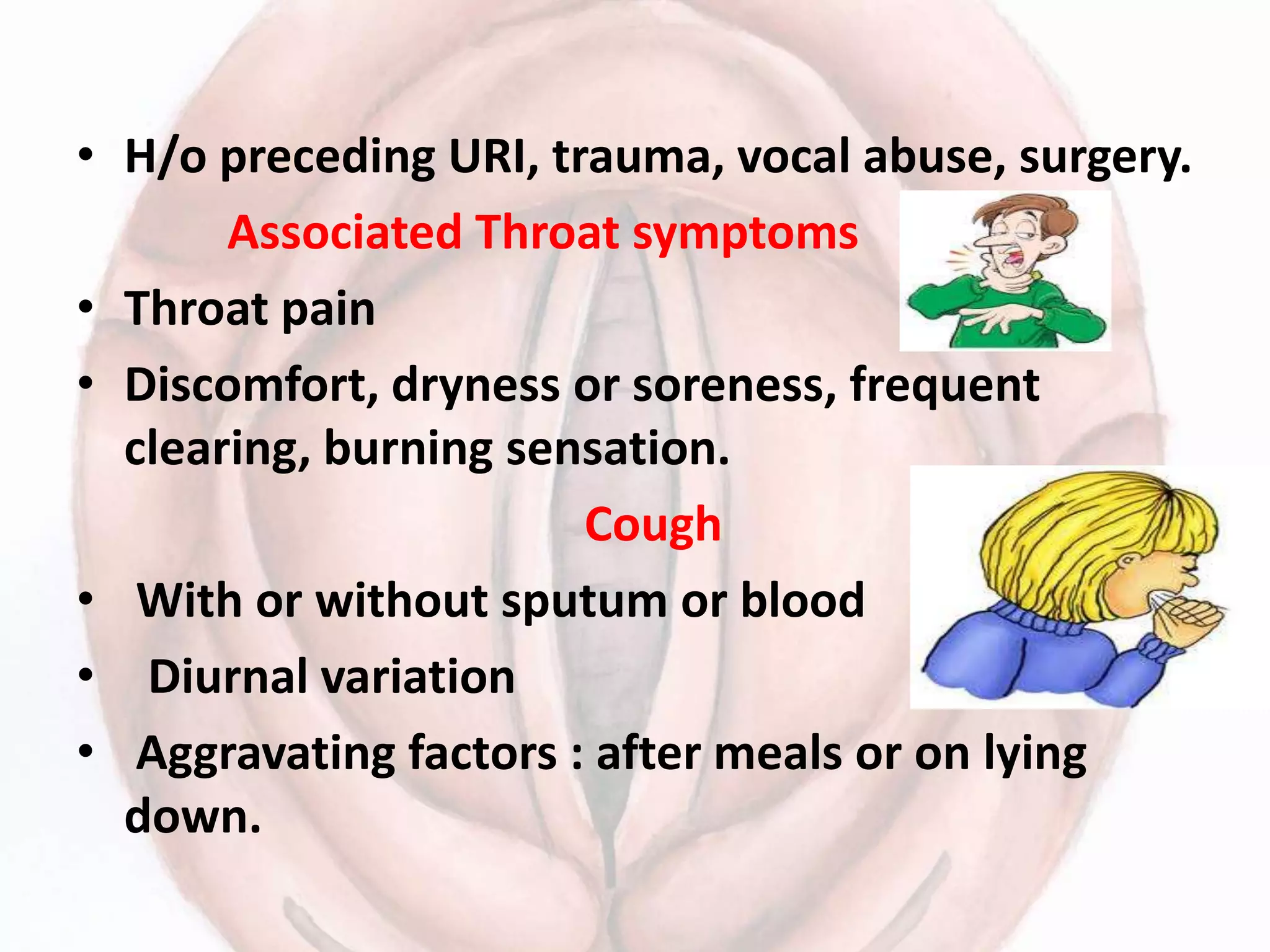
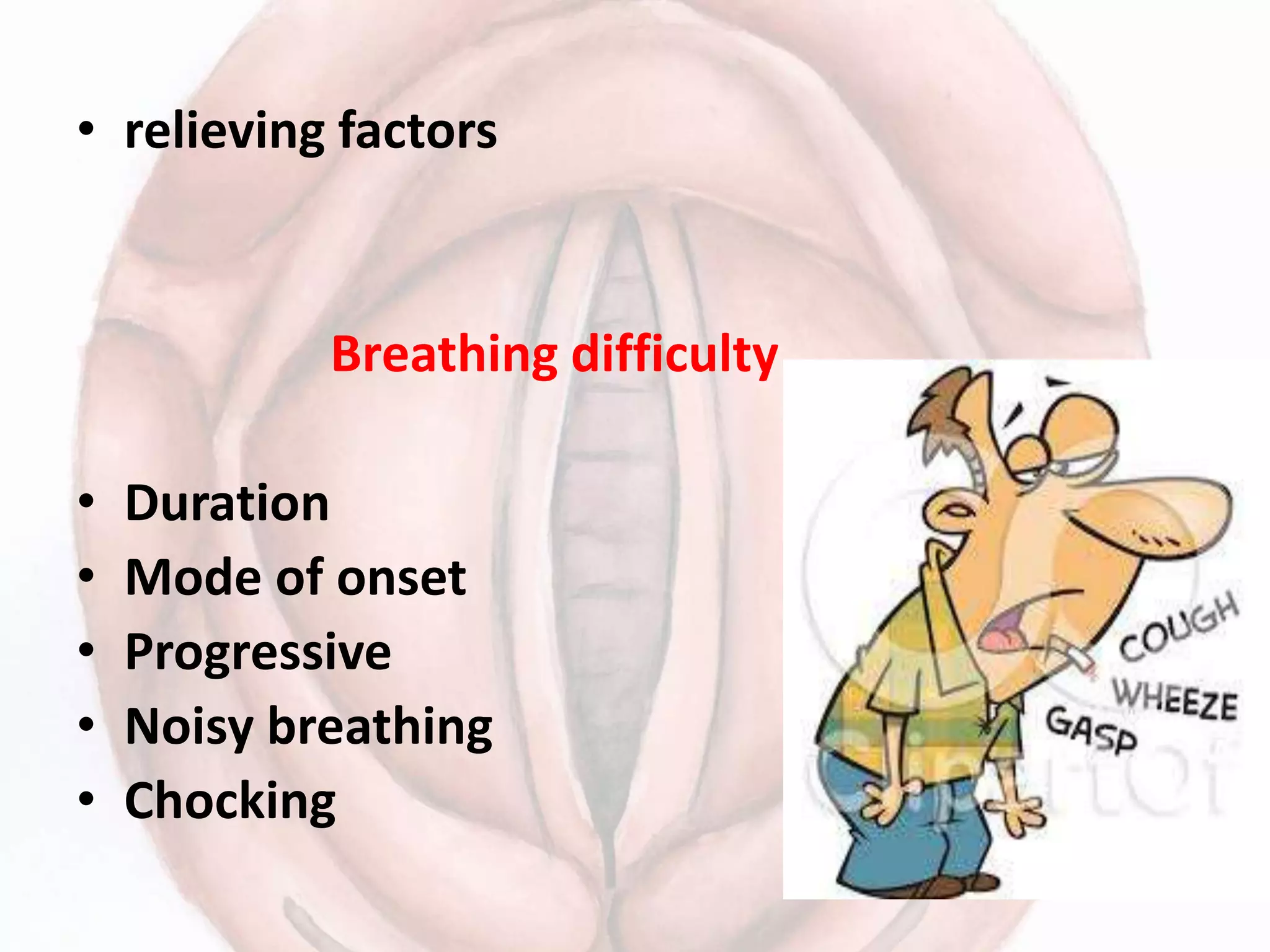
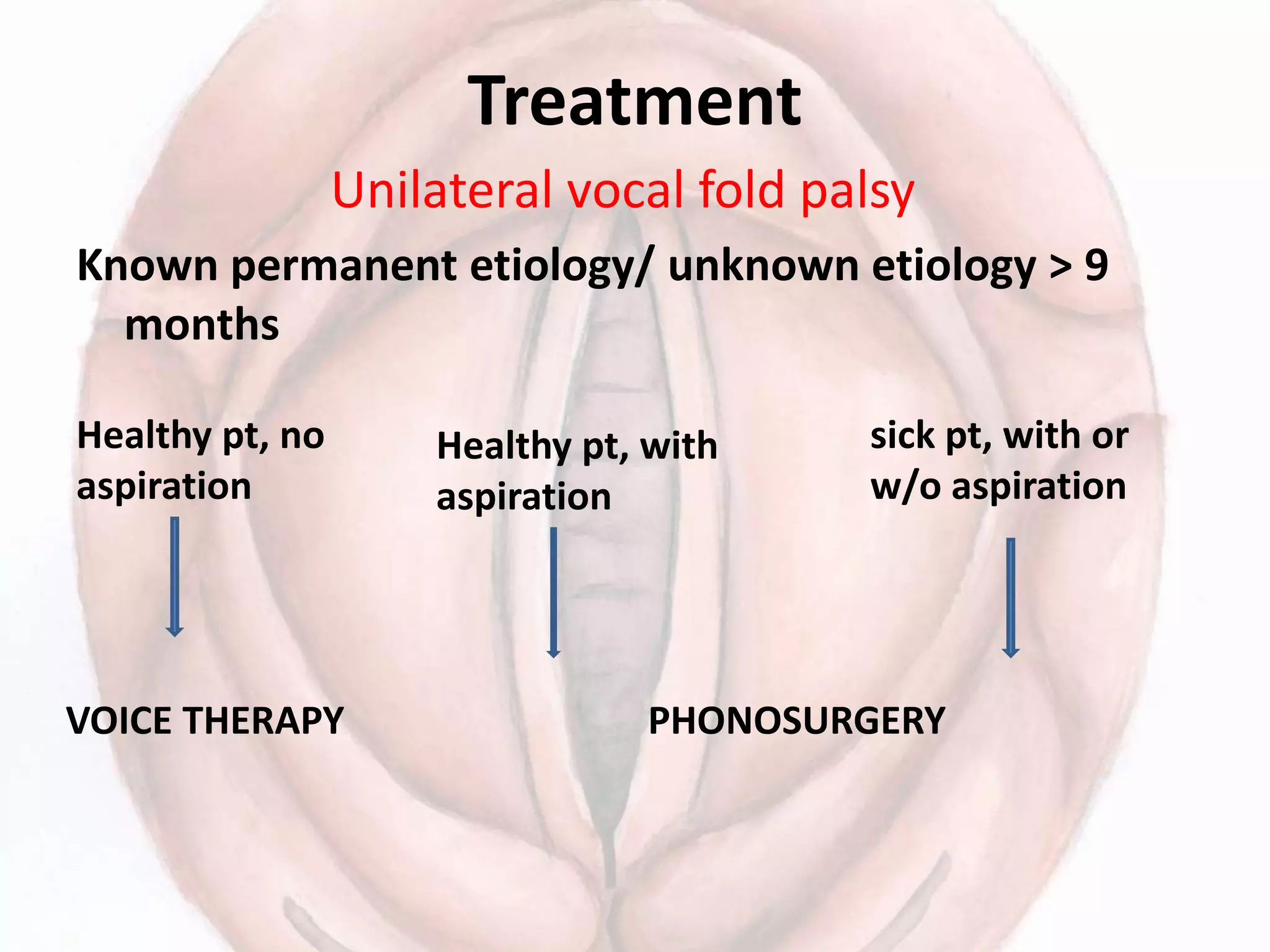
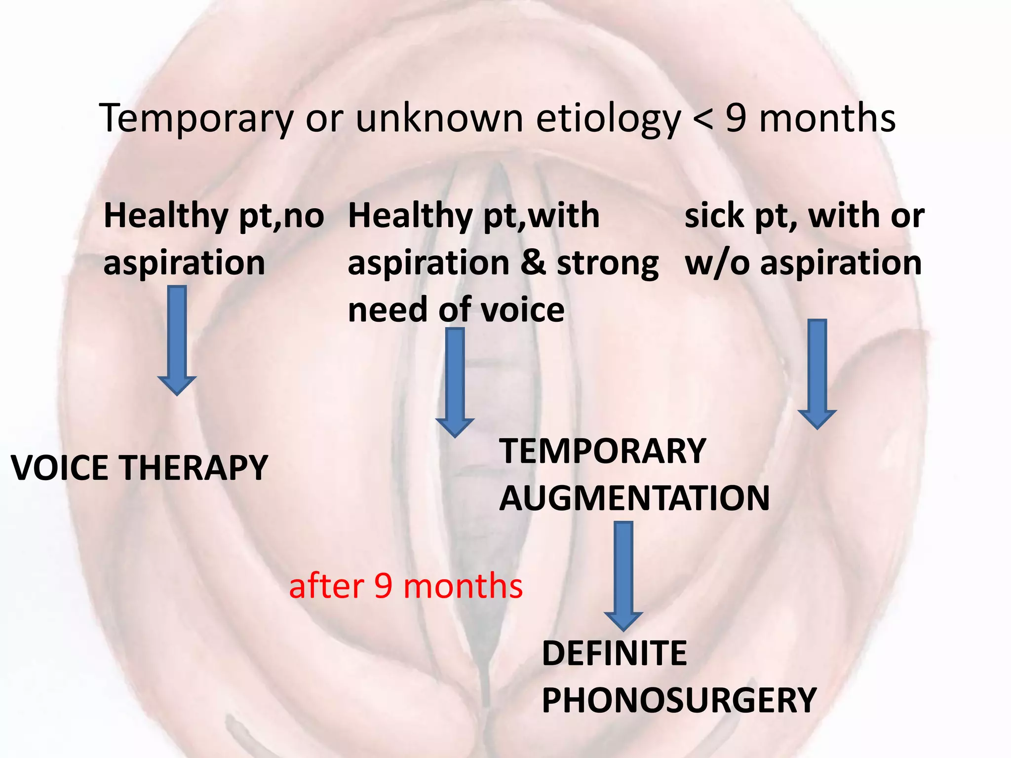
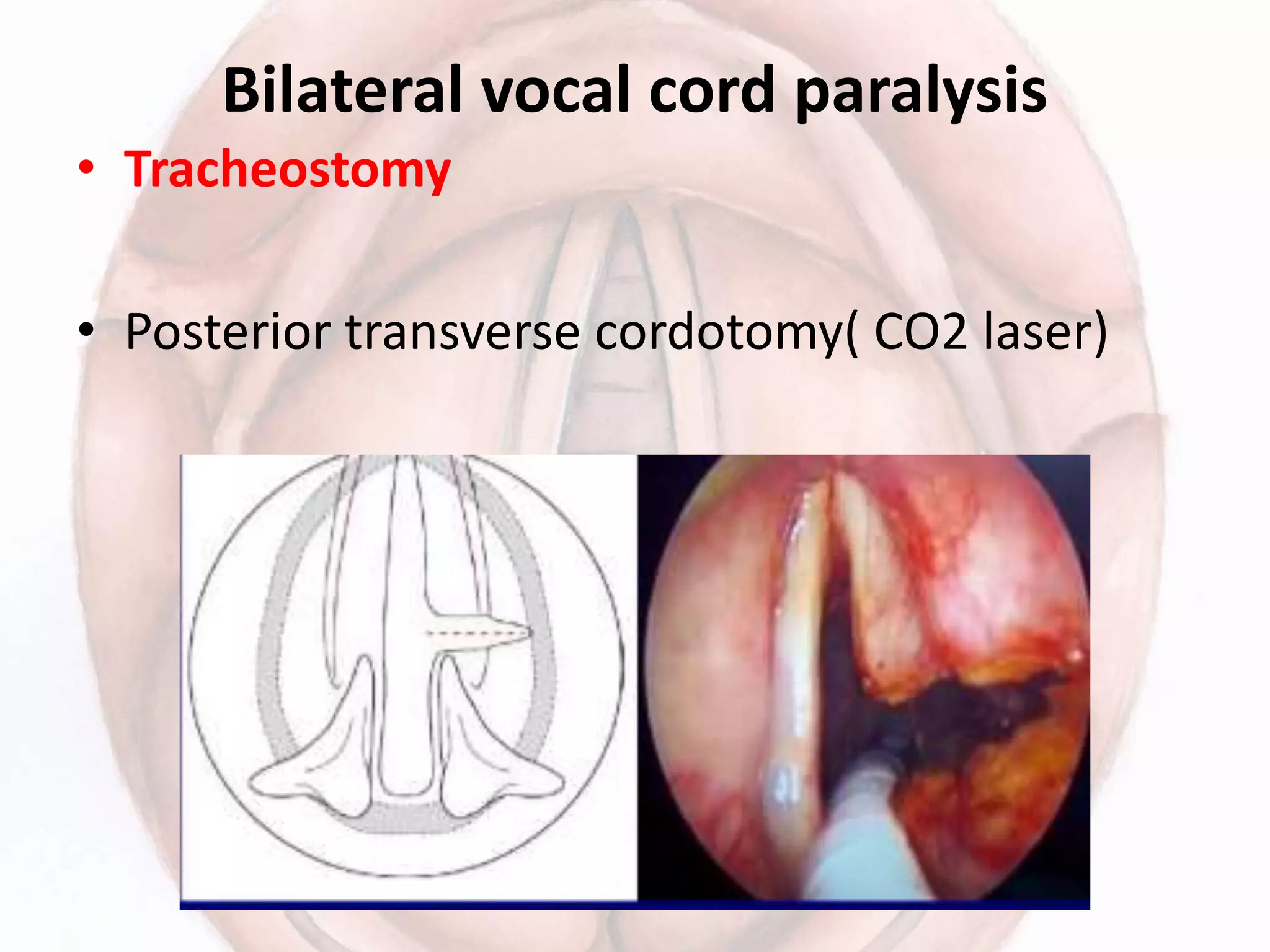
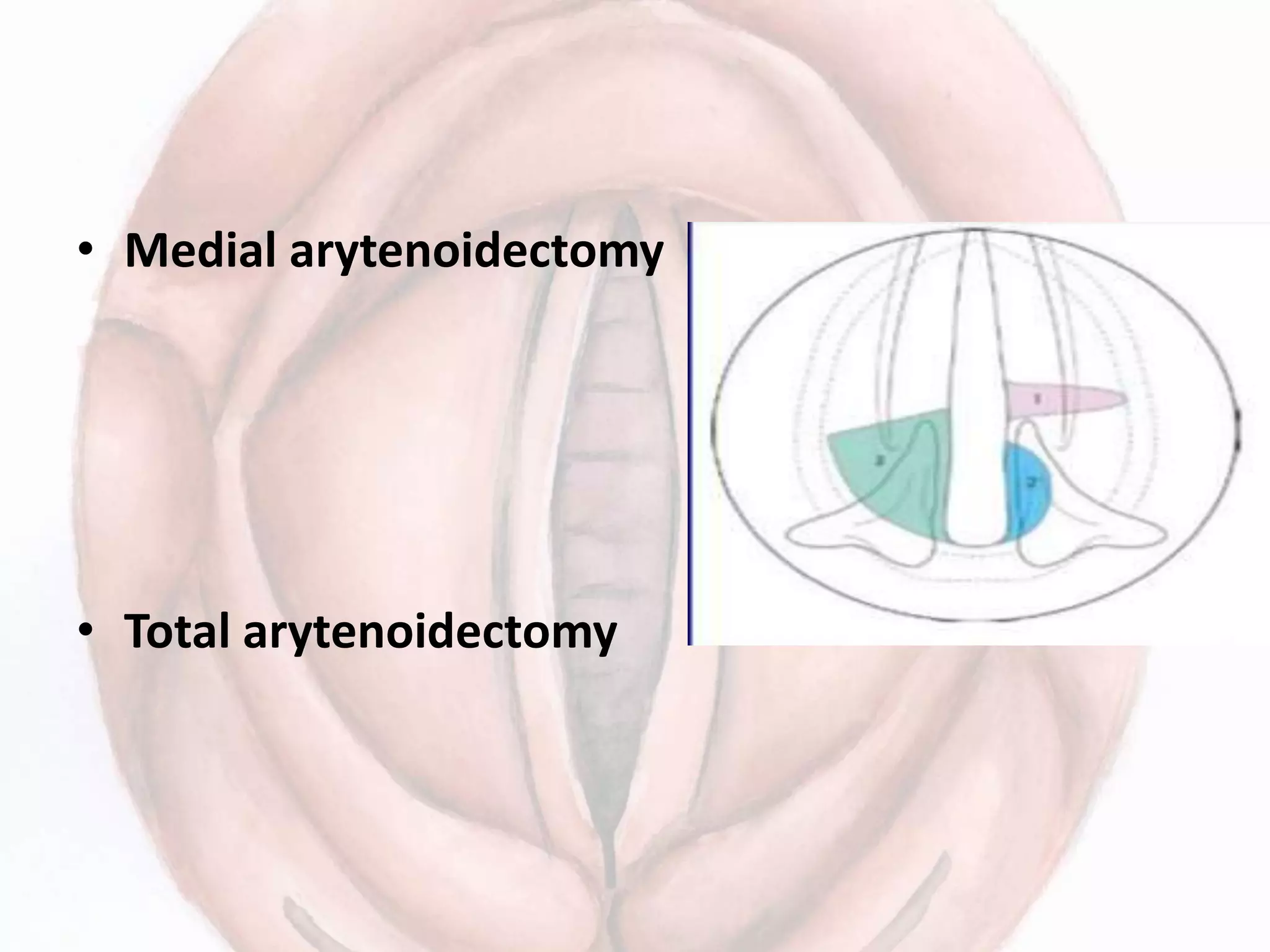
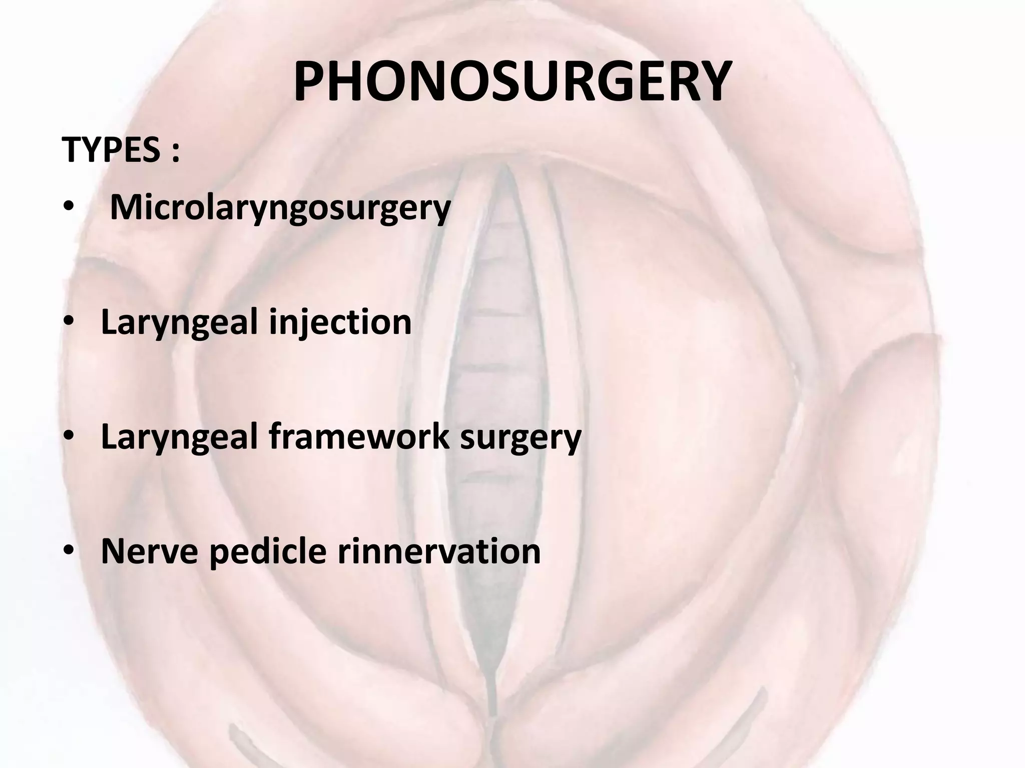
APPROACH TO A PATIENT WITH
VOCAL CORD PARALYSIS
EPIDEMIOLOGY
• Acquired and congenital
• Asymptomatic: 30-50%
• Incidence increases with age.
• left recurrent laryngeal nerve is more
frequently involved: lung ca, esophageal ca, aortic
aneurysm, tuberculosis, sarcoidosis, lymphoma,
mediastinal Tm etc.
• Surgical etiology more frequent than tumors.
Unilateral vocal fold palsy
• Iatrogenic:
Nonthyroid
thyroid
• Malignancy :
Lung
Non lung
• Idiopathic
• Neurogenic
• Intubation
• Trauma
• Aortic/cardic
• other
• 30.6%
• 15.7%
• 6.6%
• 6.9%
• 17.6%
• 7.9%
• 4.4%
• 2.2%
• .6%
• 12.6%
Bilateral vocal cord palsy
• Iatrogenic:
Thyroid
Non thyroid
• Malignancy
• Intubation
• Neurological
• Trauma
• others
• 55.5%
• 48.6%
• 6.9%
• 9.7%
• 8.3%
• 6.9%
• 1.4%
• 8.4%
Vagus nerve
Causes of laryngeal palsy
• Supranuclear
• Nuclear: vascular, neoplastic, motor, neuron
disease, polio and syringomyelia.
• High vagal lesion: skull, jugular foramen or in
parapharyngeal space
• Low vagal
• Systemic causes
• Idiopathic: 30%
Causes of combined palsy
• Intracranial: Tumors of posterior fossa, basal
meningitis .
• Skull base: fracture, nasopharyngeal Tumors
and glomus Tumors
• Neck: penetrating injury, parapharyngeal
Tumors, metastatic nodes and lymphoma.
Causes of RLN paralysis
RIGHT(15%)
• Neck trauma
• Thyroid surgery
• Ca cx esophagus
• Cx LAP
• Subclavian artery
aneurysm
• Ca apex of lung
• idiopathic
LEFT(75%)
• Neck
Mediastinal
• Bronchogenic Ca
• Ca thoracic esophagus
• Aortic aneurysm
• Mediastinal LAP
• Enlarged left
auricle(oatner’s
syndrome)
• idiopathic
Approach to patient with vocal cord
palsy
Voice change
• Mode of onset
• Duration
• Progressive, intermittent or constant.
• Aggravating and relieving factors.
• Effortful phonation
• Vocal fatigue
acute
insidious
• H/o preceding URI, trauma, vocal abuse, surgery.
Associated Throat symptoms
• Throat pain
• Discomfort, dryness or soreness, frequent
clearing, burning sensation.
Cough
• With or without sputum or blood
• Diurnal variation
• Aggravating factors : after meals or on lying
down.
• relieving factors
Breathing difficulty
• Duration
• Mode of onset
• Progressive
• Noisy breathing
• Chocking
Difficult swallowing
• Duration
• Onset
• For liquids/solids
• Pain
• Progressive or non progressive
H/o:
• trauma
• Fever with evening rise
• Weight loss
• Decrease appetite
• Swelling neck or other sites of body
• Symptoms of hyper/hypothyroidism
• Chest pain
• Weakness & numbness
Past history
• Trauma
• Viral infection or URI
• Previous surgery
• Prolonged intubation
• Drug intake
• DM/ Tuberculosis/ HT
• radiation
Family history
• DM , HT, tuberculosis
• Heart disease
• Carcinoma
• Neurogenic disorders
Personal history
• Tobacco chewing
• Smoking
• Alcohol intake
• Sleep habits
Professional history
EXAMINATION
General physical examination.
• Build and nourishment
• Vitals
• Pallor, ictreus, anemia, clubbing, LAP,JVP
• Cranial nerve examination.
• Chest examination
• CVS examination
• GIT examination
LOCAL EXAMINATION
Voice evaluation(perceptual)
Quality: - normal( 50% Pt with u/l RLN or SLN palsy)
- mild to moderately breathy( u/l SLN)
- mod to severe breathy (u/l RLN)
- hoarse
- mild to moderate or severe hypernasality
- strained
• loudness: Soft
• Pitch
• Reduced
• High ( paralytic falsetto )
• Pitch breaks
• Diplophonia ( u/l palsy)
• Weak cough
Quantitative analysis
• Magneting tape recording
• Performance assesment: MFT & range of speech
frequencies
• Phonetogram: pitch vs. intensity
• Spectogram: time, frequency and amplitude
• Aerodynamics analysis: phonatory airflow rate,
subglottic air pressure & air volume.
Fourier’s spectral analysis:
• Fundamental frequency: sustaining a single
tone at fundamental frequency.
• Shimmer: avg cycle to cycle difference in
amplitude of sound
• Jitter: avg cycle to cycle difference in pitch of
sound.
ENT examination
Nose and PNS
Lips, vestibule, oral cavity and oropharynx
Palatopharyngeal gag reflex reduced or absent,
inability to elevate soft palate.
Neck examination
Inspection: -laryngeal framework
- swelling
Palpation: - laryngeal crepitus
- swelling
- lymph nodes
Indirect laryngoscopy
• BOT, Vallecula, epiglottis, vocal
cord, arytenoids, pyriform
fossa.
• Vocal cords: appearance,
position at rest, in relation to
each other, symmetry, glottic
closure, movements in quite
breathing and vocalization.
• ee sniff test : maximum
adduction and abduction.
Features
• U/L SLN palsy: during phonation.
• Usually normal and difficult to appreciate
• Floppy, lower level of paralysed cord.
• Askew position of glottis
• Short, bowed and bulky cords
• hyperemia of hemilarynx ( loss of sympathetic
nerve supply)
• B/L SLN palsy: difficult to detect.
• Epiglottis hangs over due to anterior tilt of larynx.
• Cords are flaccid, bowed and hyperemic.
• Guttmann’s test: frontal pressure on the thyroid
cartilage will normally lower voice pitch by
counteracting cricothyroid, whereas lateral pressure
has opposite effect.
• RLN palsy ( abductor palsy):
• Cord is not mobile
• Floppy
• Flickers on phonation
• Paralysed cord balloons out on phonation
• Arytenoid crosses midline
• B/L : cord in median position
- tends to limit activity
- URTI precipitates laryngeal obstruction
Combined paralysis U/L: healthy cord not able to
approximate paralysed cord
• Glottic incompetence.
Bilateral combined:
• Cords lie in cadaveric position
• Aphonia & aspiration.
Adavntages: simple opd procedure, max
information.
Disadvantages: brief duration, anterior glottic
not seen, depth perception handicapped,
ventricles , post cricoid, apex of pyriform
sinus not seen and mucosal waves cannot be
seen
• Vocal cord position: 6 positions not valid
anymore
• Semon’s law
• Wager & grossman hypothesis
• Modern theory: final position not static
depends on – degree of muscle atrophy &
fibrosis
- degree of reinnervation
-Extent of synkinesis of muscles
Three positions: abduction, adduction and
midline
Specific investigations of cord
movement
• Rigid 70° video- telescopy.
• Fiberoptic video laryngoscopy.
• Laryngostroboscopic: glottic closure pattern
evaluation - mucosal wave in response
- of pitch and loudness
- Lesion
- Vocal fold opening and closing
pattern
- Supraglottic appearance
- Symmetry of arytenoids
LARYNGEAL ELECTROMYOGRAPHY
Gold standard
• Degree of paralysis & prognosis
• Differiating from mechanical fixation of CA joint
• Neurological diagnosis
• Site of lesion
• Synkinesis & dysfunction reinnervation
• Intaoperative nerve monitoring
• Therapeutic inspection
• Biofeedback in speech & swallowing disorder.
INVESTIGATIONS
Vocal cord palsy is not a disease per se, it’s
just a sign of underlying disease.
57% of cases can be diagnosed by taking proper
history and detailed examination
Routine : CBC , RBS, SE, VDRL and LFT, barium
swallow & thyroid scan.
low diagnostic yield ( usually not
recommended)
Radiological
chest xray:- secondaries, primary carcinoma,
apiration pneumonia, metastatic lymph nodes,
aortic arch aneurysm and TB. (54% diagnostic yield)
No other detectable lesion: contrast CT ( skull
base to aortic arch)
No mass lesion – idiopathic.
Palatal & pharyngeal paralysis and other
neuropathies: gadolinium enhanched MRI skull
base and neck.
If negative- HRCT temporal bone for bony mets
• Flexible or rigid esophagoscopy with biopsy.
Treatment
Unilateral vocal fold palsy
Known permanent etiology/ unknown etiology > 9
months
Healthy pt, no
aspiration
Healthy pt, with
aspiration
sick pt, with or
w/o aspiration
VOICE THERAPY PHONOSURGERY
Temporary or unknown etiology < 9 months
Healthy pt,no
aspiration
Healthy pt,with
aspiration & strong
need of voice
sick pt, with or
w/o aspiration
VOICE THERAPY TEMPORARY
AUGMENTATION
after 9 months
DEFINITE
PHONOSURGERY
• Educational information regarding phonation
• Vocal hygiene: voice rest, avoid shouting, talking
loudly, clearing throat
- adequate hydration
- steam inhalation
- smoking cessation, reducing alcohol,
- Diet and reflux reduction
VOICE THERAPY
• Vocal exercise : strengthening the muscle groups,
improving glottic closure and efficiency.
• Reducing excessive tension in muscles around
larynx, neck and shoulders.
• Advice on posture and breathing during speech
• Laryngeal massage
• General relaxation exercise
• Psychological counseling.
Bilateral vocal cord paralysis
• Tracheostomy
• Posterior transverse cordotomy( CO2 laser)
• Medial arytenoidectomy
• Total arytenoidectomy
• Endo-extralaryngeal suture.
• Laryngeal pacing.
In cases of contraindication
• Epiglottopexy
• Vocal cord plication
• Total laryngectomy: cause is progressive,
irreversible and speech is unservicable.
• Diversion procedures: intractable aspiration
PHONOSURGERY
TYPES :
• Microlaryngosurgery
• Laryngeal injection
• Laryngeal framework surgery
• Nerve pedicle rinnervation
• Laryngeal injection techniques:- for
phonatory gap in u/l abductor or adductor
palsy
• Teflon, fat, collagen, gelfoam, silicone etc
Laryngeal framework surgery
• THYROPLASTY: type 1( medial displacement)
• Arytenoid adduction: large posterior glottic
gap.
• Laryngeal reinnervation: nerve muscle pedicle
graft technique.
• Anterior belly of omohyoid with ansa hypoglossi
and vessels.
THANK YOU

APPROACH TO A PATIENT WITH VOCAL CORD PARALYSIS

  • 1.
    APPROACH TO APATIENT WITH VOCAL CORD PARALYSIS
  • 2.
    EPIDEMIOLOGY • Acquired andcongenital • Asymptomatic: 30-50% • Incidence increases with age. • left recurrent laryngeal nerve is more frequently involved: lung ca, esophageal ca, aortic aneurysm, tuberculosis, sarcoidosis, lymphoma, mediastinal Tm etc. • Surgical etiology more frequent than tumors.
  • 3.
    Unilateral vocal foldpalsy • Iatrogenic: Nonthyroid thyroid • Malignancy : Lung Non lung • Idiopathic • Neurogenic • Intubation • Trauma • Aortic/cardic • other • 30.6% • 15.7% • 6.6% • 6.9% • 17.6% • 7.9% • 4.4% • 2.2% • .6% • 12.6%
  • 4.
    Bilateral vocal cordpalsy • Iatrogenic: Thyroid Non thyroid • Malignancy • Intubation • Neurological • Trauma • others • 55.5% • 48.6% • 6.9% • 9.7% • 8.3% • 6.9% • 1.4% • 8.4%
  • 5.
  • 7.
    Causes of laryngealpalsy • Supranuclear • Nuclear: vascular, neoplastic, motor, neuron disease, polio and syringomyelia. • High vagal lesion: skull, jugular foramen or in parapharyngeal space • Low vagal • Systemic causes • Idiopathic: 30%
  • 8.
    Causes of combinedpalsy • Intracranial: Tumors of posterior fossa, basal meningitis . • Skull base: fracture, nasopharyngeal Tumors and glomus Tumors • Neck: penetrating injury, parapharyngeal Tumors, metastatic nodes and lymphoma.
  • 9.
    Causes of RLNparalysis RIGHT(15%) • Neck trauma • Thyroid surgery • Ca cx esophagus • Cx LAP • Subclavian artery aneurysm • Ca apex of lung • idiopathic LEFT(75%) • Neck Mediastinal • Bronchogenic Ca • Ca thoracic esophagus • Aortic aneurysm • Mediastinal LAP • Enlarged left auricle(oatner’s syndrome) • idiopathic
  • 11.
    Approach to patientwith vocal cord palsy Voice change • Mode of onset • Duration • Progressive, intermittent or constant. • Aggravating and relieving factors. • Effortful phonation • Vocal fatigue acute insidious
  • 12.
    • H/o precedingURI, trauma, vocal abuse, surgery. Associated Throat symptoms • Throat pain • Discomfort, dryness or soreness, frequent clearing, burning sensation. Cough • With or without sputum or blood • Diurnal variation • Aggravating factors : after meals or on lying down.
  • 13.
    • relieving factors Breathingdifficulty • Duration • Mode of onset • Progressive • Noisy breathing • Chocking
  • 14.
    Difficult swallowing • Duration •Onset • For liquids/solids • Pain • Progressive or non progressive
  • 15.
    H/o: • trauma • Feverwith evening rise • Weight loss • Decrease appetite • Swelling neck or other sites of body • Symptoms of hyper/hypothyroidism • Chest pain • Weakness & numbness
  • 16.
    Past history • Trauma •Viral infection or URI • Previous surgery • Prolonged intubation • Drug intake • DM/ Tuberculosis/ HT • radiation
  • 17.
    Family history • DM, HT, tuberculosis • Heart disease • Carcinoma • Neurogenic disorders Personal history • Tobacco chewing • Smoking • Alcohol intake • Sleep habits Professional history
  • 18.
    EXAMINATION General physical examination. •Build and nourishment • Vitals • Pallor, ictreus, anemia, clubbing, LAP,JVP • Cranial nerve examination. • Chest examination • CVS examination • GIT examination
  • 19.
    LOCAL EXAMINATION Voice evaluation(perceptual) Quality:- normal( 50% Pt with u/l RLN or SLN palsy) - mild to moderately breathy( u/l SLN) - mod to severe breathy (u/l RLN) - hoarse - mild to moderate or severe hypernasality - strained
  • 21.
    • loudness: Soft •Pitch • Reduced • High ( paralytic falsetto ) • Pitch breaks • Diplophonia ( u/l palsy) • Weak cough
  • 22.
    Quantitative analysis • Magnetingtape recording • Performance assesment: MFT & range of speech frequencies • Phonetogram: pitch vs. intensity • Spectogram: time, frequency and amplitude • Aerodynamics analysis: phonatory airflow rate, subglottic air pressure & air volume.
  • 23.
    Fourier’s spectral analysis: •Fundamental frequency: sustaining a single tone at fundamental frequency. • Shimmer: avg cycle to cycle difference in amplitude of sound • Jitter: avg cycle to cycle difference in pitch of sound.
  • 24.
    ENT examination Nose andPNS Lips, vestibule, oral cavity and oropharynx Palatopharyngeal gag reflex reduced or absent, inability to elevate soft palate.
  • 25.
    Neck examination Inspection: -laryngealframework - swelling Palpation: - laryngeal crepitus - swelling - lymph nodes
  • 26.
    Indirect laryngoscopy • BOT,Vallecula, epiglottis, vocal cord, arytenoids, pyriform fossa. • Vocal cords: appearance, position at rest, in relation to each other, symmetry, glottic closure, movements in quite breathing and vocalization. • ee sniff test : maximum adduction and abduction.
  • 27.
    Features • U/L SLNpalsy: during phonation. • Usually normal and difficult to appreciate • Floppy, lower level of paralysed cord. • Askew position of glottis • Short, bowed and bulky cords • hyperemia of hemilarynx ( loss of sympathetic nerve supply)
  • 28.
    • B/L SLNpalsy: difficult to detect. • Epiglottis hangs over due to anterior tilt of larynx. • Cords are flaccid, bowed and hyperemic. • Guttmann’s test: frontal pressure on the thyroid cartilage will normally lower voice pitch by counteracting cricothyroid, whereas lateral pressure has opposite effect.
  • 30.
    • RLN palsy( abductor palsy): • Cord is not mobile • Floppy • Flickers on phonation • Paralysed cord balloons out on phonation • Arytenoid crosses midline • B/L : cord in median position - tends to limit activity - URTI precipitates laryngeal obstruction
  • 34.
    Combined paralysis U/L:healthy cord not able to approximate paralysed cord • Glottic incompetence. Bilateral combined: • Cords lie in cadaveric position • Aphonia & aspiration.
  • 35.
    Adavntages: simple opdprocedure, max information. Disadvantages: brief duration, anterior glottic not seen, depth perception handicapped, ventricles , post cricoid, apex of pyriform sinus not seen and mucosal waves cannot be seen
  • 36.
    • Vocal cordposition: 6 positions not valid anymore • Semon’s law • Wager & grossman hypothesis
  • 37.
    • Modern theory:final position not static depends on – degree of muscle atrophy & fibrosis - degree of reinnervation -Extent of synkinesis of muscles Three positions: abduction, adduction and midline
  • 38.
    Specific investigations ofcord movement • Rigid 70° video- telescopy. • Fiberoptic video laryngoscopy. • Laryngostroboscopic: glottic closure pattern evaluation - mucosal wave in response - of pitch and loudness - Lesion - Vocal fold opening and closing pattern - Supraglottic appearance - Symmetry of arytenoids
  • 40.
    LARYNGEAL ELECTROMYOGRAPHY Gold standard •Degree of paralysis & prognosis • Differiating from mechanical fixation of CA joint • Neurological diagnosis • Site of lesion • Synkinesis & dysfunction reinnervation • Intaoperative nerve monitoring • Therapeutic inspection • Biofeedback in speech & swallowing disorder.
  • 41.
    INVESTIGATIONS Vocal cord palsyis not a disease per se, it’s just a sign of underlying disease. 57% of cases can be diagnosed by taking proper history and detailed examination Routine : CBC , RBS, SE, VDRL and LFT, barium swallow & thyroid scan. low diagnostic yield ( usually not recommended)
  • 42.
    Radiological chest xray:- secondaries,primary carcinoma, apiration pneumonia, metastatic lymph nodes, aortic arch aneurysm and TB. (54% diagnostic yield) No other detectable lesion: contrast CT ( skull base to aortic arch) No mass lesion – idiopathic. Palatal & pharyngeal paralysis and other neuropathies: gadolinium enhanched MRI skull base and neck. If negative- HRCT temporal bone for bony mets
  • 43.
    • Flexible orrigid esophagoscopy with biopsy.
  • 44.
    Treatment Unilateral vocal foldpalsy Known permanent etiology/ unknown etiology > 9 months Healthy pt, no aspiration Healthy pt, with aspiration sick pt, with or w/o aspiration VOICE THERAPY PHONOSURGERY
  • 45.
    Temporary or unknownetiology < 9 months Healthy pt,no aspiration Healthy pt,with aspiration & strong need of voice sick pt, with or w/o aspiration VOICE THERAPY TEMPORARY AUGMENTATION after 9 months DEFINITE PHONOSURGERY
  • 46.
    • Educational informationregarding phonation • Vocal hygiene: voice rest, avoid shouting, talking loudly, clearing throat - adequate hydration - steam inhalation - smoking cessation, reducing alcohol, - Diet and reflux reduction
  • 47.
    VOICE THERAPY • Vocalexercise : strengthening the muscle groups, improving glottic closure and efficiency. • Reducing excessive tension in muscles around larynx, neck and shoulders. • Advice on posture and breathing during speech • Laryngeal massage • General relaxation exercise • Psychological counseling.
  • 48.
    Bilateral vocal cordparalysis • Tracheostomy • Posterior transverse cordotomy( CO2 laser)
  • 49.
    • Medial arytenoidectomy •Total arytenoidectomy
  • 50.
  • 51.
    In cases ofcontraindication • Epiglottopexy • Vocal cord plication • Total laryngectomy: cause is progressive, irreversible and speech is unservicable. • Diversion procedures: intractable aspiration
  • 52.
    PHONOSURGERY TYPES : • Microlaryngosurgery •Laryngeal injection • Laryngeal framework surgery • Nerve pedicle rinnervation
  • 53.
    • Laryngeal injectiontechniques:- for phonatory gap in u/l abductor or adductor palsy • Teflon, fat, collagen, gelfoam, silicone etc
  • 55.
    Laryngeal framework surgery •THYROPLASTY: type 1( medial displacement)
  • 57.
    • Arytenoid adduction:large posterior glottic gap. • Laryngeal reinnervation: nerve muscle pedicle graft technique. • Anterior belly of omohyoid with ansa hypoglossi and vessels.
  • 58.