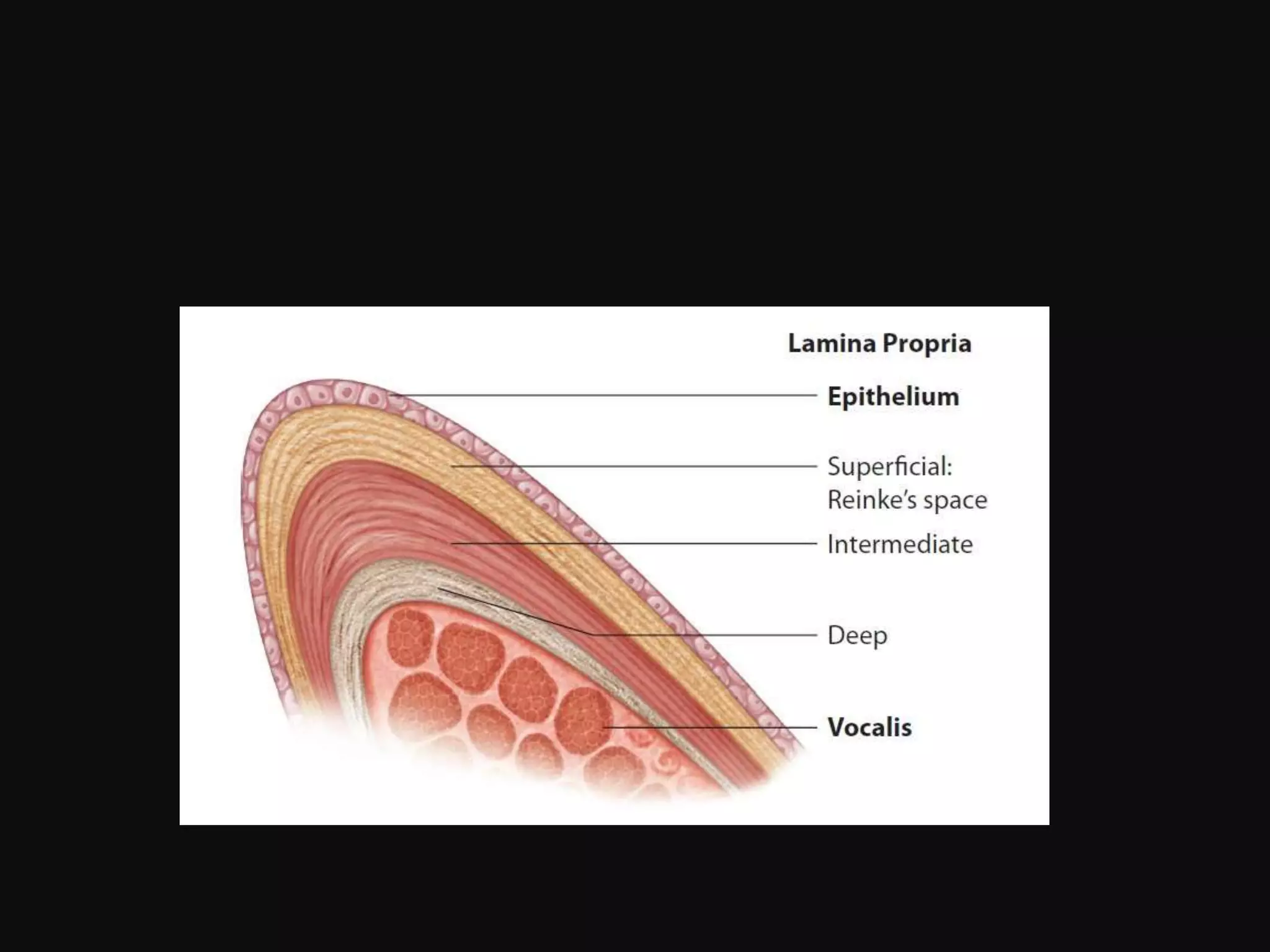
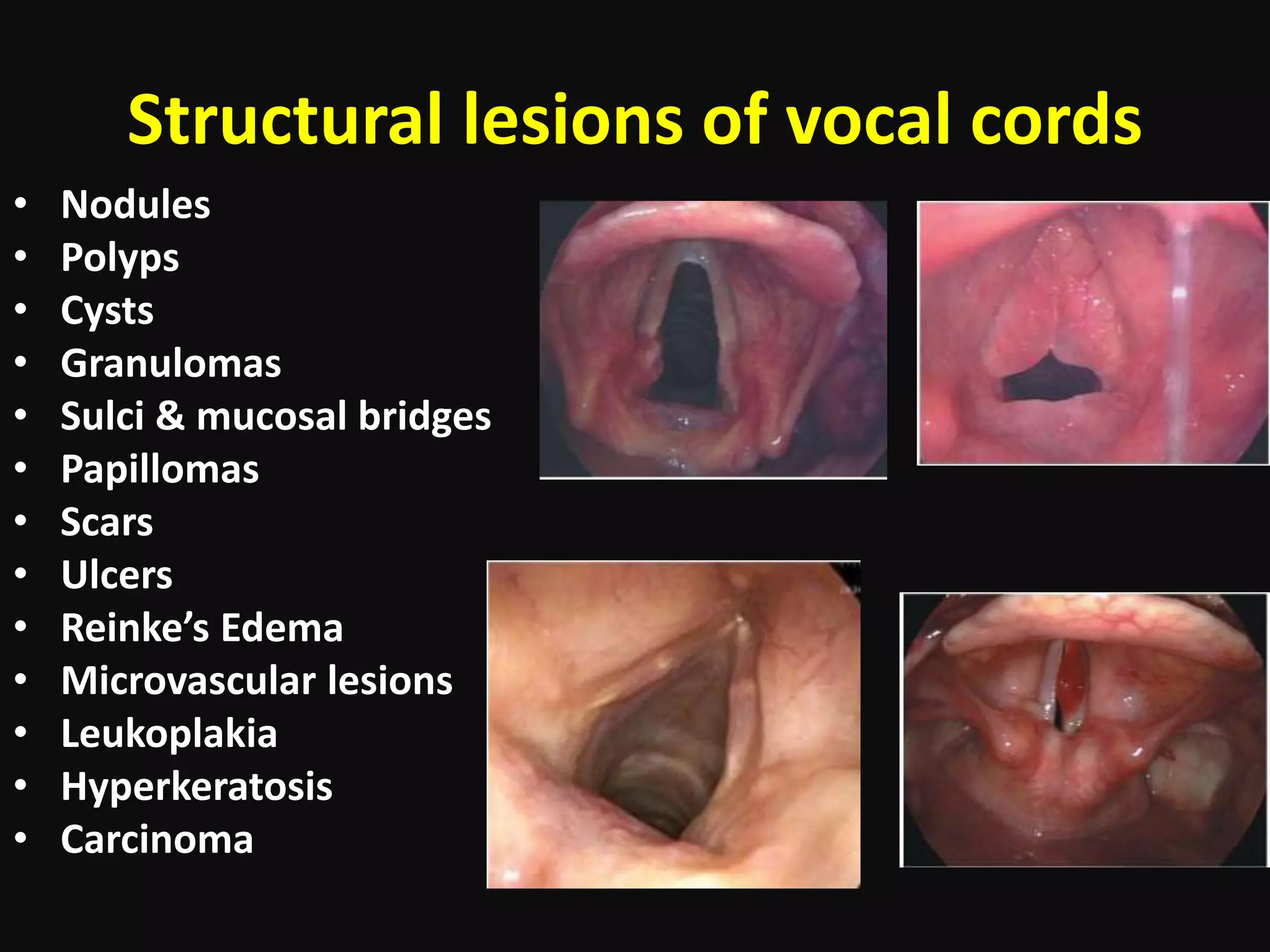
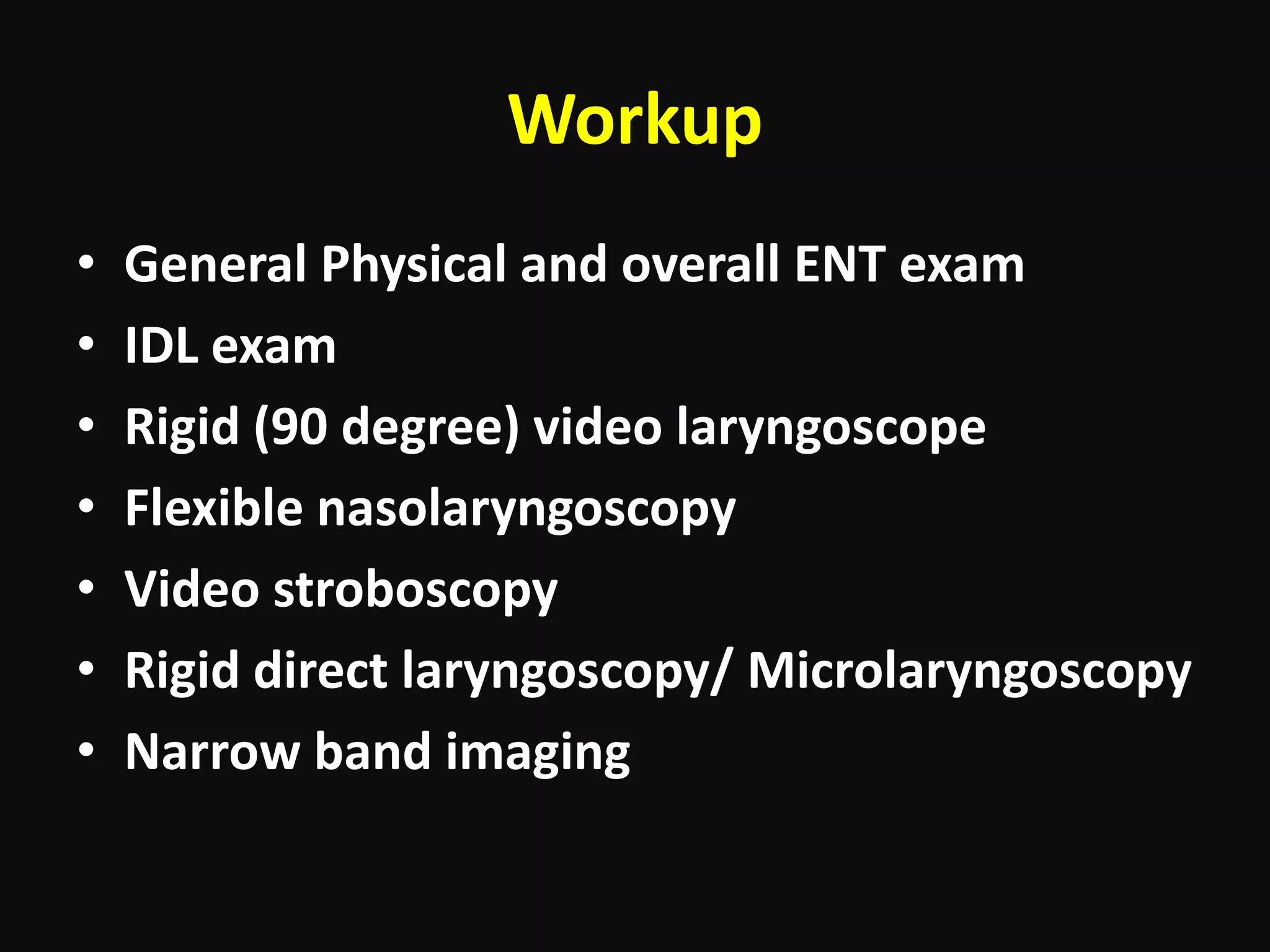
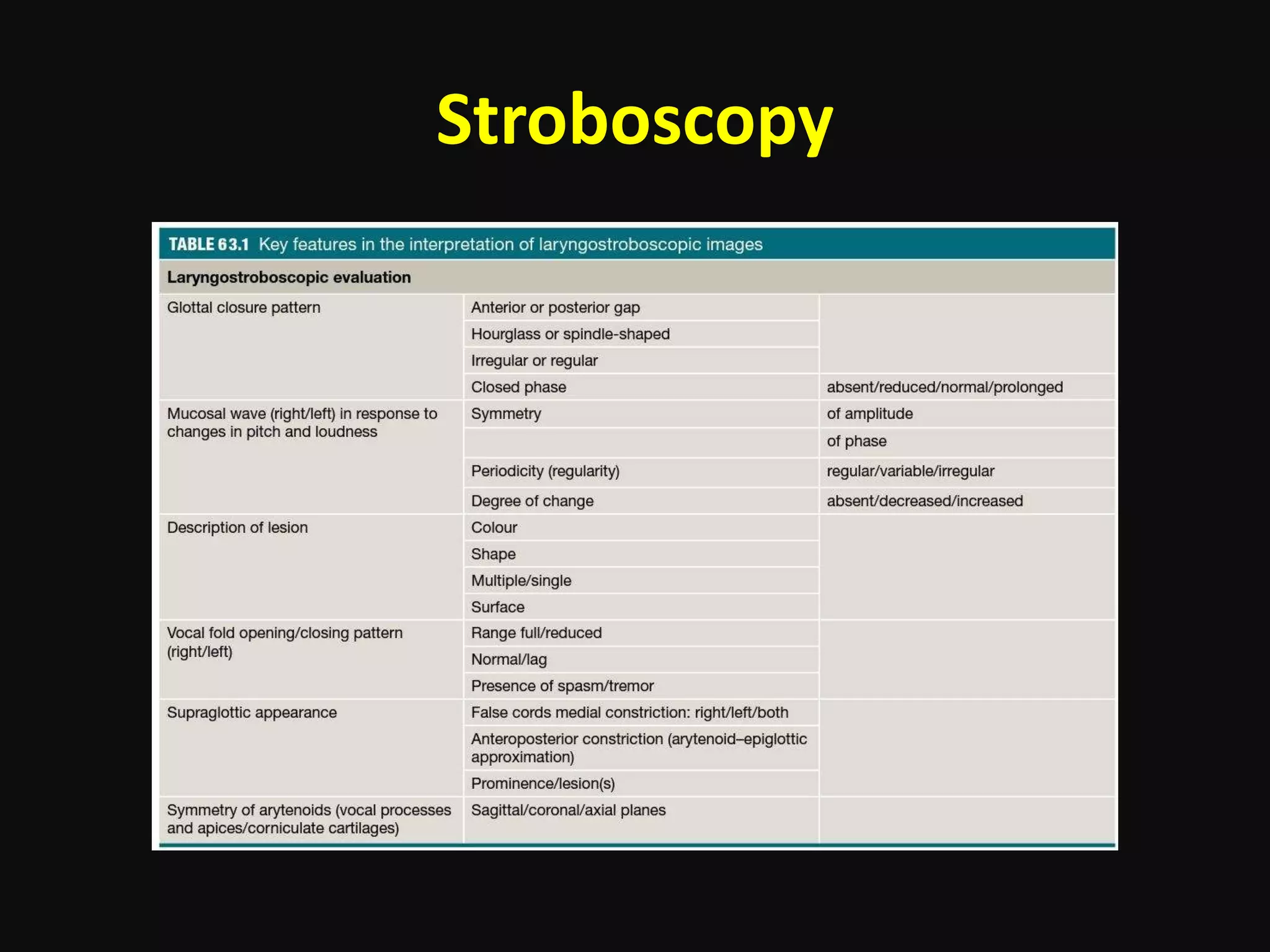
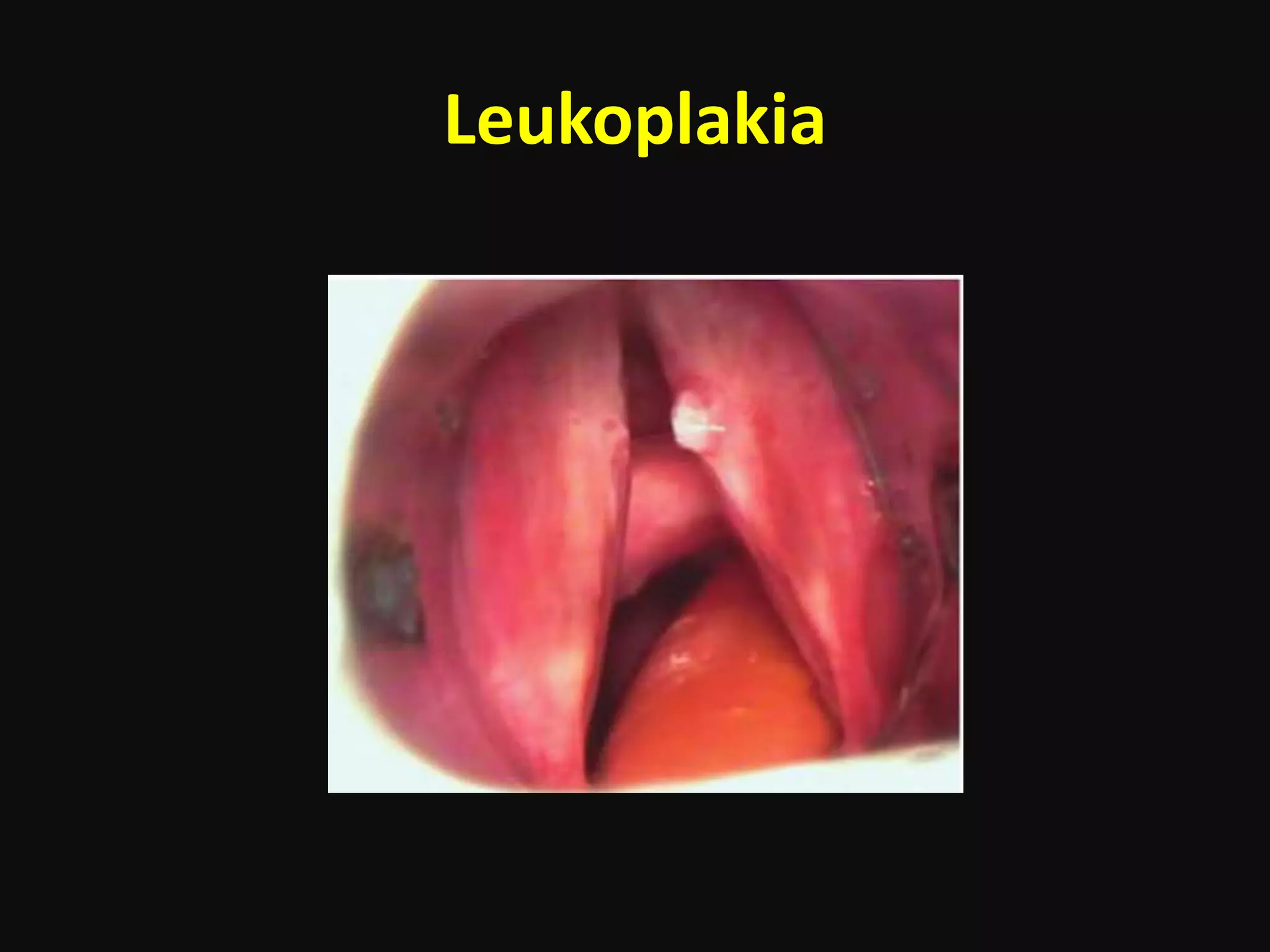
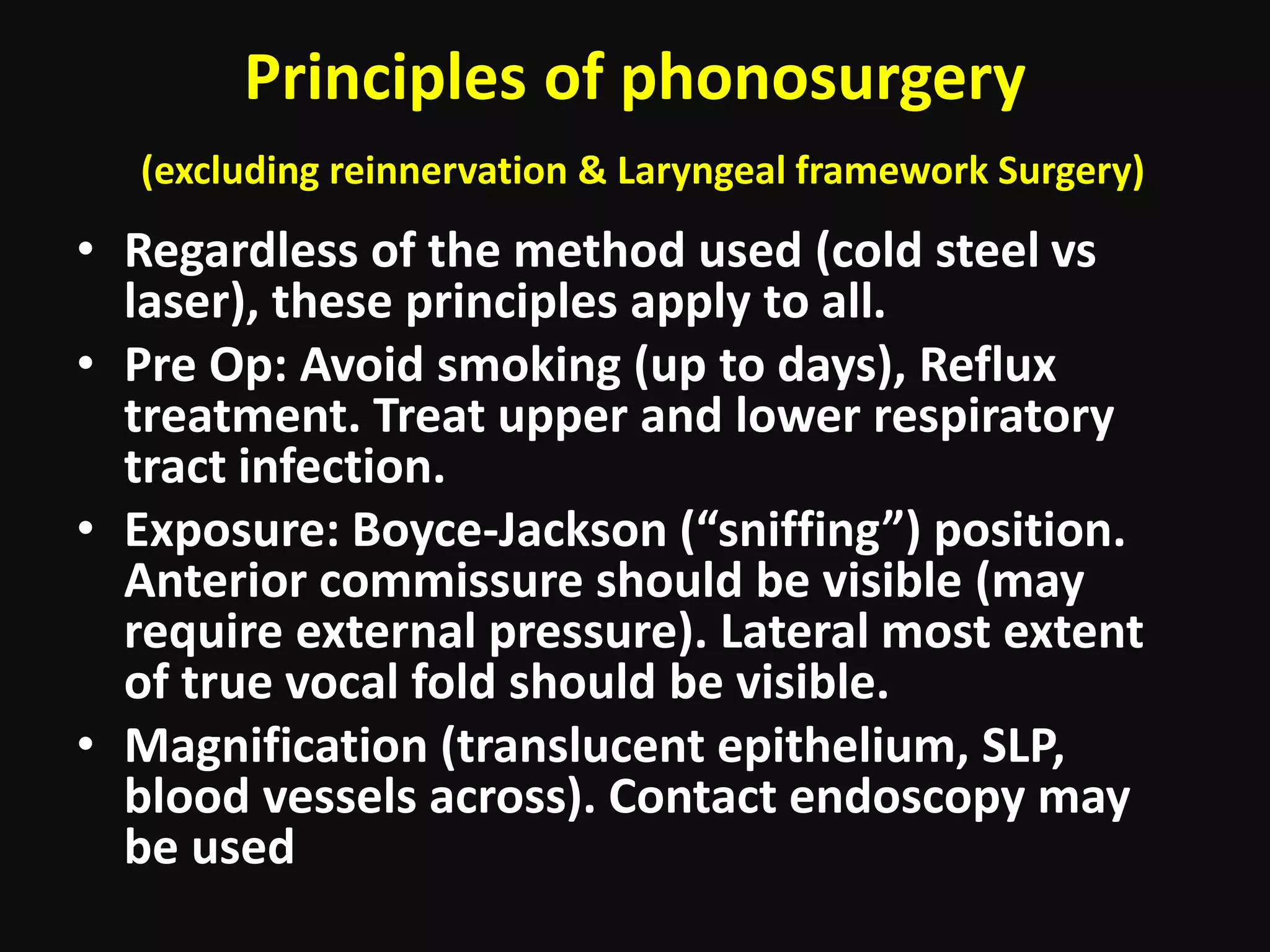
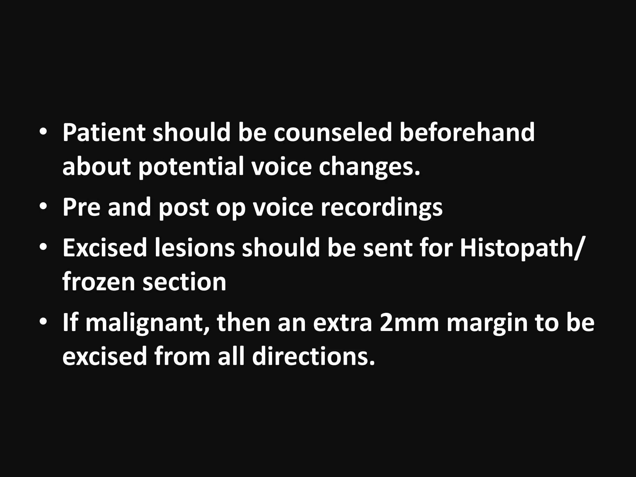
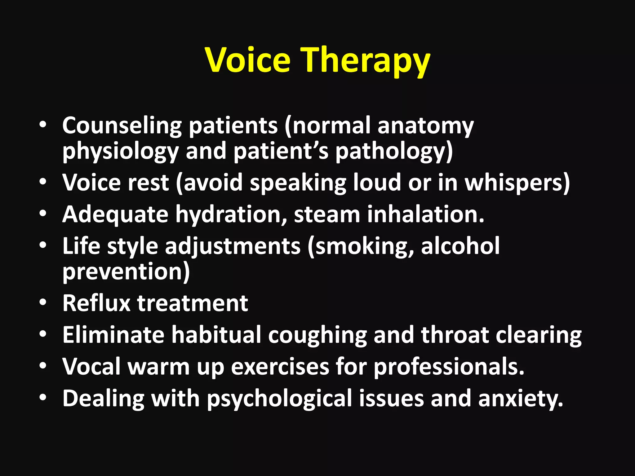
Pre malignant lesions of vocal cords
and principles of phonomicrosurgery
Dr. Uzair Mushahid
Outline
• Anatomy of the vocal cords
• Common differential diagnosis and causes
• Pre malignant conditions
• Clinical presentation, assessment & workup
• Surgical procedures
• Post op care
• Voice therapy and rehab
Physiology of vocal cords and voice
production
Laryngeal lesions
• Four main causes of voice disorders:
• Inflammatory
• Structural or neoplastic
• Neuromuscular
• Muscle tension imbalance.
Signs and symptoms
• Knowing the terminologies :
– Dysphonia, Dysarthria, Dysarthrophonia, Dysphasia,
Hoarseness, Odynophonia.
• Disordered voice:
– It is not audible, clear or stable in a wide range of acoustic
settings
– It is not appropriate for the gender and age of the speaker.
– It is not capable of fulfilling its linguistic and paralinguistic
functions.
– It fatigues easily.
– It is associated with discomfort and pain on phonation.
– Severity of disability ( various scales and questionarres)
Structural lesions of vocal cords
• Nodules
• Polyps
• Cysts
• Granulomas
• Sulci & mucosal bridges
• Papillomas
• Scars
• Ulcers
• Reinke’s Edema
• Microvascular lesions
• Leukoplakia
• Hyperkeratosis
• Carcinoma
Workup
• General Physical and overall ENT exam
• IDL exam
• Rigid (90 degree) video laryngoscope
• Flexible nasolaryngoscopy
• Video stroboscopy
• Rigid direct laryngoscopy/ Microlaryngoscopy
• Narrow band imaging
Stroboscopy
Pre malignant lesions
• Morphological alterations of the mucosa (caused
by chronic local irritation factors or local
expression of a generalized illness) presenting a
higher probability of degeneration into carcinoma
with respect to surrounding mucosa.
• Must be based on histopathological characteristics.
• 90% of laryngeal malignancies develop from pre
malignant lesions.
Hyperkeratosis
Leukoplakia
Erythroplakia
Grading of lesion
• Many grading options proposed (Ljubljana
Classification 1997)
– Simple hyperplasia (SH) Benign
– Abnormal Hyperplasia (AbH) Benign
– Atypical Hyperplasia (AtH) Potentially malignant
(19-28%)
– Carcinoma in Situ.
General treatment options
• Vocal hygiene, lifestyle and dietary advice
• Voice (speech) therapy
• Specialist therapy, e.g. singing therapy,
osteopathy
• Medical treatment (treat reflux and irritants)
• Phonosurgery
• Regular follow up (especially high risk
individuals)
Principles of phonosurgery
(excluding reinnervation & Laryngeal framework Surgery)
• Regardless of the method used (cold steel vs
laser), these principles apply to all.
• Pre Op: Avoid smoking (up to days), Reflux
treatment. Treat upper and lower respiratory
tract infection.
• Exposure: Boyce-Jackson (“sniffing”) position.
Anterior commissure should be visible (may
require external pressure). Lateral most extent
of true vocal fold should be visible.
• Magnification (translucent epithelium, SLP,
blood vessels across). Contact endoscopy may
be used
• Subepithelial infiltration (provides plane of
dissection, differentiates epithelial from
infiltrating lesions).
• Incisions: lateral to the lesion and superior
surface of cords. Avoid giving incision on
medial vibrating edge or on corresponding
points (synechiae formation). Sharp
instruments.
• Microflap elevation (avoid tears). Avoid undue
traction or abrasions of lamina propria.
• Medial vibrating edge to be preserved ( to
preserve voice clarity). Lamina propria to be
preserved (as it cannot regenerate, may require
graft if damaged)
• Anterior commissure, more at risk for synechiae
formation. May require silicone keel or
delayed/staged procedure.
• Hemostasis.
• Patient should be counseled beforehand
about potential voice changes.
• Pre and post op voice recordings
• Excised lesions should be sent for Histopath/
frozen section
• If malignant, then an extra 2mm margin to be
excised from all directions.
CO2 laser
Post op
• Complications:
– Trauma to lips, teeth, tongue base, tonsillar pillars, pharynx.
– Laser fires.
– Laryngeal webs, vocal fold adhesions (Role of Mitomycin C)
– Delayed healing, excessive scar
– Edema and respiratory distress
– Shared airway issues with anesthesia team.
• Post op voice rest
• Hydration, steam
• Reflux treatment, nasal decongestants.
• Steroids
Voice Therapy
• Counseling patients (normal anatomy
physiology and patient’s pathology)
• Voice rest (avoid speaking loud or in whispers)
• Adequate hydration, steam inhalation.
• Life style adjustments (smoking, alcohol
prevention)
• Reflux treatment
• Eliminate habitual coughing and throat clearing
• Vocal warm up exercises for professionals.
• Dealing with psychological issues and anxiety.
Facilitative techniques
• yawn–sigh
• chewing
• pitch variation and control
• reduction of vocal loudness
• elimination of hard glottal attack
• specific laryngeal relaxation
• voice ‘placing’
• pushing exercises for glottal incompetence
• confidential voice.
Voice Therapy programs
• Accent method (improving coordination between breathing,
voicing, articulation, body movement and language for
individuals. Emphasis on producing easy voice with abdominal
engagement in breath flow. Effective for vocal fold lesions and
muscle tension dysphonias)
• Estill voice training and Alison Bagnall’s
voicecraft: (isolated control over individual structures in voice
production apparatus. More common among SLTs of UK, more
published data in support).
• Lee silverman voice therapy (LSVT®)
• Lessac–Madsen resonance Voice therapy
(LMRVT)
• Semi-occluded vocal tract therapy (SOVT)
Summary
• Structural lesions of vocal cords present with
dysfunctional voice.
• 90% of malignancies of larynx arise from pre
malignant conditions.
• Removal of Pre malignant lesions reduces the
chances of progression to cancer.
• Conservative treatment should be tried before
opting for phonosurgery.
• Microsurgery principles should be adhered to
for proper lesion clearance while preserving
voice quality.

Vocal cord lesions and Phonomicrosurgery

  • 1.
    Pre malignant lesionsof vocal cords and principles of phonomicrosurgery Dr. Uzair Mushahid
  • 2.
    Outline • Anatomy ofthe vocal cords • Common differential diagnosis and causes • Pre malignant conditions • Clinical presentation, assessment & workup • Surgical procedures • Post op care • Voice therapy and rehab
  • 6.
    Physiology of vocalcords and voice production
  • 8.
    Laryngeal lesions • Fourmain causes of voice disorders: • Inflammatory • Structural or neoplastic • Neuromuscular • Muscle tension imbalance.
  • 9.
    Signs and symptoms •Knowing the terminologies : – Dysphonia, Dysarthria, Dysarthrophonia, Dysphasia, Hoarseness, Odynophonia. • Disordered voice: – It is not audible, clear or stable in a wide range of acoustic settings – It is not appropriate for the gender and age of the speaker. – It is not capable of fulfilling its linguistic and paralinguistic functions. – It fatigues easily. – It is associated with discomfort and pain on phonation. – Severity of disability ( various scales and questionarres)
  • 11.
    Structural lesions ofvocal cords • Nodules • Polyps • Cysts • Granulomas • Sulci & mucosal bridges • Papillomas • Scars • Ulcers • Reinke’s Edema • Microvascular lesions • Leukoplakia • Hyperkeratosis • Carcinoma
  • 12.
    Workup • General Physicaland overall ENT exam • IDL exam • Rigid (90 degree) video laryngoscope • Flexible nasolaryngoscopy • Video stroboscopy • Rigid direct laryngoscopy/ Microlaryngoscopy • Narrow band imaging
  • 13.
  • 17.
    Pre malignant lesions •Morphological alterations of the mucosa (caused by chronic local irritation factors or local expression of a generalized illness) presenting a higher probability of degeneration into carcinoma with respect to surrounding mucosa. • Must be based on histopathological characteristics. • 90% of laryngeal malignancies develop from pre malignant lesions.
  • 18.
  • 19.
  • 20.
  • 21.
    Grading of lesion •Many grading options proposed (Ljubljana Classification 1997) – Simple hyperplasia (SH) Benign – Abnormal Hyperplasia (AbH) Benign – Atypical Hyperplasia (AtH) Potentially malignant (19-28%) – Carcinoma in Situ.
  • 22.
    General treatment options •Vocal hygiene, lifestyle and dietary advice • Voice (speech) therapy • Specialist therapy, e.g. singing therapy, osteopathy • Medical treatment (treat reflux and irritants) • Phonosurgery • Regular follow up (especially high risk individuals)
  • 23.
    Principles of phonosurgery (excludingreinnervation & Laryngeal framework Surgery) • Regardless of the method used (cold steel vs laser), these principles apply to all. • Pre Op: Avoid smoking (up to days), Reflux treatment. Treat upper and lower respiratory tract infection. • Exposure: Boyce-Jackson (“sniffing”) position. Anterior commissure should be visible (may require external pressure). Lateral most extent of true vocal fold should be visible. • Magnification (translucent epithelium, SLP, blood vessels across). Contact endoscopy may be used
  • 24.
    • Subepithelial infiltration(provides plane of dissection, differentiates epithelial from infiltrating lesions). • Incisions: lateral to the lesion and superior surface of cords. Avoid giving incision on medial vibrating edge or on corresponding points (synechiae formation). Sharp instruments.
  • 25.
    • Microflap elevation(avoid tears). Avoid undue traction or abrasions of lamina propria. • Medial vibrating edge to be preserved ( to preserve voice clarity). Lamina propria to be preserved (as it cannot regenerate, may require graft if damaged) • Anterior commissure, more at risk for synechiae formation. May require silicone keel or delayed/staged procedure. • Hemostasis.
  • 26.
    • Patient shouldbe counseled beforehand about potential voice changes. • Pre and post op voice recordings • Excised lesions should be sent for Histopath/ frozen section • If malignant, then an extra 2mm margin to be excised from all directions.
  • 28.
  • 29.
    Post op • Complications: –Trauma to lips, teeth, tongue base, tonsillar pillars, pharynx. – Laser fires. – Laryngeal webs, vocal fold adhesions (Role of Mitomycin C) – Delayed healing, excessive scar – Edema and respiratory distress – Shared airway issues with anesthesia team. • Post op voice rest • Hydration, steam • Reflux treatment, nasal decongestants. • Steroids
  • 30.
    Voice Therapy • Counselingpatients (normal anatomy physiology and patient’s pathology) • Voice rest (avoid speaking loud or in whispers) • Adequate hydration, steam inhalation. • Life style adjustments (smoking, alcohol prevention) • Reflux treatment • Eliminate habitual coughing and throat clearing • Vocal warm up exercises for professionals. • Dealing with psychological issues and anxiety.
  • 31.
    Facilitative techniques • yawn–sigh •chewing • pitch variation and control • reduction of vocal loudness • elimination of hard glottal attack • specific laryngeal relaxation • voice ‘placing’ • pushing exercises for glottal incompetence • confidential voice.
  • 32.
    Voice Therapy programs •Accent method (improving coordination between breathing, voicing, articulation, body movement and language for individuals. Emphasis on producing easy voice with abdominal engagement in breath flow. Effective for vocal fold lesions and muscle tension dysphonias) • Estill voice training and Alison Bagnall’s voicecraft: (isolated control over individual structures in voice production apparatus. More common among SLTs of UK, more published data in support). • Lee silverman voice therapy (LSVT®) • Lessac–Madsen resonance Voice therapy (LMRVT) • Semi-occluded vocal tract therapy (SOVT)
  • 33.
    Summary • Structural lesionsof vocal cords present with dysfunctional voice. • 90% of malignancies of larynx arise from pre malignant conditions. • Removal of Pre malignant lesions reduces the chances of progression to cancer. • Conservative treatment should be tried before opting for phonosurgery. • Microsurgery principles should be adhered to for proper lesion clearance while preserving voice quality.

Editor's Notes

  • #6 Intermediate and deep layer make up the vocal ligament. Vocalis is the medial fibres of thyroarytenoid.
  • #7 Closing phase: The vocal folds begin to close rapidly from their lower margin Closed phase: The medial edges of the vocal folds are in full contact Opening phase: The vocal folds begin to separate from their lower margin and gradually peel apart. The superior margin remains in contact until the end of this phase. Open phase: The vocal folds are separated. (The longest part of a normal vibratory cycle.)
  • #8 • A pressure gradient across the vocal folds created by the flow of expired air from the lungs against the partly close vocal folds • Vocal folds of appropriate structure, mass and elasticity that approximate with appropriate tension to allow them to vibrate at a range of frequencies • A resonating chamber, the vocal tract, whose size and shape can be changed to modulate the acoustic properties of sound generated by the vocal folds.
  • #14 How does stroboscopy work?
  • #23 Indications for phonosurgery: Any white lesion that does not resolve with voice therapy and conservative therapy.
  • #24 Indications: Any white lesion that does not resolve with voice therapy and conservative therapy. High quality microscope, and specialised high quality instruments.
  • #30 Add pics of each complication
  • #31 Minimum eight sessions recommended to assess benefit. What is rationale of voice therapy
  • #32 No thorough evidence base
  • #34 Add on: post op care of microlaryngeal surgery: Increase hydration, steam, risek, decongestants, voice rest (up to 20 words in a day), Steroids. Pre op prep: no smoking for 10days, no reflux, cater upper respiratory and lower resp tract infection.