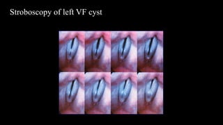
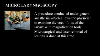
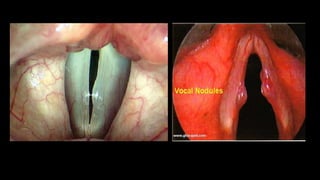
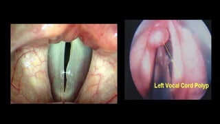
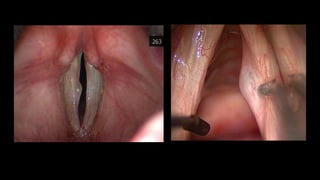
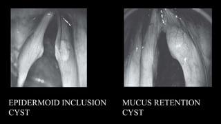
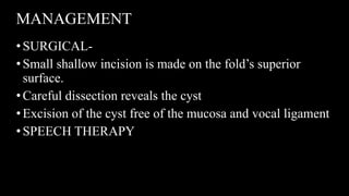
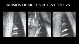
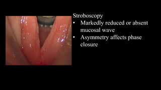
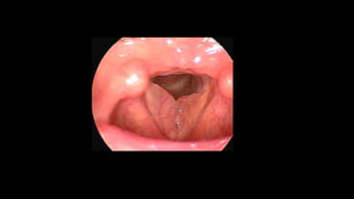
BENIGN MUCOSAL
LESIONS OF LARYNX
DR. YASHA C.
Benign epithelial lesions- caused by
•Infectious processes (fungal)
•Keratosis of the epithelium
•Papillomatous growth of the epithelium
•Malignant degeneration
Benign lesions of lamina propria
•Vocal fold nodules
•Laryngeal polyps
•Intrachordal cyst
•Fibrous mass
•Reactive vocal fold lesion
•Nonspecific vocal fold lesion
•Sulcus vocalis
Miscelleneous lesions
•Rheumatologic lesions
•Vocal fold granuloma
•Vascular lesions
•Reinke oedema
ANATOMY OF LARYNX
•The cartilages of the larynx- thyroid cartilage, the
epiglottis, the cricoid cartilage, and the arytenoid,
corniculate & cuneiform cartilages.
•Functionally, there are three groups of intrinsic laryngeal
muscles – the abductors, adductors, and tensors.
•Anatomy of the vagus nerve is important because
branches of the vagus nerve are responsible for
innervation of the larynx.
The vocal cords are
apart for breathing
in. The rings of the
trachea (wind pipe)
can be seen. They
are at their widest
apart during a sniff.
The vocal cords blur
once they are vibrating,
but at the instant before
they vibrate, the vocal
cords can be seen in
this shortened position,
slightly bowed.
The arytenoids have
come together
At a high pitch, at
the onset of
phonation, the vocal
cords are stretched.
HISTOLOGY OF VOCAL FOLDS
•Vocal folds epithelial lining- pseudostratified squamous
on the superior and inferior aspects of the fold and
nonkeratinizing squamous epithelium on the medial
contact surface.
•The subepithelial tissues are composed of a three-layered
lamina propria
•Deep to the lamina propria is the vocalis muscle.
MECHANISM OF INJURY
•Vocal fold lesions disrupt closure and vibration.
•Regardless of type, benign vocal fold lesions –
nodules, polyps, or cysts – cause hoarseness by
disrupting the vocal fold closure and vibration pattern.
ETIOLOGY
•Vibratory injury -excessive amount or manner of voice
use
•Single episode of voice strain
•Cigarette smoking
•Allergy
•LPR
•Medical conditions- AR, GERD, asthma, bronchitis,
sinusitis
•Medications
LPR
•Prevalence of LPR in patients of laryngeal & voice
disorders is about 50%
•78% with nodules had LPR
•When gastric refluxate reaches larynx or pharynx- is
called LPR
•Synonyms- extraesophageal reflux, atypical reflux,
gastropharyngeal reflux, pharyngoesophageal reflux,
supraesophageal reflux
•1 or 2 episodes of acid exposure (pH<4) at the proximal
probe located above the upper esophageal sphincter-
diagnostic of LPR
•Mucosa of larynx & pharynx is more susceptible to acid
attack than esophagus
PATHOPHYSIOLOGY
•Upper esophageal sphincter dysfunction
•Dysmotility
•Lower oesophageal sphincter dysfunction
•Action of acid & activated pepsin mucosal damage
•Laryngeal mucosa shows irreversible damage on
repeated refluxate exposure
SYMPTOMS
•Hoarseness
•Chronic cough
•Clearing of throat
•Globus pharyngeus
•Dysphagia
•Laryngospasm
•Postnasal drip
•Heartburn
REFLUX SYMPTOM INDEX
• Score >5 favors LPR
• Validated tool for LPR
PHYSICAL FINDINGS
•Most commonly related to laryngeal mucosal oedema &
injury
•Laryngoscopy
o Subglottic oedema posterior to vocal process of arytenoid
oVentricular obliteration
oInterarytenoid erythema
oVocal cord oedema
REFLUX FINDINGS SCORE
•Subglottic oedema
•Ventricular obliteration
•Laryngeal erythema/ hyperemia
•Vocal cord edema
•Diffuse laryngeal oedema
•Posterior commissure hypertrophy
•Granuloma / granulations
•Thick endolaryngeal mucus
Score >11 is in
favour of LPR
INVESTIGATIONS
•Dual pH probe monitoring- most sensitive
•Patient monitered for 18-24 hours
•Lower probe- 5cm above LES
•Upper probe- just above UES
•pH< 4 - >1 episode in 24hr- suggestive of LPR
COMPLICATIONS
•Granulomas/ granulations
•Ulcerations
•Subglottic stenosis
•Larygeal Ca.
•Laryngospasm
MANAGEMENT
•MINOR- PPI once daily for 6 months
•MAJOR- PPI bd for 6 months
•LIFE THREATENING- Fundoplication
ALLERGY
•Pts treated for allergic rhinitis had better outcome for
treatment of laryngitis
•Hypersensitivity makes laryngeal mucosa more
susceptible to stress
PATHOPHYSIOLOGY
•Mechanical stress least at midpoint of membranous VF
during phonation
•During hyperfunctioning dysphonia increased stress at
midpoint
•Increased stiffness in body of VF at midpoint results in
higher shearing stresses, worse if nodule or mass
present
•Mass adds wt to VF decreasing vibratory qualities and
frequency on stroboscopy
•Decrease in pitch range and impaired closure leads to
breathy voice and fatigue
•Asymmetry adds grainy quality to voice
•Once initiated, can cause compensatory muscle tension
to reduce air flow through glottis
SYMPTOMS OF BENIGN LESIONS
•Hoarse and rough voice
quality
•Vocal fatigue
•Unreliable voice
•Delayed voice initiation
•Low, gravelly voice
•Low pitch
•Voice breaks in first
passages of sentences
•Airy or breathy voice
•Inability to sing in high,
soft voice
•Increased effort to speak or
sing
•Frequent throat clearing
•Extra force needed
for voice
PHYSICAL EXAMINATION
•The indirect mirror exam is the initial procedure used to
view the larynx
•Gross abnormalities may be detected quickly, but subtle
abnormalities may be missed.
•Disadvantages- larynx not being in physiologic
phonation position (the tongue is extended and the larynx
is elevated), some anatomic features limit the exam, and
a hyper-reflexive gag
RIGID LARYNGOSCOPE
• Rigid laryngeal endoscopy is performed in the office using 70
or 90 degree telescopes passed through the mouth to obtain
images of the larynx and pharynx
• High quality images with excellent magnification.
• The patients are viewed in a nonphysiologic phonation
position. Anatomic factors and hyper-reflexive gags can limit
the results
FLEXIBLE LARYNGOSCOPE
•Most relied upon tool for evaluation of dysphonia
•It is the sole method that allows examination of the
nasopharynx, palate, larynx, and pharynx in a near
physiologic position.
•It can be performed relatively easily even in patients with
hyper-responsive gags and pediatric patients.
VIDEO STROBOSCOPY
•A specialized diagnostic procedure in which a
stroboscopic light is used in conjunction with a
laryngoscope to electronically slow down the motion of
the vocal folds
• To identify subtle changes in vibratory patterns that are
diagnostically significant
•Video stroboscopy is used to differentiate vocal fold
nodules from vocal fold polyps and cysts
• Videostroboscopy provides a
highly detailed view of the vocal
folds
• Can be performed via a nasal or
oral pathway
• Stroboscopic examination of the
vocal fold lesion is essential for
accurate diagnosis and successful
treatment.
Stroboscopy of left VF cyst
MICROLARYNGOSCOPY
A procedure conducted under general
anesthesia which allows the physician
to examine the vocal folds of the
larynx with magnification tools.
Microsurgical and laser removal of
lesions is done at this time
BEHAVIOURAL TREATMENT
•Behavioral modification is the primary treatment of
mucosal lesions
•Likely a lifelong treatment of the problem
•This may reduce a vocal fold swelling to an acceptable
size such that the voice becomes dependable and
acceptable to the patient.
•Hemorrhage can generally be managed behaviorally,
particularly if it was from a one time indiscretion
•Traditional voice therapy consists of two primary
avenues-
•Vocal hygiene is a daily regimen to achieve and maintain
a healthy voice. It includes maintaining adequate
hydration, minimizing exposure to noxious chemicals, no
smoking, and the avoidance of loud voice use.
•Voice therapy is a behavioral intervention technique that
makes use of vocal exercises, speaker awareness and
proper postures and alignment when using the voice.
MEDICAL TREATMENT
•Reflux may be a contributing factor- two week trial of
medication, dietary and life-style adjustments and bed
positioning
•Granulomas should be managed medically as they nearly
always spontaneously decapitate in 4 to 6 months. They
tend to recur because they are located in an area of
constant movement. The first line of treatment should be
an antireflux regimen
•Steroid treatment reduces the overlying and sometimes
camouflaging inflammation and swelling while
leaving the cyst unchanged, thus making its diagnosis
easier.
•Patients may be placed on a 2-week period of vocal
rest, perhaps accompanied by a high-dose
corticosteroid taper
SURGICAL MANAGEMENT
•Surgery is directed at removing only the mucosal lesion
and preserving as much of the intermediate and deep
layers of the vocal fold as possible.
•Surgery is performed when the nodules or polyps are
very large or have existed for a long time and if the
lesion extends deeper into the layers of the vocal fold.
•MICROFLAP TECHNIQUES
• The use of microflap techniques avoids a raw mucosal
surface that heals by secondary intention
• Avoidance of the deeper layers of the lamina propria and
vocal ligament minimizes the fibroblastic response
SURGICAL COMPLICATIONS
• Complications are related either to laryngoscopy or to vocal fold
mucosal injury.
• Pressure effects from suspension laryngoscopy- tongue
numbness, altered taste, and oropharyngeal, mucosal, and dental
injuries.
•Deep-plane dissection or exposure of the vocal
ligament can result in scarring and fibrosis of the
mucosa with loss of mucosal wave and glottal
insufficiency.
•Injudicious use of the laser can result in a wide zone of
thermal damage with mucosal scarring and fibrosis,
unintended burn injuries, and endotracheal tube fires.
VOCAL NODULES
•Bilateral small swellings (<3mm) that develop on the
free edge of vocal fold at membranous portion
ETIOLOGY
•Hyperkinetic phonation
•Chronic cough
•GERD
•Infections- nasal, throat, chest
•Allergy
•Cleft palate
•PATHOPHYSIOLOGY
SYMPTOMS
•Hoarseness
•Loss of the ability to sing high notes softly
•Delayed phonatory onset
•Increased breathiness (air escape), roughness, and
harshness
•Reduced vocal endurance
CLINICAL FEATURES
•Bilateral
•At the junction of anterior one thirds and posterior two
thirds of free edge of vocal cords
•Varies in size, symmetry, color
•Stroboscopic pattern shows normal or minimal reduction
of the mucosal wave activity
MANAGEMENT
•MEDICAL- good hydration, management of allergy &
reflux
•BEHAVIOURAL- Speech therapy
•SURGICAL- Laryngeal microsurgery with precise
excision
VOCAL FOLD POLYPS
•Benign swelling > 3 mm that arise from free edge of
vocal fold
•Unilateral or bilateral lesions
•Typically exophytic lesions
•Associated with vocal fold haemorrhage
ETIOPATHOGENESIS
•ETIOLOGY- Intermittent voice abuse, sudden shouting,
aspirin
•PATHOGENESIS- Vocal exertion Shearing forces
act on capillaries of mucosa capillary rupture
•Extravasation of blood focal accumulation of blood
hemorrhagic polyp
SYMPTOMS
•Abrupt onset of hoarseness during extreme vocal effort
•Intermittent subtle aberrant sounds
•Impaired falsetto register
•Chronic vocal huskiness
CLINICAL FEATURES
•Unilateral
•At the junction of anterior one thirds and posterior two
thirds of free edge of vocal cords
•May appear dark & blood filled in early stages
•May become pedunculated- moving in and out of the
glottis with inspiration and expiration
MANAGEMENT
•Microlaryngeal precise excision
•Speech therapy
INTRACORDAL CYSTS
• Classified as either mucus retention cysts or epidermoid
inclusion cysts
•PATHOPHYSIOLOGY-
• MUCUS RETENTION CYST- arise when the duct of a
mucus gland becomes plugged and glandular secretions
•EPIERMOID INCLUSION CYST-
contain accumulated keratin
Healing of injured mucosa
Epithelial cell rests in
subepithelial layer
CLINICAL FEATURES
•Voice abuse- in epidermoid cyst
•Husky, breathy or harsh voice
•Mucus retention cyst- less vocal limitations
•Originate just below the free margin of the vocal fold
with medial projection
•Stroboscopy- mucosa overlying cysts stops vibrating
before mucosa surrounding the cyst
EPIDERMOID INCLUSION
CYST
MUCUS RETENTION
CYST
MANAGEMENT
•SURGICAL-
•Small shallow incision is made on the fold’s superior
surface.
•Careful dissection reveals the cyst
•Excision of the cyst free of the mucosa and vocal ligament
•SPEECH THERAPY
EXCISION OF MUCUS RETENTION CYST
FIBROUS MASS OF VOCAL CORD
•unilateral or bilateral
•located in the subepithelial or ligamentous area
•do not respond to voice therapy
•impairment in the mucosal wave activity during
stroboscopy
•Intraoperatively- fibrous gray, well organized,
encapsulated lesion in the midmembranous vocal fold
•Rx- microflap excision
B/L subepithelial fibrous mass Microflap excision
REACTIVE VOCAL FOLD LESION
•Occurs in response to a contralateral lesion
•Has an indentation where the contralateral lesion has
created a concavity due to repeated trauma during vocal
fold vibration
•The two lesions interdigitating with each other during
vocal fold closure and vibration is called Cup and Saucer
deformity
•Reduces in size with behavioral therapy.
Reactive vocal fold lesion of left true vocal fold
NON SPECIFIC VOCAL FOLD LESIONS
•Unilateral or bilateral
•Subepithelial or ligamentous pathology
•Not responsive to non surgical interventions- voice
rest, medical therapy of comorbidities,voice therapy
•But does not impair function sufficiently to necessitate
phonomicrosurgical removal
INTRACORDAL SCARRING
•Scarring in Reinke space after repeated inflammation,
trauma or vocal hemorrhage
•Subepithelial scar
oDisorganized collagen
oLoss of ECM
oDistinguish from epithelial scarring or vocal sulcus
•VF appears stiff, white or opaque
•Hoarseness, vocal fatigue, breathiness
SULCUS VOCALIS
CAUSES OF INTRACORDAL SCARRING
• Cysts predispose to scar formation (ruptured, epidermoid
origin)
• Trauma
oVocal fold surgery involving lamina propria
oRepeated epithelial procedures
oBiopsy, stripping
oInhalational
oIntubation
• CO2 laser
• Radiation
• Rheumatologic disease
Stroboscopy
• Markedly reduced or absent
mucosal wave
• Asymmetry affects phase
closure
TREATMENT
•Microflap to remove cyst elements and adynamic
fibrous components
•Medialization thyroplasty for glottic gaps
•Replacement soft tissue (Fillers)
oCollagen
oFat
oHyaluronic acid
REINKE OEDEMA
•Polypoid corditis
•Proliferation of superficial lamina propria
•Chronic irritant exposure
•Smoke, LPR, occupational exposures
•Water-balloon outpouching from membranous VF
•Ball-valving effect
TREATMENT
•Surgery
•Airway compromise
•Preserve some superficial lamina propria and overlying
epithelium to preserve mucosal wave
•Stage for bilateral disease to prevent anterior web
•Remove irritants and treat LPR
VOCAL PROCESS GRANULOMA
GRANULOMAS
•Response to trauma
•LPR, throat clearing, chronic cough
•Intubation
•Compensatory forceful glottic closure
oVF paresis
oPresbylarynges
•Does not affect mucosal wave or phase closure
TREATMENT
•LPR treatment
•Speech therapy
•Botox to thyroarytenoid muscle
•Surgery
oCompromise voice, breathing or swallowing
oSuspicion for malignancy
oHigh recurrence rate
PAPILLOMAS
PAPILLOMAS
• HPV (Strain 6 and 11 most common)
• 2% malignant transformation (HPV 16 and 18)
• 10% rate of spread to other sites (trachea, supraglottis,
NP)
• Most commonly found at columnar and squamous
junction
• Host immune recognition
oHPV 11 growth more aggressive during pregnancy
o40% HPV+ larynx without RRP
JUVENILE PAPILLOMA
Squamous papillomas the most common (80%) benign
tumors of larynx.
Juvenile papillomas are seen in children
Associated with Human Papilloma Virus types 6 and 11
infection
They are usually multiple, and well known for recurrence
after removal
Symptoms: Hoarseness and stridor
ADULT PAPILLOMA
•They are single
•Less aggressive
•Less prone for recurrence
SIGNS
•Signs: Warty masses which may be
sessile/pedunculated, and friable to touch.
TREATMENT
• Medical: Inteferons; anti virals like cedofovir, acyclovir
and ribavirin; and immunomodulator therapy.
• Surgical: Endoscopic removal using KTP-532/CO2
LASER, forceps, cryotherapy electrocautery, or
microshaver
• Excision is followed by interferon therapy to prevent
recurrence
• Vaccine
LEUKOPLAKIA
LEUKOPLAKIA
• Spectrum of change in epithelium
• Hyperkeratosis Dysplasia (mild, moderate)
CIS/ severe dysplasia
•Pattern of growth
oSuperficial, broad
oVerrucous, exophytic with surrounding erythema
•Appearance does not correlate with degree of dysplasia
• 8% to 14% rate of malignant transformation
TREATMENT
• CO2 laser
• PDL
• Microflap excision
• Preservation of normal mucosal wave for mild dysplasia
•More aggressive excision with increasing dysplasia
FUNGAL LARYNGITIS
•Disease of both immunocompromised and
immunocompetent hosts
•May mimic leukoplakia or malignancy
oWhite or gray pseudomembrane on mucosa
oMucosal erythema and edema (focal or diffuse) surrounding
white plaques
oMucosal ulcerations
oContact bleeding
FUNGAL LARYNGITIS
RISK FACTORS
•LPR, smoking, inhaled steroids, prolonged antibiotic
use
•DM, immunosuppressants, CA, nutritional deficits
•Compromise mucosal barrier
DIAGNOSIS
•Suspicion and response to empiric therapy
•Any question can culture by laryngeal brushing or biopsy
•Dysphagia may also have esophageal involvement
•Candida species most commonly cultured
•Blastomyces (Eastern US and Midwest)
•Histoplasma (Ohio and Mississippi River Valleys)
•Coccidioides (Southwestern US)
•Bacterial superinfection
oHoney-colored crusts
TREATMENT
•Fluconazole x 3wks
•Nystatin swish and swallow (100,000 units/ml, 10ml
tid)
•Prevention
ospacers for inhaled steroids
ooral rinse, gargle with water after use
SACCULAR CYST
•The laryngeal saccule is a membranous sac in
the larynx located between the false vocal cords and
the inner surface of the thyroid cartilage
•The saccule contains mucus-secreting glands
•Saccular cysts are retention cysts due to obstruction to
opening of the saccule
SACCULAR CYST
LARYNGOCELE
•A laryngocele is an air filled cystic dilatation of the
saccule or appendix of the ventricle.
•It occurs in people with congenitally large ventricular
appendix.
•It is predisposed by activities which increase the
intralaryngeal pressure, like straining (weight lifters),
glass blowers and trumpet players
Pathological types:
•External-swelling lateral to thyrohyoid membrane
•Internal-swelling of false cords and aryepiglottic folds
•Combined/Mixed
•Symptoms:
•Internal & Mixed: Hoarseness and cough. Stridor if
large.
•External: Compressible mass in the neck, that
increases on coughing or Valsalva manoeuvre.
•Bryce’s sign: Gurgling and hissing sound when the
neck mass is compressed.
•Investigations: X-Ray during Valsalva, CT scan,
Laryngoscopy to rule out malignancy
TREATMENT:
•External/mixed:
Transcervical neck
dissection with
removal of part of
thyroid lamina.
•Excision of the cyst
or marsupialisation
THANK YOU

BENIGN_MUCOSAL_LESIONS_OF_LARYNX.pptx

  • 1.
    BENIGN MUCOSAL LESIONS OFLARYNX DR. YASHA C.
  • 2.
    Benign epithelial lesions-caused by •Infectious processes (fungal) •Keratosis of the epithelium •Papillomatous growth of the epithelium •Malignant degeneration
  • 3.
    Benign lesions oflamina propria •Vocal fold nodules •Laryngeal polyps •Intrachordal cyst •Fibrous mass •Reactive vocal fold lesion •Nonspecific vocal fold lesion •Sulcus vocalis
  • 4.
    Miscelleneous lesions •Rheumatologic lesions •Vocalfold granuloma •Vascular lesions •Reinke oedema
  • 5.
    ANATOMY OF LARYNX •Thecartilages of the larynx- thyroid cartilage, the epiglottis, the cricoid cartilage, and the arytenoid, corniculate & cuneiform cartilages. •Functionally, there are three groups of intrinsic laryngeal muscles – the abductors, adductors, and tensors. •Anatomy of the vagus nerve is important because branches of the vagus nerve are responsible for innervation of the larynx.
  • 7.
    The vocal cordsare apart for breathing in. The rings of the trachea (wind pipe) can be seen. They are at their widest apart during a sniff. The vocal cords blur once they are vibrating, but at the instant before they vibrate, the vocal cords can be seen in this shortened position, slightly bowed. The arytenoids have come together At a high pitch, at the onset of phonation, the vocal cords are stretched.
  • 8.
    HISTOLOGY OF VOCALFOLDS •Vocal folds epithelial lining- pseudostratified squamous on the superior and inferior aspects of the fold and nonkeratinizing squamous epithelium on the medial contact surface. •The subepithelial tissues are composed of a three-layered lamina propria •Deep to the lamina propria is the vocalis muscle.
  • 10.
    MECHANISM OF INJURY •Vocalfold lesions disrupt closure and vibration. •Regardless of type, benign vocal fold lesions – nodules, polyps, or cysts – cause hoarseness by disrupting the vocal fold closure and vibration pattern.
  • 11.
    ETIOLOGY •Vibratory injury -excessiveamount or manner of voice use •Single episode of voice strain •Cigarette smoking •Allergy •LPR •Medical conditions- AR, GERD, asthma, bronchitis, sinusitis •Medications
  • 12.
    LPR •Prevalence of LPRin patients of laryngeal & voice disorders is about 50% •78% with nodules had LPR •When gastric refluxate reaches larynx or pharynx- is called LPR •Synonyms- extraesophageal reflux, atypical reflux, gastropharyngeal reflux, pharyngoesophageal reflux, supraesophageal reflux
  • 13.
    •1 or 2episodes of acid exposure (pH<4) at the proximal probe located above the upper esophageal sphincter- diagnostic of LPR •Mucosa of larynx & pharynx is more susceptible to acid attack than esophagus
  • 14.
    PATHOPHYSIOLOGY •Upper esophageal sphincterdysfunction •Dysmotility •Lower oesophageal sphincter dysfunction •Action of acid & activated pepsin mucosal damage •Laryngeal mucosa shows irreversible damage on repeated refluxate exposure
  • 15.
    SYMPTOMS •Hoarseness •Chronic cough •Clearing ofthroat •Globus pharyngeus •Dysphagia •Laryngospasm •Postnasal drip •Heartburn REFLUX SYMPTOM INDEX • Score >5 favors LPR • Validated tool for LPR
  • 16.
    PHYSICAL FINDINGS •Most commonlyrelated to laryngeal mucosal oedema & injury •Laryngoscopy o Subglottic oedema posterior to vocal process of arytenoid oVentricular obliteration oInterarytenoid erythema oVocal cord oedema
  • 17.
    REFLUX FINDINGS SCORE •Subglotticoedema •Ventricular obliteration •Laryngeal erythema/ hyperemia •Vocal cord edema •Diffuse laryngeal oedema •Posterior commissure hypertrophy •Granuloma / granulations •Thick endolaryngeal mucus Score >11 is in favour of LPR
  • 18.
    INVESTIGATIONS •Dual pH probemonitoring- most sensitive •Patient monitered for 18-24 hours •Lower probe- 5cm above LES •Upper probe- just above UES •pH< 4 - >1 episode in 24hr- suggestive of LPR
  • 19.
  • 20.
    MANAGEMENT •MINOR- PPI oncedaily for 6 months •MAJOR- PPI bd for 6 months •LIFE THREATENING- Fundoplication
  • 21.
    ALLERGY •Pts treated forallergic rhinitis had better outcome for treatment of laryngitis •Hypersensitivity makes laryngeal mucosa more susceptible to stress
  • 22.
    PATHOPHYSIOLOGY •Mechanical stress leastat midpoint of membranous VF during phonation •During hyperfunctioning dysphonia increased stress at midpoint •Increased stiffness in body of VF at midpoint results in higher shearing stresses, worse if nodule or mass present
  • 23.
    •Mass adds wtto VF decreasing vibratory qualities and frequency on stroboscopy •Decrease in pitch range and impaired closure leads to breathy voice and fatigue •Asymmetry adds grainy quality to voice •Once initiated, can cause compensatory muscle tension to reduce air flow through glottis
  • 24.
    SYMPTOMS OF BENIGNLESIONS •Hoarse and rough voice quality •Vocal fatigue •Unreliable voice •Delayed voice initiation •Low, gravelly voice •Low pitch •Voice breaks in first passages of sentences •Airy or breathy voice •Inability to sing in high, soft voice •Increased effort to speak or sing •Frequent throat clearing •Extra force needed for voice
  • 25.
    PHYSICAL EXAMINATION •The indirectmirror exam is the initial procedure used to view the larynx •Gross abnormalities may be detected quickly, but subtle abnormalities may be missed. •Disadvantages- larynx not being in physiologic phonation position (the tongue is extended and the larynx is elevated), some anatomic features limit the exam, and a hyper-reflexive gag
  • 26.
    RIGID LARYNGOSCOPE • Rigidlaryngeal endoscopy is performed in the office using 70 or 90 degree telescopes passed through the mouth to obtain images of the larynx and pharynx • High quality images with excellent magnification. • The patients are viewed in a nonphysiologic phonation position. Anatomic factors and hyper-reflexive gags can limit the results
  • 27.
    FLEXIBLE LARYNGOSCOPE •Most reliedupon tool for evaluation of dysphonia •It is the sole method that allows examination of the nasopharynx, palate, larynx, and pharynx in a near physiologic position. •It can be performed relatively easily even in patients with hyper-responsive gags and pediatric patients.
  • 28.
    VIDEO STROBOSCOPY •A specializeddiagnostic procedure in which a stroboscopic light is used in conjunction with a laryngoscope to electronically slow down the motion of the vocal folds • To identify subtle changes in vibratory patterns that are diagnostically significant •Video stroboscopy is used to differentiate vocal fold nodules from vocal fold polyps and cysts
  • 29.
    • Videostroboscopy providesa highly detailed view of the vocal folds • Can be performed via a nasal or oral pathway • Stroboscopic examination of the vocal fold lesion is essential for accurate diagnosis and successful treatment.
  • 30.
  • 31.
    MICROLARYNGOSCOPY A procedure conductedunder general anesthesia which allows the physician to examine the vocal folds of the larynx with magnification tools. Microsurgical and laser removal of lesions is done at this time
  • 32.
    BEHAVIOURAL TREATMENT •Behavioral modificationis the primary treatment of mucosal lesions •Likely a lifelong treatment of the problem •This may reduce a vocal fold swelling to an acceptable size such that the voice becomes dependable and acceptable to the patient. •Hemorrhage can generally be managed behaviorally, particularly if it was from a one time indiscretion
  • 33.
    •Traditional voice therapyconsists of two primary avenues- •Vocal hygiene is a daily regimen to achieve and maintain a healthy voice. It includes maintaining adequate hydration, minimizing exposure to noxious chemicals, no smoking, and the avoidance of loud voice use. •Voice therapy is a behavioral intervention technique that makes use of vocal exercises, speaker awareness and proper postures and alignment when using the voice.
  • 34.
    MEDICAL TREATMENT •Reflux maybe a contributing factor- two week trial of medication, dietary and life-style adjustments and bed positioning •Granulomas should be managed medically as they nearly always spontaneously decapitate in 4 to 6 months. They tend to recur because they are located in an area of constant movement. The first line of treatment should be an antireflux regimen
  • 35.
    •Steroid treatment reducesthe overlying and sometimes camouflaging inflammation and swelling while leaving the cyst unchanged, thus making its diagnosis easier. •Patients may be placed on a 2-week period of vocal rest, perhaps accompanied by a high-dose corticosteroid taper
  • 36.
    SURGICAL MANAGEMENT •Surgery isdirected at removing only the mucosal lesion and preserving as much of the intermediate and deep layers of the vocal fold as possible. •Surgery is performed when the nodules or polyps are very large or have existed for a long time and if the lesion extends deeper into the layers of the vocal fold.
  • 37.
    •MICROFLAP TECHNIQUES • Theuse of microflap techniques avoids a raw mucosal surface that heals by secondary intention • Avoidance of the deeper layers of the lamina propria and vocal ligament minimizes the fibroblastic response
  • 38.
    SURGICAL COMPLICATIONS • Complicationsare related either to laryngoscopy or to vocal fold mucosal injury. • Pressure effects from suspension laryngoscopy- tongue numbness, altered taste, and oropharyngeal, mucosal, and dental injuries.
  • 39.
    •Deep-plane dissection orexposure of the vocal ligament can result in scarring and fibrosis of the mucosa with loss of mucosal wave and glottal insufficiency. •Injudicious use of the laser can result in a wide zone of thermal damage with mucosal scarring and fibrosis, unintended burn injuries, and endotracheal tube fires.
  • 40.
    VOCAL NODULES •Bilateral smallswellings (<3mm) that develop on the free edge of vocal fold at membranous portion ETIOLOGY •Hyperkinetic phonation •Chronic cough •GERD •Infections- nasal, throat, chest •Allergy •Cleft palate
  • 41.
  • 42.
    SYMPTOMS •Hoarseness •Loss of theability to sing high notes softly •Delayed phonatory onset •Increased breathiness (air escape), roughness, and harshness •Reduced vocal endurance
  • 43.
    CLINICAL FEATURES •Bilateral •At thejunction of anterior one thirds and posterior two thirds of free edge of vocal cords •Varies in size, symmetry, color •Stroboscopic pattern shows normal or minimal reduction of the mucosal wave activity
  • 45.
    MANAGEMENT •MEDICAL- good hydration,management of allergy & reflux •BEHAVIOURAL- Speech therapy •SURGICAL- Laryngeal microsurgery with precise excision
  • 46.
    VOCAL FOLD POLYPS •Benignswelling > 3 mm that arise from free edge of vocal fold •Unilateral or bilateral lesions •Typically exophytic lesions •Associated with vocal fold haemorrhage
  • 47.
    ETIOPATHOGENESIS •ETIOLOGY- Intermittent voiceabuse, sudden shouting, aspirin •PATHOGENESIS- Vocal exertion Shearing forces act on capillaries of mucosa capillary rupture •Extravasation of blood focal accumulation of blood hemorrhagic polyp
  • 48.
    SYMPTOMS •Abrupt onset ofhoarseness during extreme vocal effort •Intermittent subtle aberrant sounds •Impaired falsetto register •Chronic vocal huskiness
  • 49.
    CLINICAL FEATURES •Unilateral •At thejunction of anterior one thirds and posterior two thirds of free edge of vocal cords •May appear dark & blood filled in early stages •May become pedunculated- moving in and out of the glottis with inspiration and expiration
  • 52.
  • 53.
    INTRACORDAL CYSTS • Classifiedas either mucus retention cysts or epidermoid inclusion cysts •PATHOPHYSIOLOGY- • MUCUS RETENTION CYST- arise when the duct of a mucus gland becomes plugged and glandular secretions •EPIERMOID INCLUSION CYST- contain accumulated keratin Healing of injured mucosa Epithelial cell rests in subepithelial layer
  • 54.
    CLINICAL FEATURES •Voice abuse-in epidermoid cyst •Husky, breathy or harsh voice •Mucus retention cyst- less vocal limitations •Originate just below the free margin of the vocal fold with medial projection •Stroboscopy- mucosa overlying cysts stops vibrating before mucosa surrounding the cyst
  • 56.
  • 57.
    MANAGEMENT •SURGICAL- •Small shallow incisionis made on the fold’s superior surface. •Careful dissection reveals the cyst •Excision of the cyst free of the mucosa and vocal ligament •SPEECH THERAPY
  • 58.
    EXCISION OF MUCUSRETENTION CYST
  • 59.
    FIBROUS MASS OFVOCAL CORD •unilateral or bilateral •located in the subepithelial or ligamentous area •do not respond to voice therapy •impairment in the mucosal wave activity during stroboscopy •Intraoperatively- fibrous gray, well organized, encapsulated lesion in the midmembranous vocal fold •Rx- microflap excision
  • 60.
    B/L subepithelial fibrousmass Microflap excision
  • 61.
    REACTIVE VOCAL FOLDLESION •Occurs in response to a contralateral lesion •Has an indentation where the contralateral lesion has created a concavity due to repeated trauma during vocal fold vibration •The two lesions interdigitating with each other during vocal fold closure and vibration is called Cup and Saucer deformity •Reduces in size with behavioral therapy.
  • 62.
    Reactive vocal foldlesion of left true vocal fold
  • 63.
    NON SPECIFIC VOCALFOLD LESIONS •Unilateral or bilateral •Subepithelial or ligamentous pathology •Not responsive to non surgical interventions- voice rest, medical therapy of comorbidities,voice therapy •But does not impair function sufficiently to necessitate phonomicrosurgical removal
  • 64.
    INTRACORDAL SCARRING •Scarring inReinke space after repeated inflammation, trauma or vocal hemorrhage •Subepithelial scar oDisorganized collagen oLoss of ECM oDistinguish from epithelial scarring or vocal sulcus •VF appears stiff, white or opaque •Hoarseness, vocal fatigue, breathiness
  • 65.
  • 66.
    CAUSES OF INTRACORDALSCARRING • Cysts predispose to scar formation (ruptured, epidermoid origin) • Trauma oVocal fold surgery involving lamina propria oRepeated epithelial procedures oBiopsy, stripping oInhalational oIntubation • CO2 laser • Radiation • Rheumatologic disease
  • 67.
    Stroboscopy • Markedly reducedor absent mucosal wave • Asymmetry affects phase closure
  • 68.
    TREATMENT •Microflap to removecyst elements and adynamic fibrous components •Medialization thyroplasty for glottic gaps •Replacement soft tissue (Fillers) oCollagen oFat oHyaluronic acid
  • 69.
    REINKE OEDEMA •Polypoid corditis •Proliferationof superficial lamina propria •Chronic irritant exposure •Smoke, LPR, occupational exposures •Water-balloon outpouching from membranous VF •Ball-valving effect
  • 71.
    TREATMENT •Surgery •Airway compromise •Preserve somesuperficial lamina propria and overlying epithelium to preserve mucosal wave •Stage for bilateral disease to prevent anterior web •Remove irritants and treat LPR
  • 72.
  • 73.
    GRANULOMAS •Response to trauma •LPR,throat clearing, chronic cough •Intubation •Compensatory forceful glottic closure oVF paresis oPresbylarynges •Does not affect mucosal wave or phase closure
  • 74.
    TREATMENT •LPR treatment •Speech therapy •Botoxto thyroarytenoid muscle •Surgery oCompromise voice, breathing or swallowing oSuspicion for malignancy oHigh recurrence rate
  • 75.
  • 76.
    PAPILLOMAS • HPV (Strain6 and 11 most common) • 2% malignant transformation (HPV 16 and 18) • 10% rate of spread to other sites (trachea, supraglottis, NP) • Most commonly found at columnar and squamous junction • Host immune recognition oHPV 11 growth more aggressive during pregnancy o40% HPV+ larynx without RRP
  • 77.
    JUVENILE PAPILLOMA Squamous papillomasthe most common (80%) benign tumors of larynx. Juvenile papillomas are seen in children Associated with Human Papilloma Virus types 6 and 11 infection They are usually multiple, and well known for recurrence after removal Symptoms: Hoarseness and stridor
  • 78.
    ADULT PAPILLOMA •They aresingle •Less aggressive •Less prone for recurrence SIGNS •Signs: Warty masses which may be sessile/pedunculated, and friable to touch.
  • 79.
    TREATMENT • Medical: Inteferons;anti virals like cedofovir, acyclovir and ribavirin; and immunomodulator therapy. • Surgical: Endoscopic removal using KTP-532/CO2 LASER, forceps, cryotherapy electrocautery, or microshaver • Excision is followed by interferon therapy to prevent recurrence • Vaccine
  • 80.
  • 81.
    LEUKOPLAKIA • Spectrum ofchange in epithelium • Hyperkeratosis Dysplasia (mild, moderate) CIS/ severe dysplasia •Pattern of growth oSuperficial, broad oVerrucous, exophytic with surrounding erythema •Appearance does not correlate with degree of dysplasia • 8% to 14% rate of malignant transformation
  • 82.
    TREATMENT • CO2 laser •PDL • Microflap excision • Preservation of normal mucosal wave for mild dysplasia •More aggressive excision with increasing dysplasia
  • 83.
    FUNGAL LARYNGITIS •Disease ofboth immunocompromised and immunocompetent hosts •May mimic leukoplakia or malignancy oWhite or gray pseudomembrane on mucosa oMucosal erythema and edema (focal or diffuse) surrounding white plaques oMucosal ulcerations oContact bleeding
  • 84.
  • 85.
    RISK FACTORS •LPR, smoking,inhaled steroids, prolonged antibiotic use •DM, immunosuppressants, CA, nutritional deficits •Compromise mucosal barrier
  • 86.
    DIAGNOSIS •Suspicion and responseto empiric therapy •Any question can culture by laryngeal brushing or biopsy •Dysphagia may also have esophageal involvement
  • 87.
    •Candida species mostcommonly cultured •Blastomyces (Eastern US and Midwest) •Histoplasma (Ohio and Mississippi River Valleys) •Coccidioides (Southwestern US) •Bacterial superinfection oHoney-colored crusts
  • 88.
    TREATMENT •Fluconazole x 3wks •Nystatinswish and swallow (100,000 units/ml, 10ml tid) •Prevention ospacers for inhaled steroids ooral rinse, gargle with water after use
  • 89.
    SACCULAR CYST •The laryngealsaccule is a membranous sac in the larynx located between the false vocal cords and the inner surface of the thyroid cartilage •The saccule contains mucus-secreting glands •Saccular cysts are retention cysts due to obstruction to opening of the saccule
  • 90.
  • 91.
    LARYNGOCELE •A laryngocele isan air filled cystic dilatation of the saccule or appendix of the ventricle. •It occurs in people with congenitally large ventricular appendix. •It is predisposed by activities which increase the intralaryngeal pressure, like straining (weight lifters), glass blowers and trumpet players
  • 92.
    Pathological types: •External-swelling lateralto thyrohyoid membrane •Internal-swelling of false cords and aryepiglottic folds •Combined/Mixed
  • 95.
    •Symptoms: •Internal & Mixed:Hoarseness and cough. Stridor if large. •External: Compressible mass in the neck, that increases on coughing or Valsalva manoeuvre. •Bryce’s sign: Gurgling and hissing sound when the neck mass is compressed. •Investigations: X-Ray during Valsalva, CT scan, Laryngoscopy to rule out malignancy
  • 97.
    TREATMENT: •External/mixed: Transcervical neck dissection with removalof part of thyroid lamina. •Excision of the cyst or marsupialisation
  • 98.