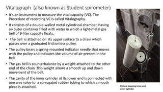
DETERMINATION OF VITAL CAPACITY
Dr. Ravi Dutt (MBBS, MD)
Senior Resident
Department of physiology
MAMC, Delhi
Learning objectives
After completing this practical, Student should be able to:
1. Define vital capacity
2. Record the vital capacity and explain the effect of posture on it.
3. Describe the physiological and pathological factors that affect vital
capacity.
AIM
• To measure the vital capacity using Vitalograph
• To study the effect of posture on the vital capacity using
Vitalograph
APPARATUS
• Vitalograph
• Nose clips
• Potassium permanganate
solution/Spirit to clean
mouth piece
Pulley
chain
Hollow tube
for chain
Aluminium
cylinder( the bell)
Outer cylinder
Corrugated Rubber tube with
mouth piece
Levelling screws
Needle
pointer
Counter
weight
Vital Capacity
It is the largest volume of air a person can expel from the lungs with maximum
effort after first filling the lungs fully by a deepest possible inspiration.
 It varies between 3.5 to 5.5 litres, the value being 20% lower in females. In
general, the vital capacity is more in males, taller persons, and in younger adults.
The VC provides useful information about lung function and the strength of
respiratory muscles.
Normal values
• In males :
• In Females:
Vitalograph (also known as Student spirometer)
• It’s an instrument to measure the vital capacity (VC). The
Procedure of recording VC is called Vitalography.
• It consists of a double-walled metal cylindrical chamber, having
an outer container filled with water in which a light-metal gas
bell of 9-liter capacity floats.
• The bell is attached on its upper surface to a chain which
passes over a graduated frictionless pulley.
• The pulley bears a spring-mounted indicator needle that moves
with the pulley and indicates the volume of air present in the
bell.
• The gas bell is counterbalance by a weight attached to the other
end of the chain .This weight allows a smooth up and down
movement of the bell.
• The cavity of the inner cylinder at its lower end is connected with
one way valve to a corrugated rubber tubing to which a mouth
piece is attached. Picture showing inner and
outer cylinder
Video description of Vitalograph
Procedure
Measuring VC by collecting expired air in the spirometer (Standard Method).
1. Bring the bell to its lowest position by gently pushing it down. Adjust the
pointer needle at zero, which indicates that the bell is completely empty.
2. Ask the subject to stand comfortably, facing the spirometer so that she/he
can see the movement of the bell.
3. Tell subject to breathe normally (quietly) for a minute or so. Now direct
subject to inspire as deeply and as fully as possible to fill the lungs.
4. Then, while keeping the nostrils closed with a thumb and fingers or with
nose clip, and the mouthpiece held firmly between the lips, tell the subject
to expel all the air that he can with maximum effort into the spirometer.
Procedure cont.
5. The bell moves up and the pointer on the pulley indicates the volume of
expired air. The forced expiration should be deep and quick but without haste.
6. Take 2 more readings at intervals of 5 minutes in the standing position as
before. Make a note of the maximum value of the 3 readings to get the best
result.
Effect of posture.
• Sitting posture: Ask the subject to sit comfortably on a stool and record the
VC 3 times as before, at intervals of 2 minutes.
• Supine posture: Ask the subject to lie down on the couch in supine position
(face up) and record the VC 3 times as before.
Results
Position First Reading Second reading Third recording Maximum of 3
recordings
Standing
Siting
Supine
Physiological factors affecting VC
1. Relation to body surface area (BSA): This is called vital index.
• Males = 2.5 liters/m2 BSA
• Females = 2.1 liters/m2 BSA
2. Gender: VC is more in males because of larger chest size, more
muscle power and more body surface area.
3. Age: VC decreases in old age due to the loss of the elasticity of lungs.
4. Strength od respiratory muscles: The vital capacity is high in athletes,
swimmers, divers, etc., but is low in persons who have sedentary
habits.
Effect of posture on VC
The VC is maximum in the standing position, less in the sitting position, and least in
the supine position.
This effect of posture is due to the following factors:
1. In the sitting and supine positions, the muscles of respiration (both primary and
accessory) cannot be employed as forcefully and effectively for the expansion and
compression of lungs and chest.
2. In the supine position, the abdominal viscera push the diaphragm up and
interfere with its movements. The mobility of the chest is also reduced by the
contact of the back with the bed.
3. There is accumulation of more blood in the blood vessels of the lungs (especially
veins) in the supine position. This decreases the total lung capacity, and hence the
vital capacity.
Clinical importance
The VC provides useful information about lung function and the
strength of respiratory muscles. It also provides useful information
about abnormal ventilation
• due to airway obstruction
• fibrosis of the lungs
• mechanical interference with chest expansion and compression
• strength of respiratory muscles
• However, it cannot help in differentiating between obstructive and
restrictive lung diseases, where timed VC is of greater help.
THANK YOU

Vitalography: Physiology practical

  • 1.
    DETERMINATION OF VITALCAPACITY Dr. Ravi Dutt (MBBS, MD) Senior Resident Department of physiology MAMC, Delhi
  • 2.
    Learning objectives After completingthis practical, Student should be able to: 1. Define vital capacity 2. Record the vital capacity and explain the effect of posture on it. 3. Describe the physiological and pathological factors that affect vital capacity.
  • 3.
    AIM • To measurethe vital capacity using Vitalograph • To study the effect of posture on the vital capacity using Vitalograph
  • 4.
    APPARATUS • Vitalograph • Noseclips • Potassium permanganate solution/Spirit to clean mouth piece Pulley chain Hollow tube for chain Aluminium cylinder( the bell) Outer cylinder Corrugated Rubber tube with mouth piece Levelling screws Needle pointer Counter weight
  • 5.
    Vital Capacity It isthe largest volume of air a person can expel from the lungs with maximum effort after first filling the lungs fully by a deepest possible inspiration.  It varies between 3.5 to 5.5 litres, the value being 20% lower in females. In general, the vital capacity is more in males, taller persons, and in younger adults. The VC provides useful information about lung function and the strength of respiratory muscles. Normal values • In males : • In Females:
  • 6.
    Vitalograph (also knownas Student spirometer) • It’s an instrument to measure the vital capacity (VC). The Procedure of recording VC is called Vitalography. • It consists of a double-walled metal cylindrical chamber, having an outer container filled with water in which a light-metal gas bell of 9-liter capacity floats. • The bell is attached on its upper surface to a chain which passes over a graduated frictionless pulley. • The pulley bears a spring-mounted indicator needle that moves with the pulley and indicates the volume of air present in the bell. • The gas bell is counterbalance by a weight attached to the other end of the chain .This weight allows a smooth up and down movement of the bell. • The cavity of the inner cylinder at its lower end is connected with one way valve to a corrugated rubber tubing to which a mouth piece is attached. Picture showing inner and outer cylinder
  • 7.
  • 8.
    Procedure Measuring VC bycollecting expired air in the spirometer (Standard Method). 1. Bring the bell to its lowest position by gently pushing it down. Adjust the pointer needle at zero, which indicates that the bell is completely empty. 2. Ask the subject to stand comfortably, facing the spirometer so that she/he can see the movement of the bell. 3. Tell subject to breathe normally (quietly) for a minute or so. Now direct subject to inspire as deeply and as fully as possible to fill the lungs. 4. Then, while keeping the nostrils closed with a thumb and fingers or with nose clip, and the mouthpiece held firmly between the lips, tell the subject to expel all the air that he can with maximum effort into the spirometer.
  • 9.
    Procedure cont. 5. Thebell moves up and the pointer on the pulley indicates the volume of expired air. The forced expiration should be deep and quick but without haste. 6. Take 2 more readings at intervals of 5 minutes in the standing position as before. Make a note of the maximum value of the 3 readings to get the best result. Effect of posture. • Sitting posture: Ask the subject to sit comfortably on a stool and record the VC 3 times as before, at intervals of 2 minutes. • Supine posture: Ask the subject to lie down on the couch in supine position (face up) and record the VC 3 times as before.
  • 10.
    Results Position First ReadingSecond reading Third recording Maximum of 3 recordings Standing Siting Supine
  • 11.
    Physiological factors affectingVC 1. Relation to body surface area (BSA): This is called vital index. • Males = 2.5 liters/m2 BSA • Females = 2.1 liters/m2 BSA 2. Gender: VC is more in males because of larger chest size, more muscle power and more body surface area. 3. Age: VC decreases in old age due to the loss of the elasticity of lungs. 4. Strength od respiratory muscles: The vital capacity is high in athletes, swimmers, divers, etc., but is low in persons who have sedentary habits.
  • 12.
    Effect of postureon VC The VC is maximum in the standing position, less in the sitting position, and least in the supine position. This effect of posture is due to the following factors: 1. In the sitting and supine positions, the muscles of respiration (both primary and accessory) cannot be employed as forcefully and effectively for the expansion and compression of lungs and chest. 2. In the supine position, the abdominal viscera push the diaphragm up and interfere with its movements. The mobility of the chest is also reduced by the contact of the back with the bed. 3. There is accumulation of more blood in the blood vessels of the lungs (especially veins) in the supine position. This decreases the total lung capacity, and hence the vital capacity.
  • 13.
    Clinical importance The VCprovides useful information about lung function and the strength of respiratory muscles. It also provides useful information about abnormal ventilation • due to airway obstruction • fibrosis of the lungs • mechanical interference with chest expansion and compression • strength of respiratory muscles • However, it cannot help in differentiating between obstructive and restrictive lung diseases, where timed VC is of greater help.
  • 14.