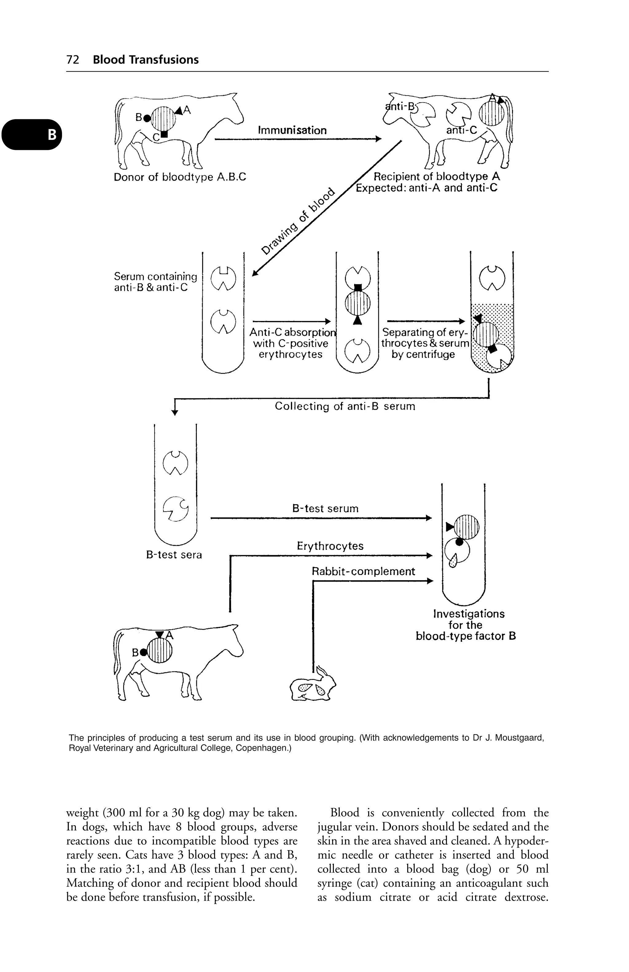
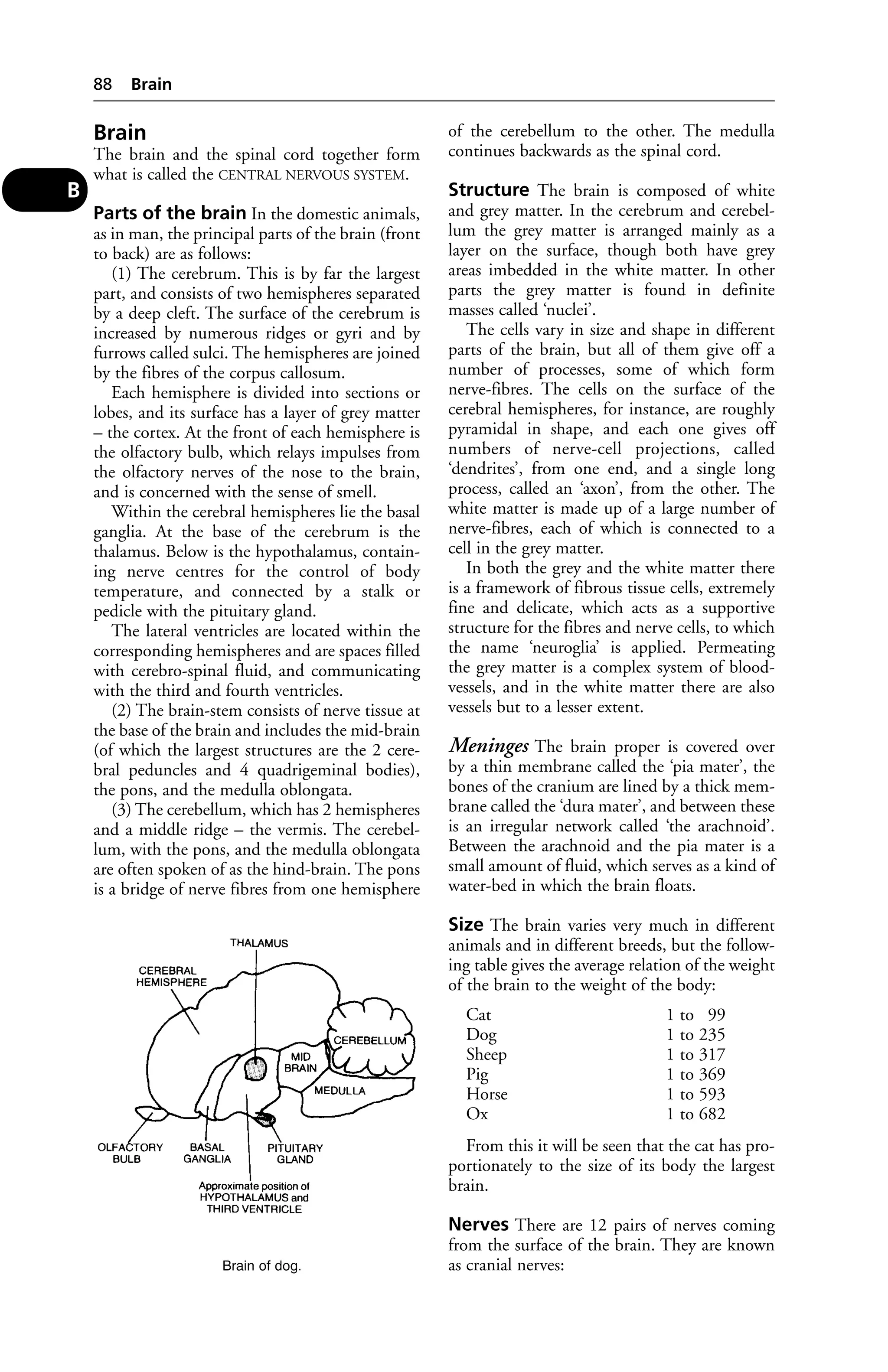
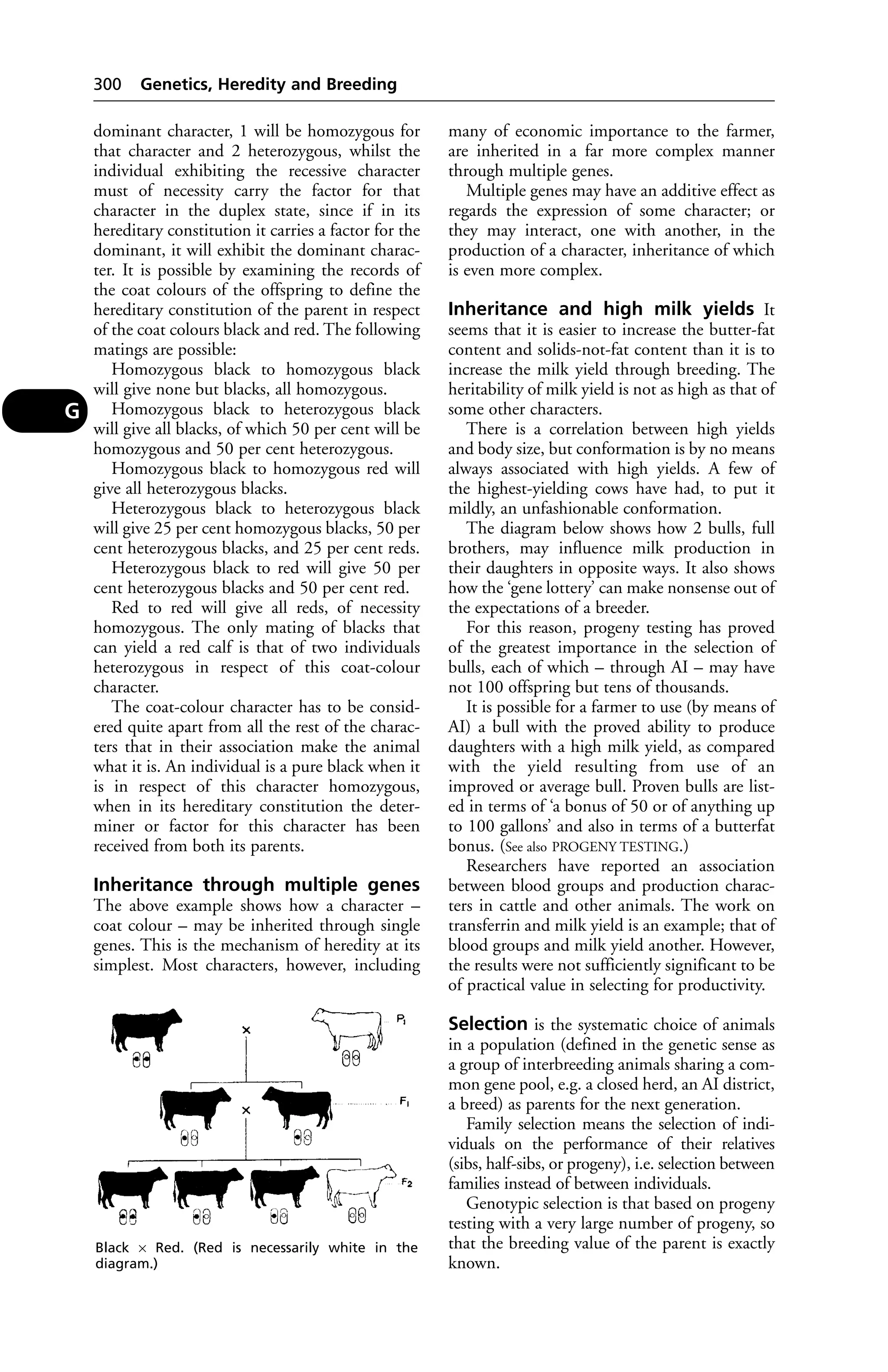
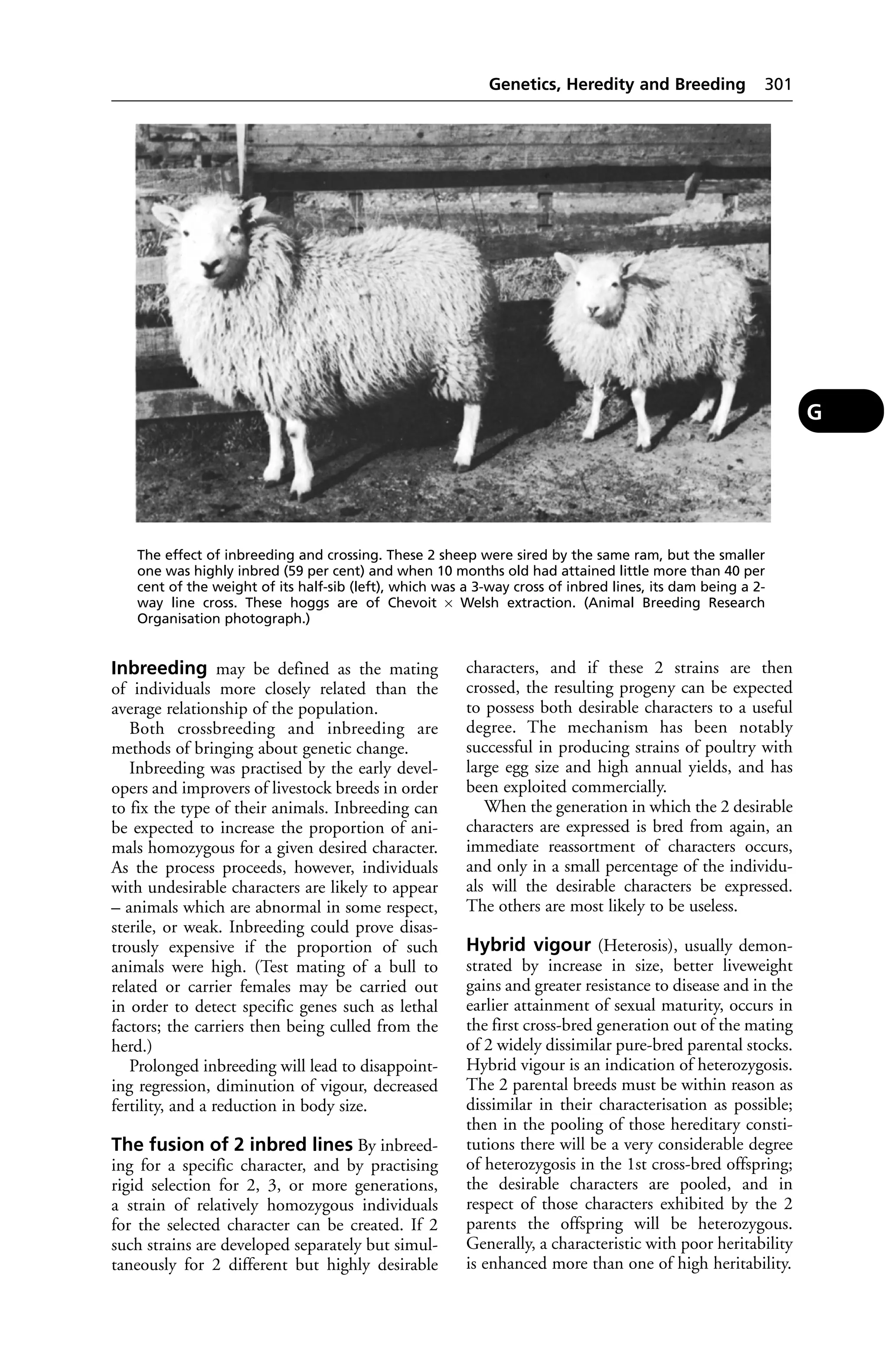
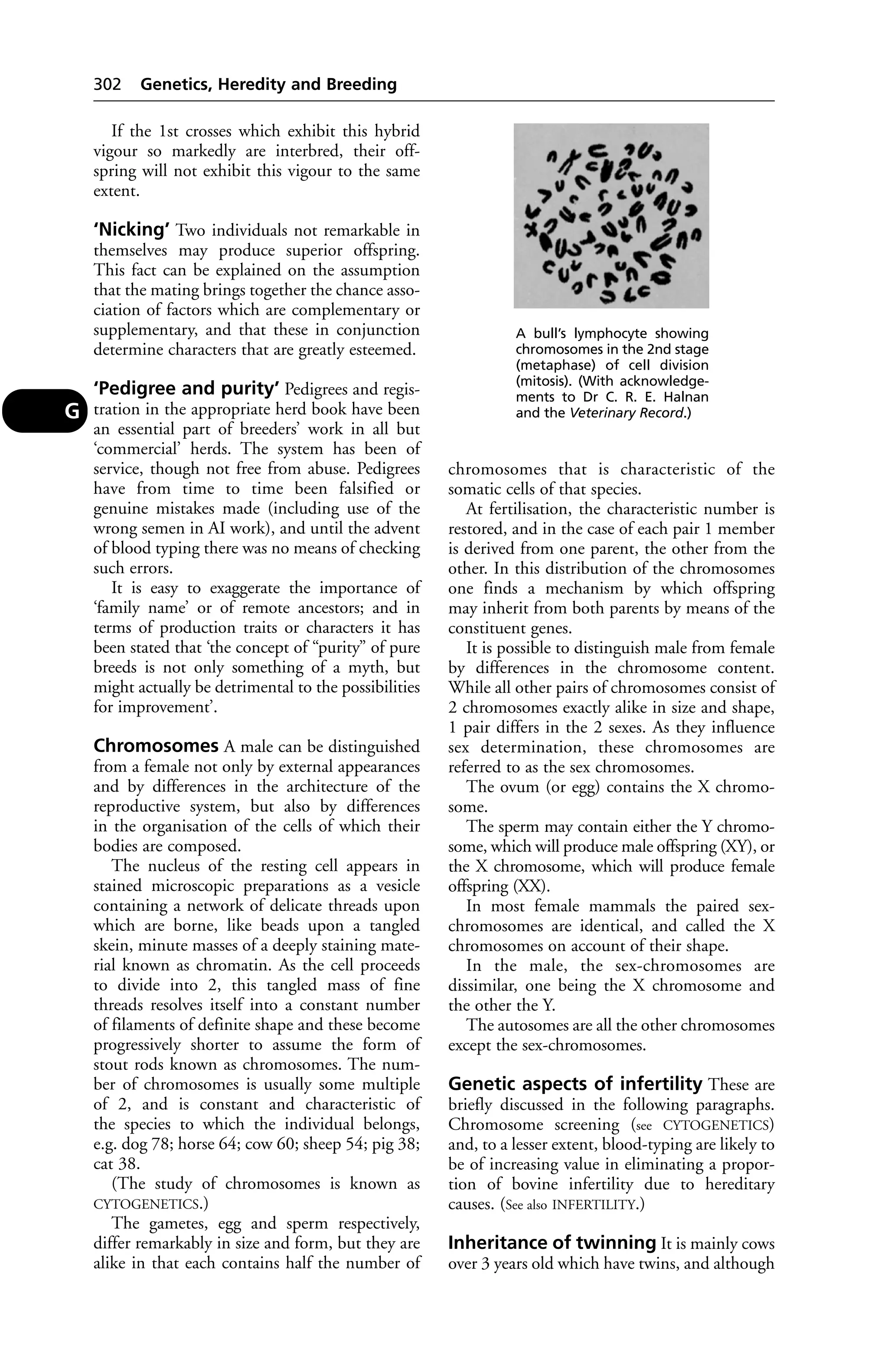
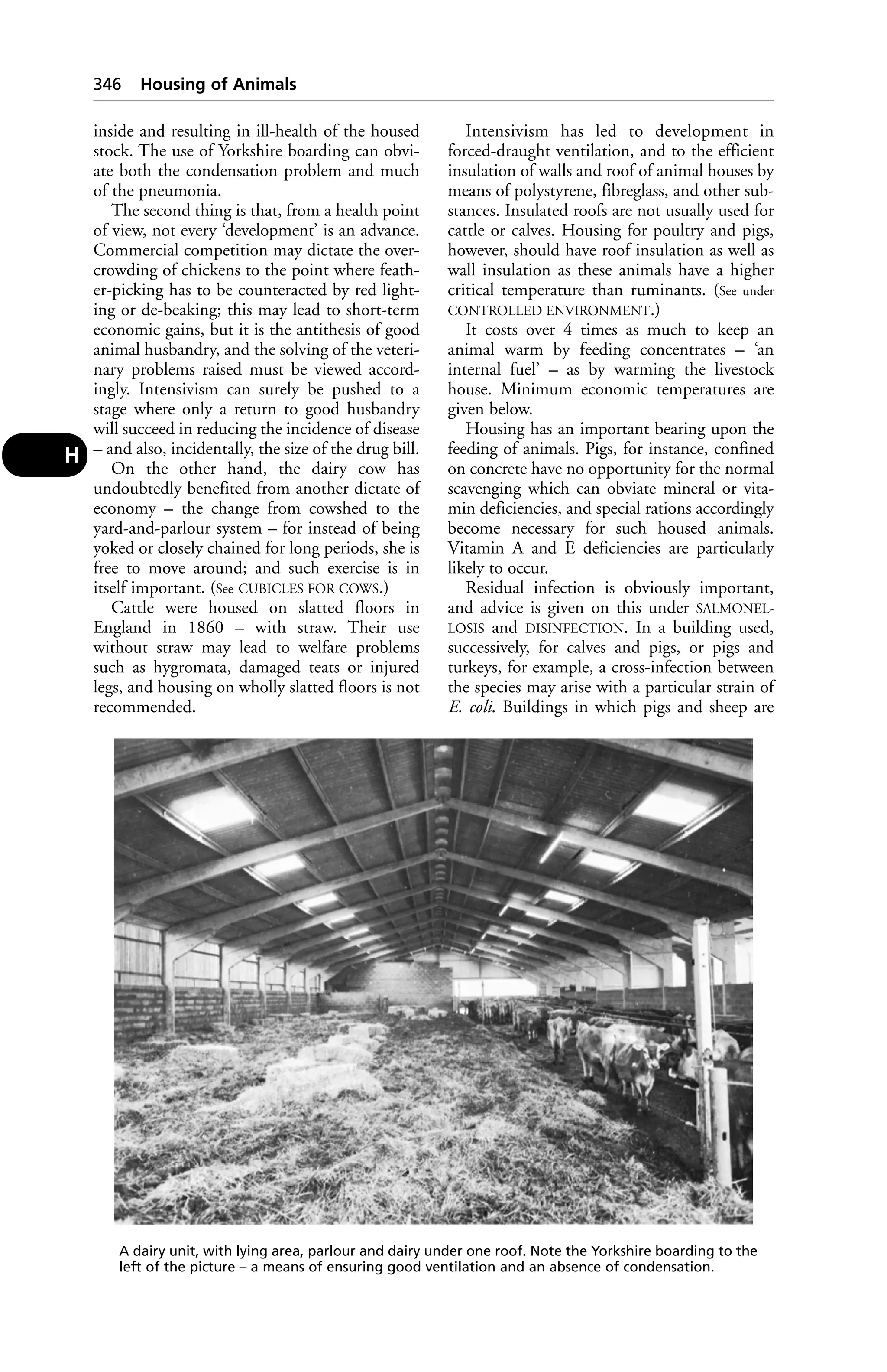
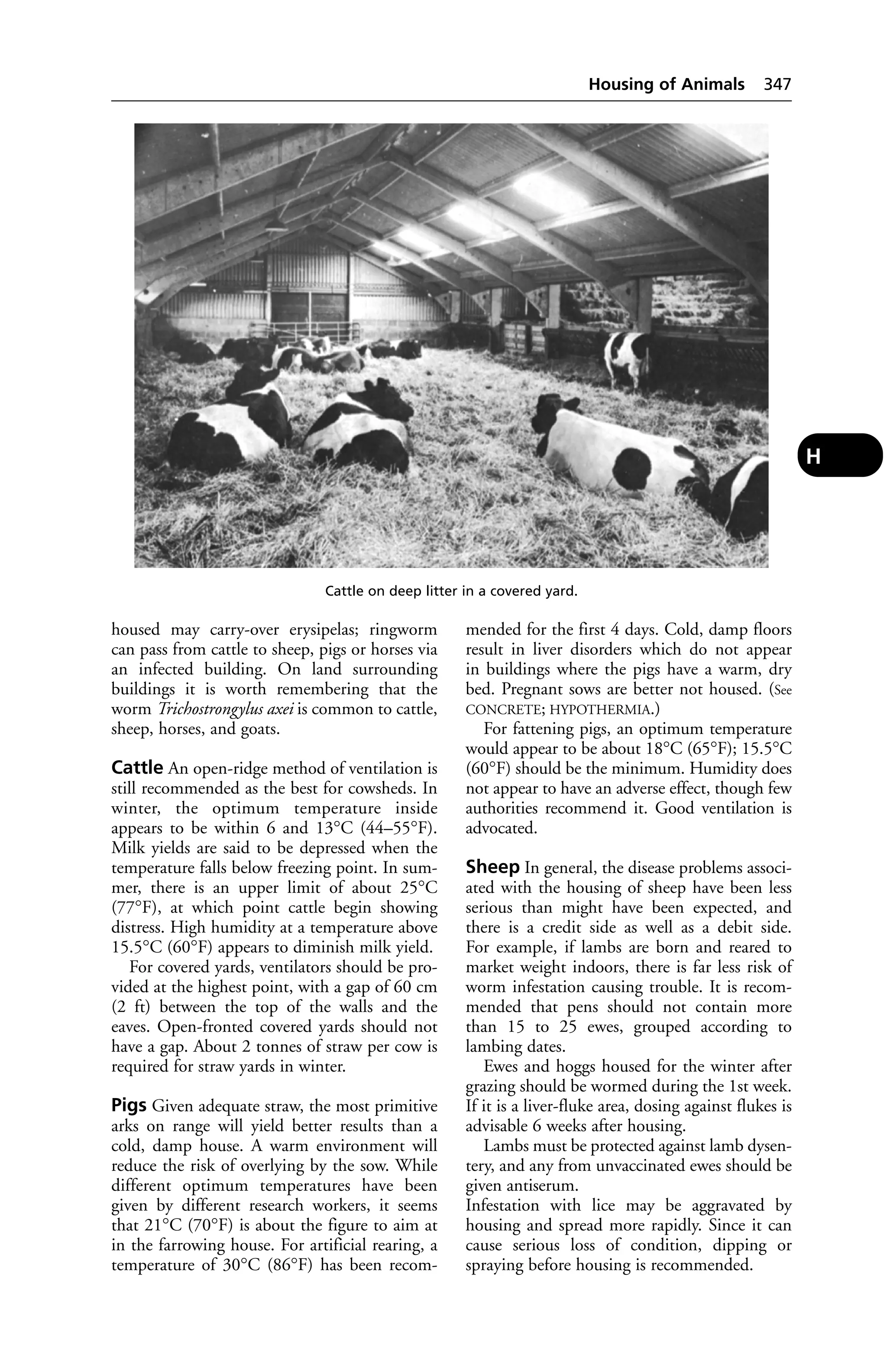
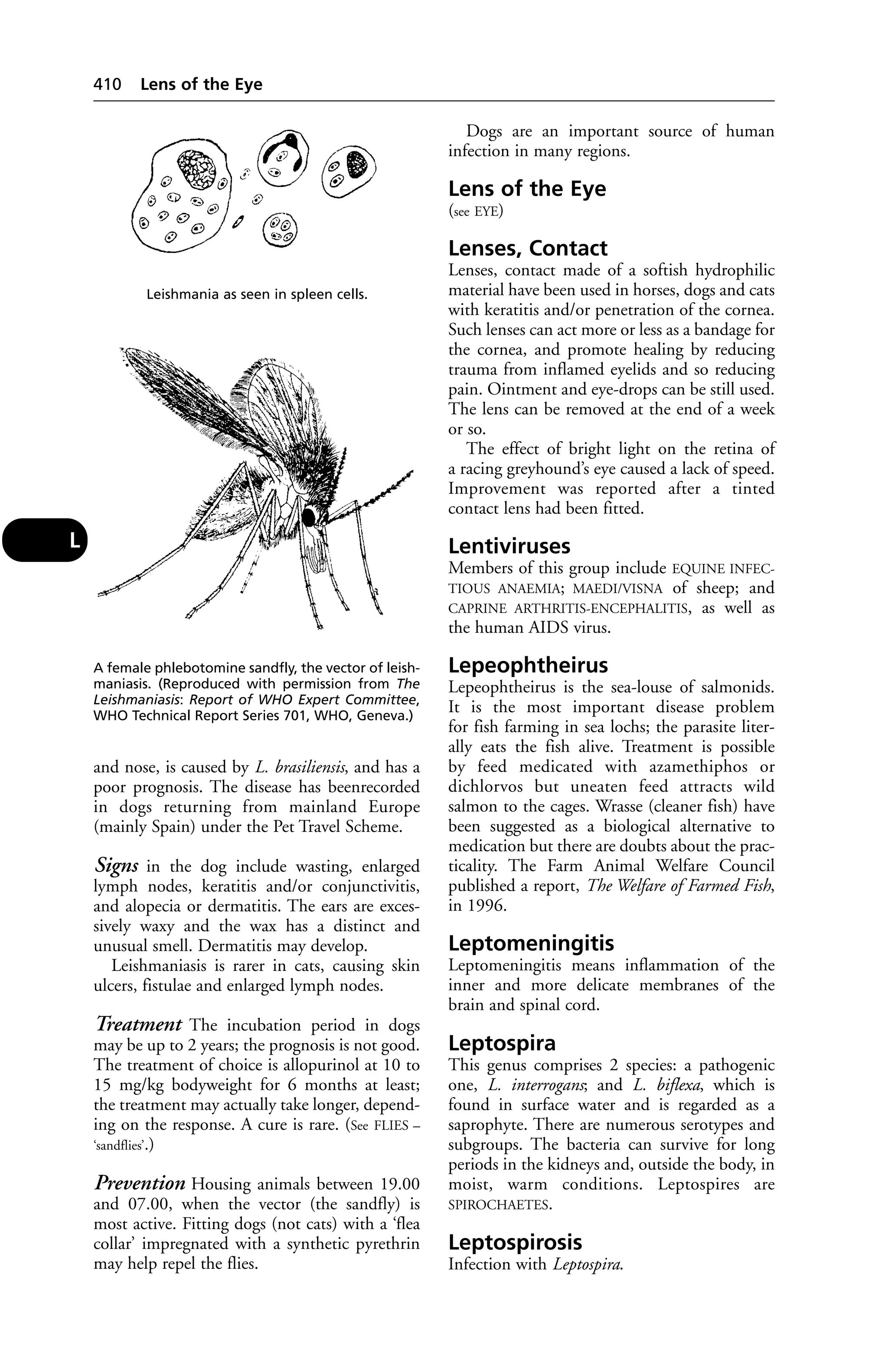
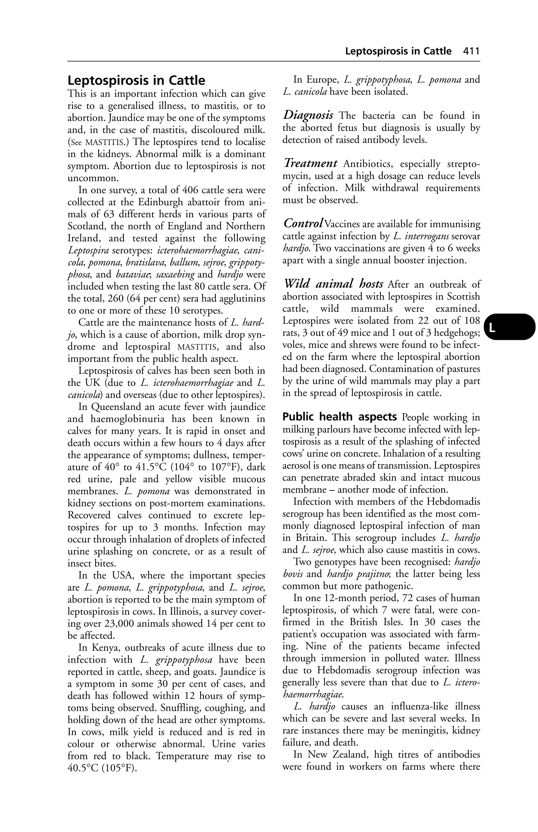
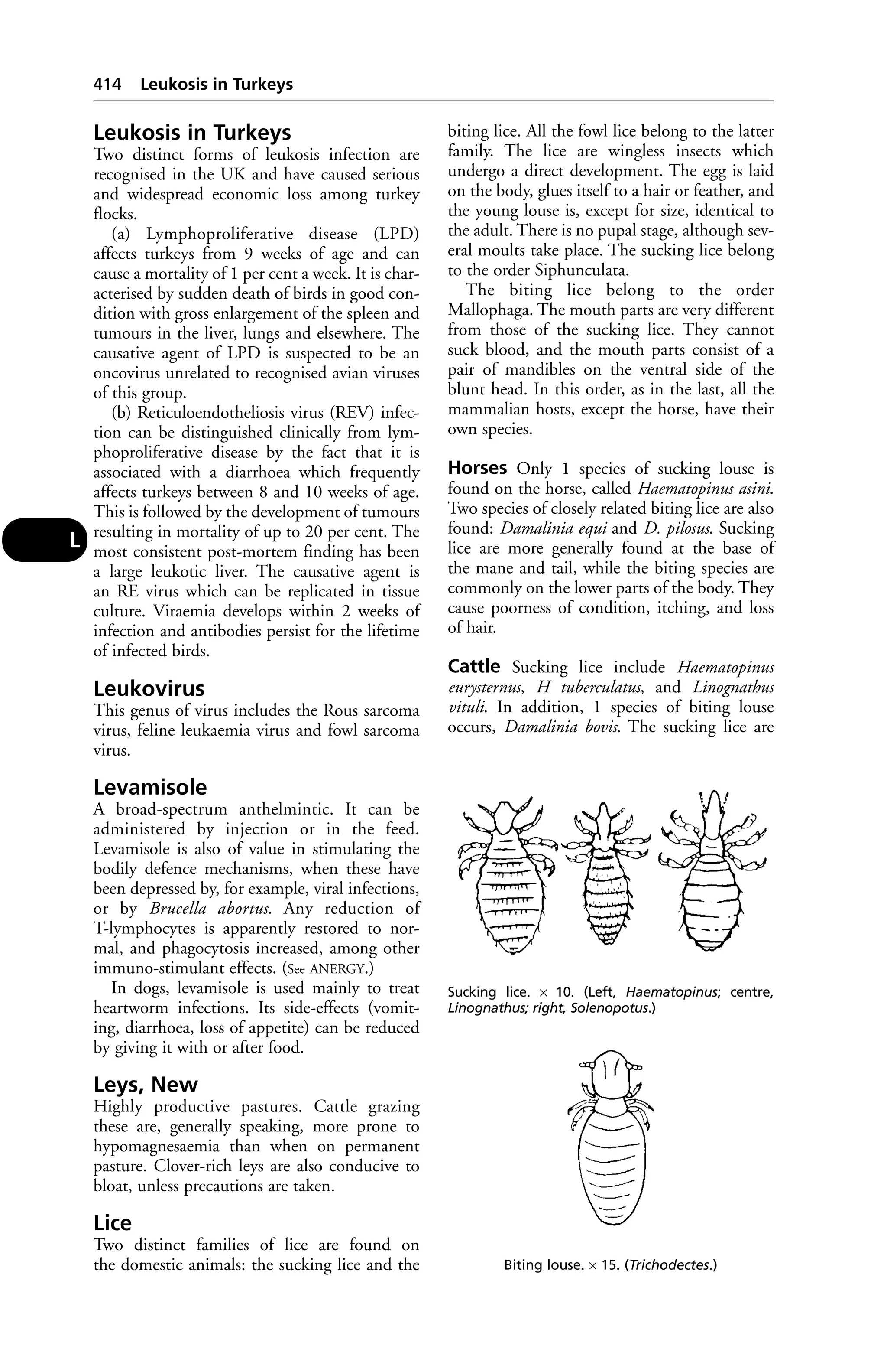
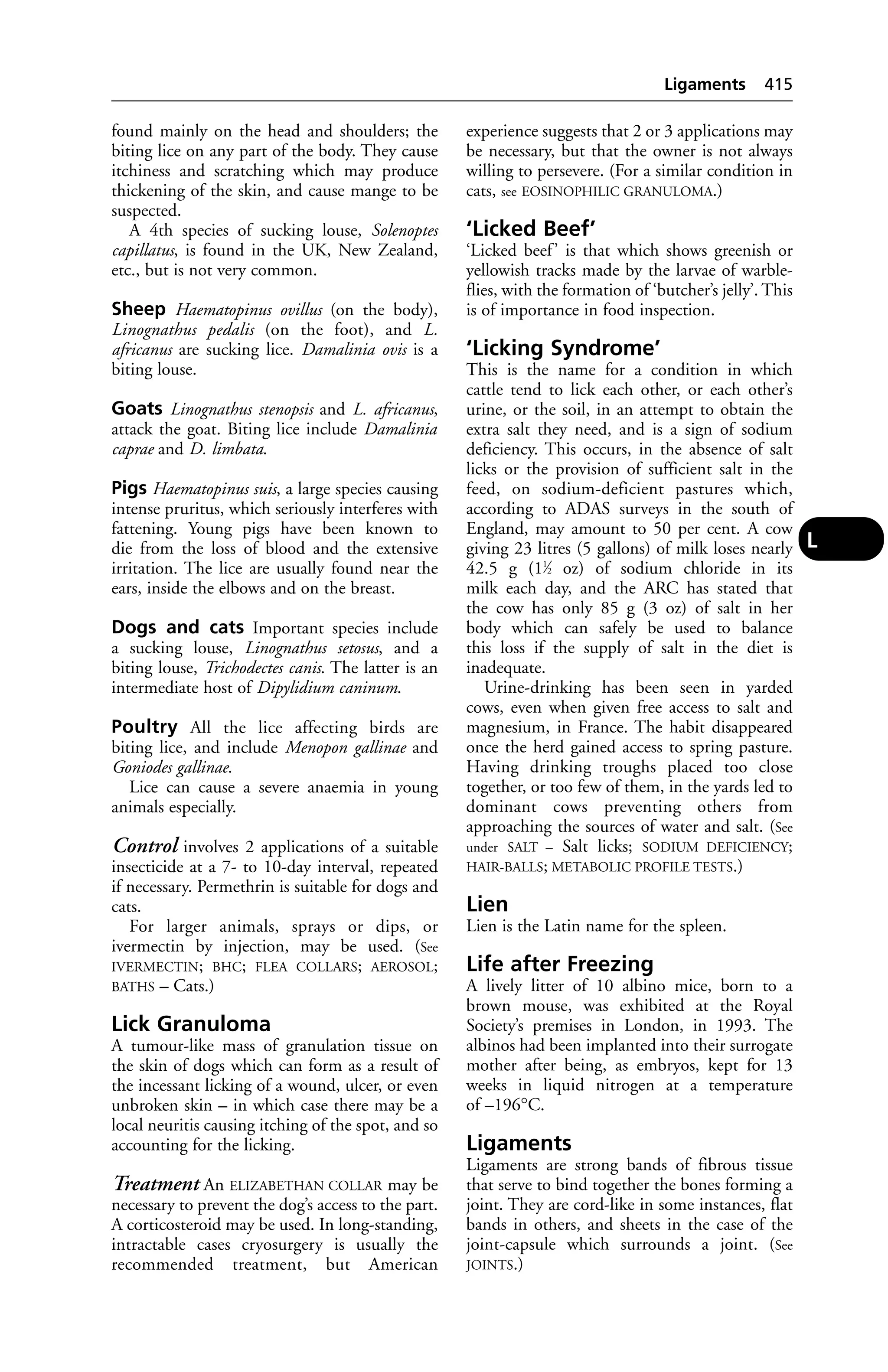
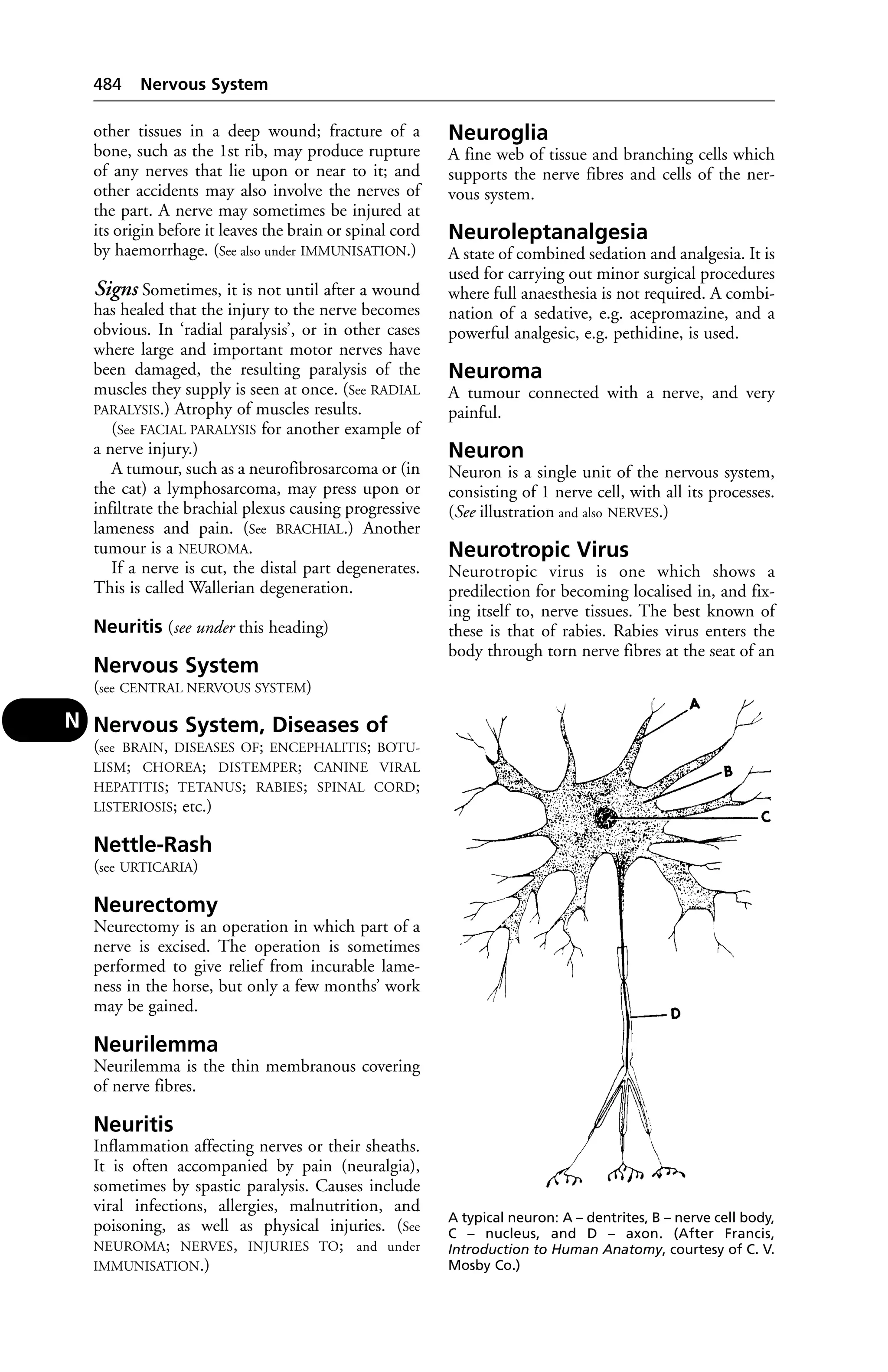
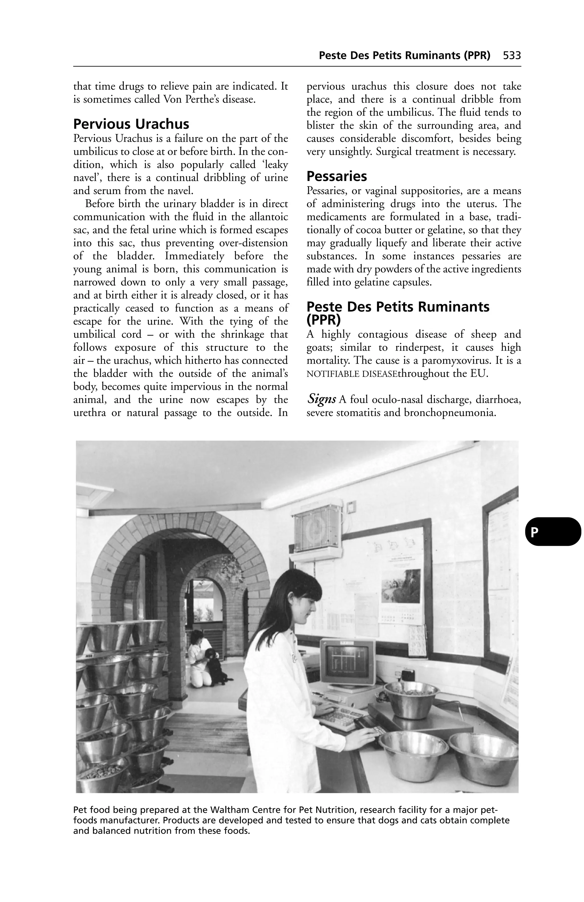
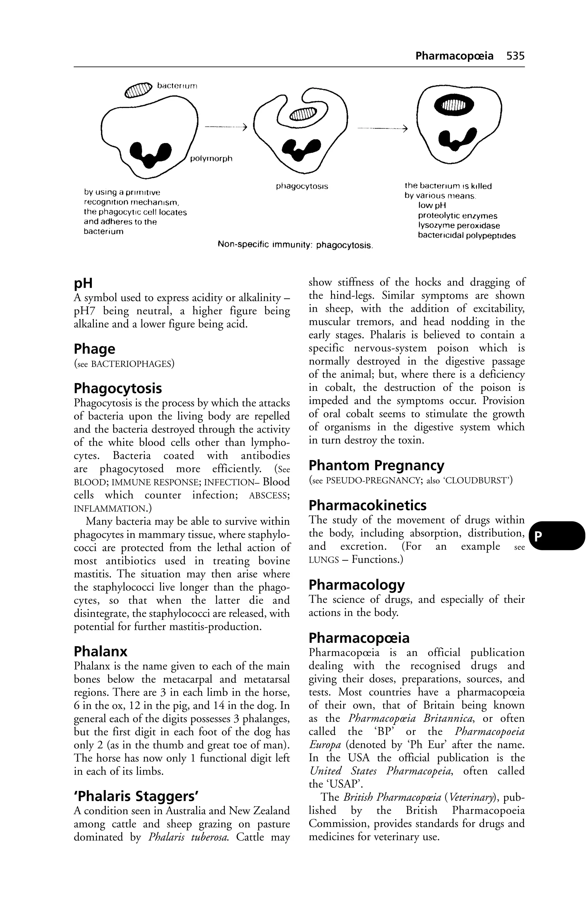
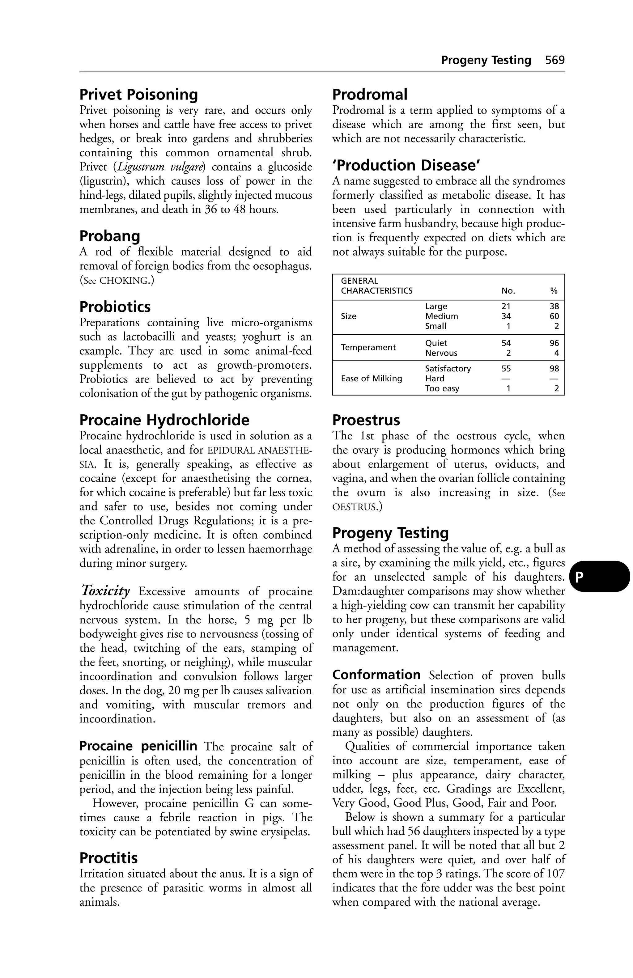
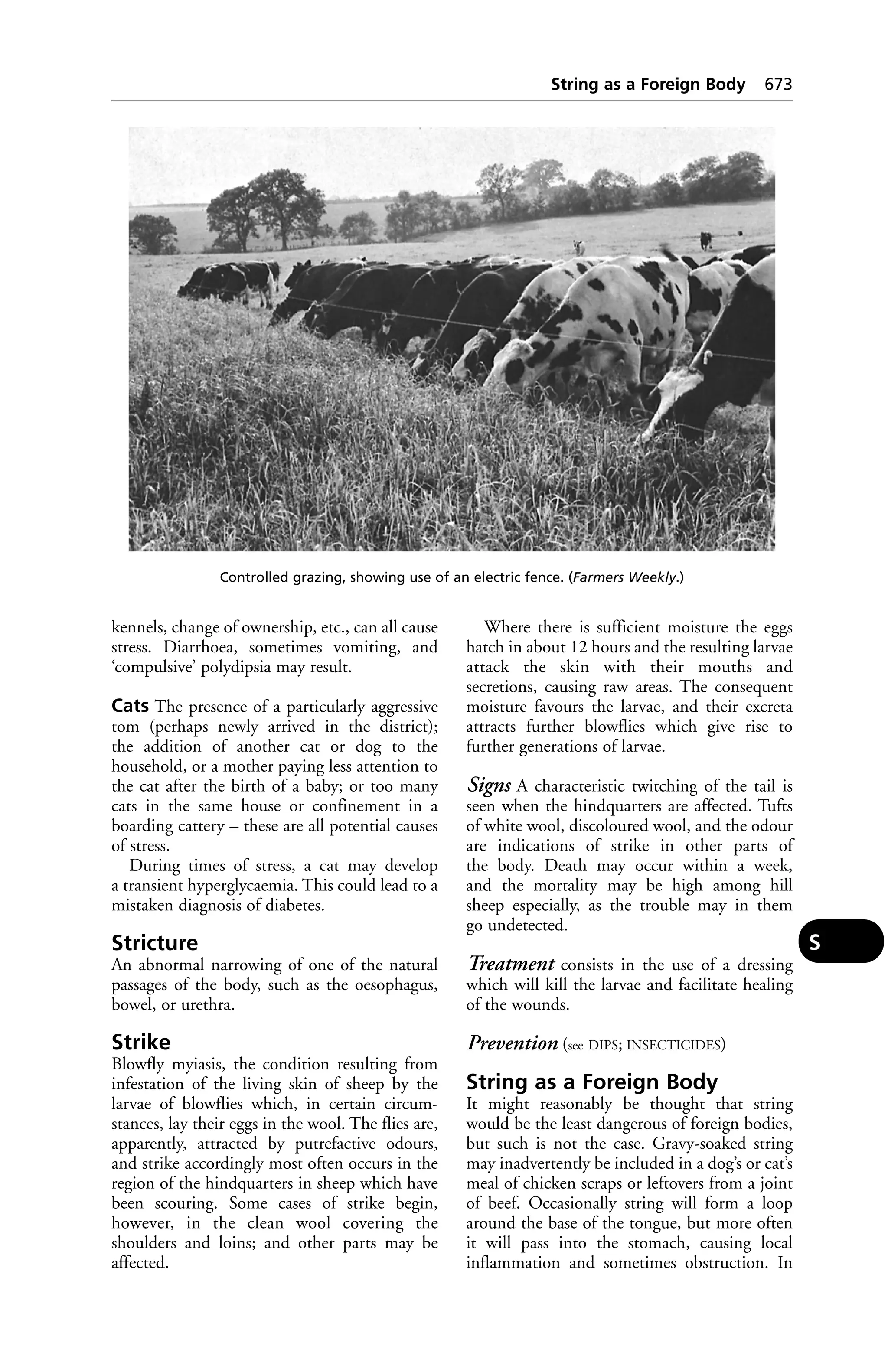
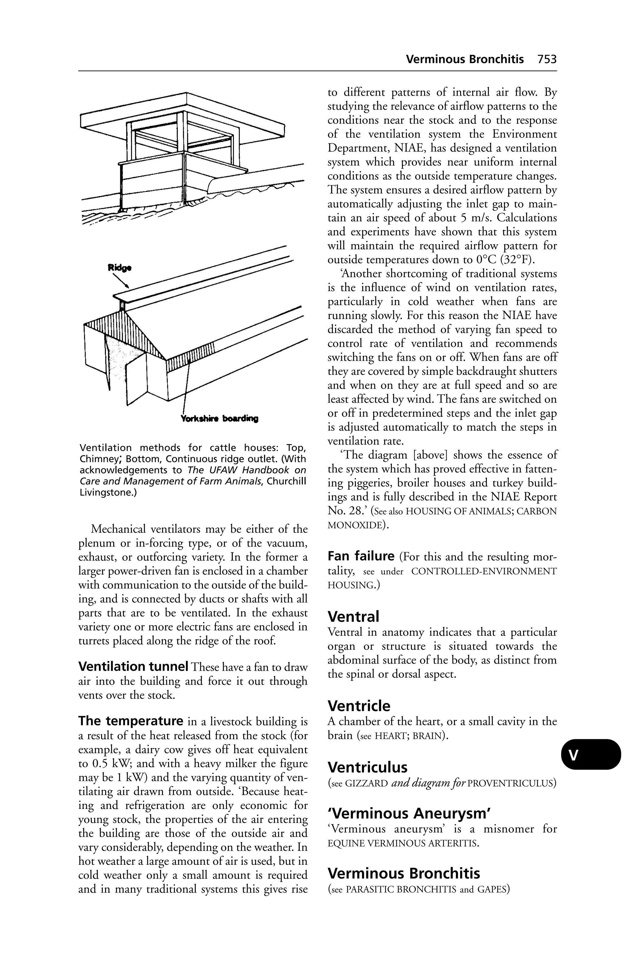
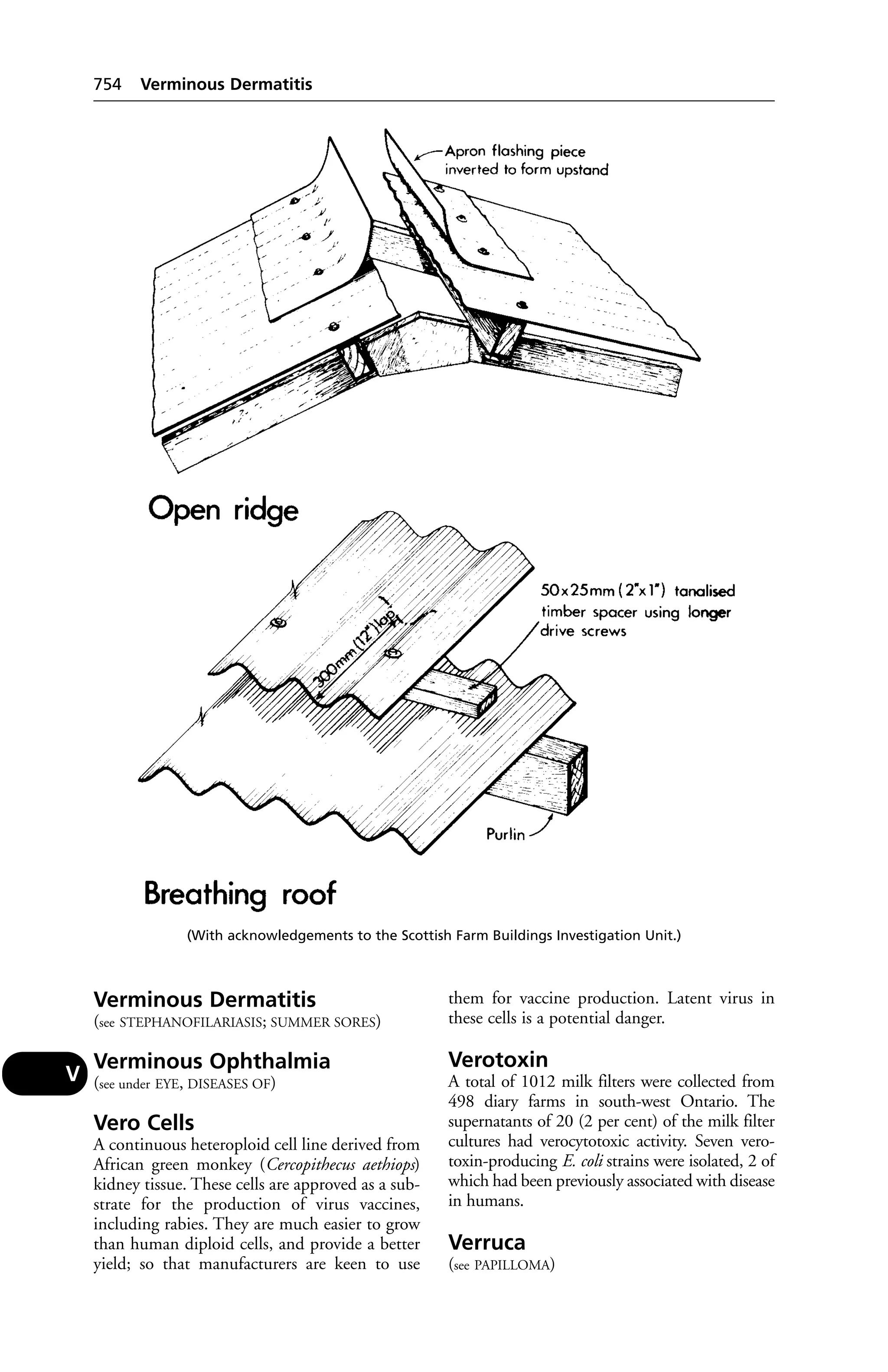
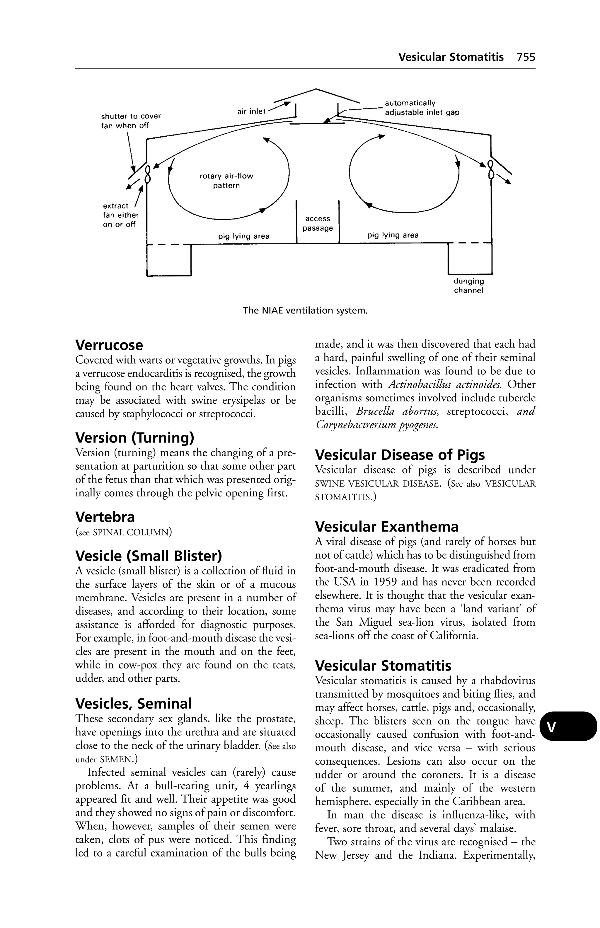
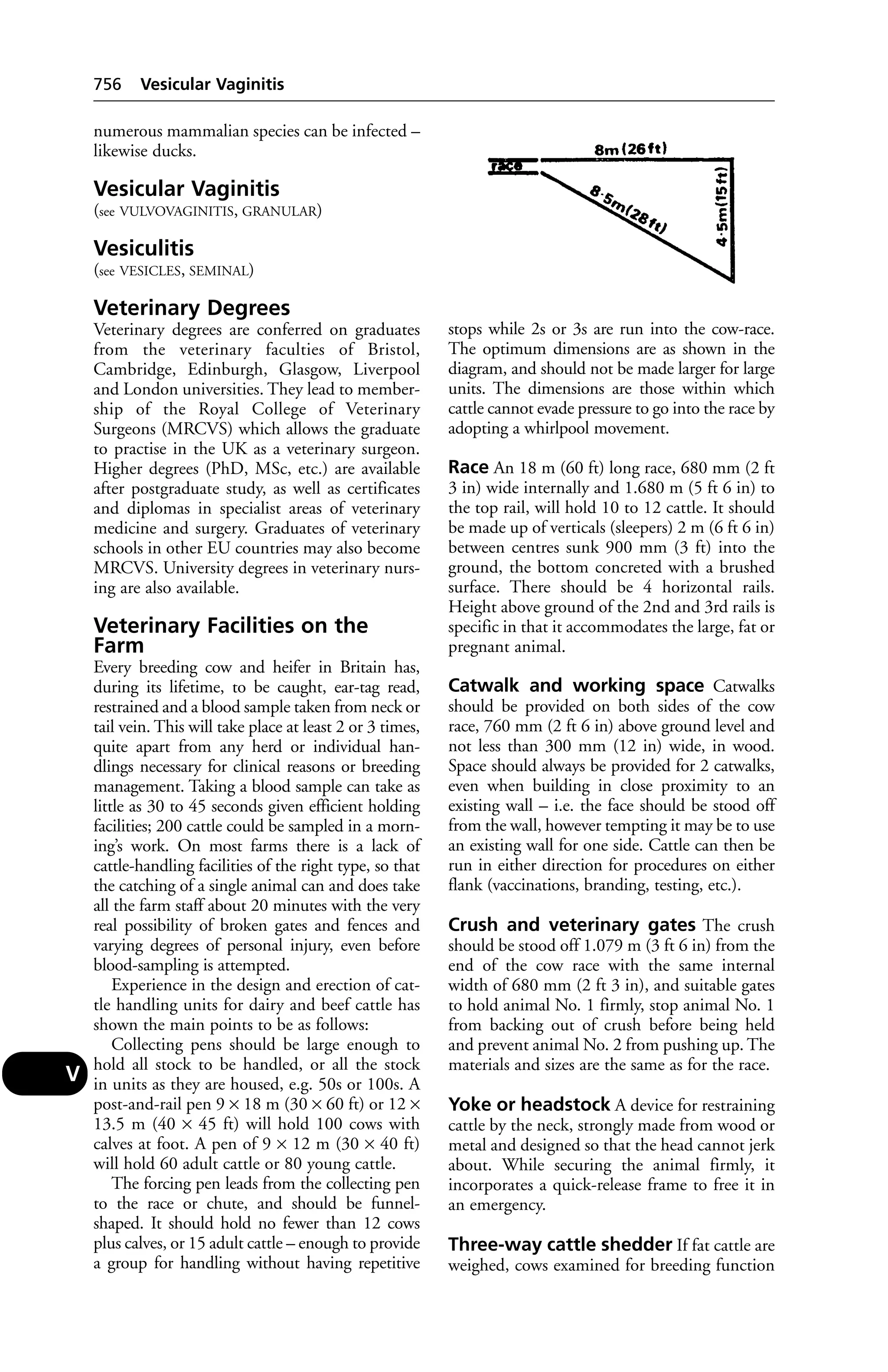
Downloaded 148 times


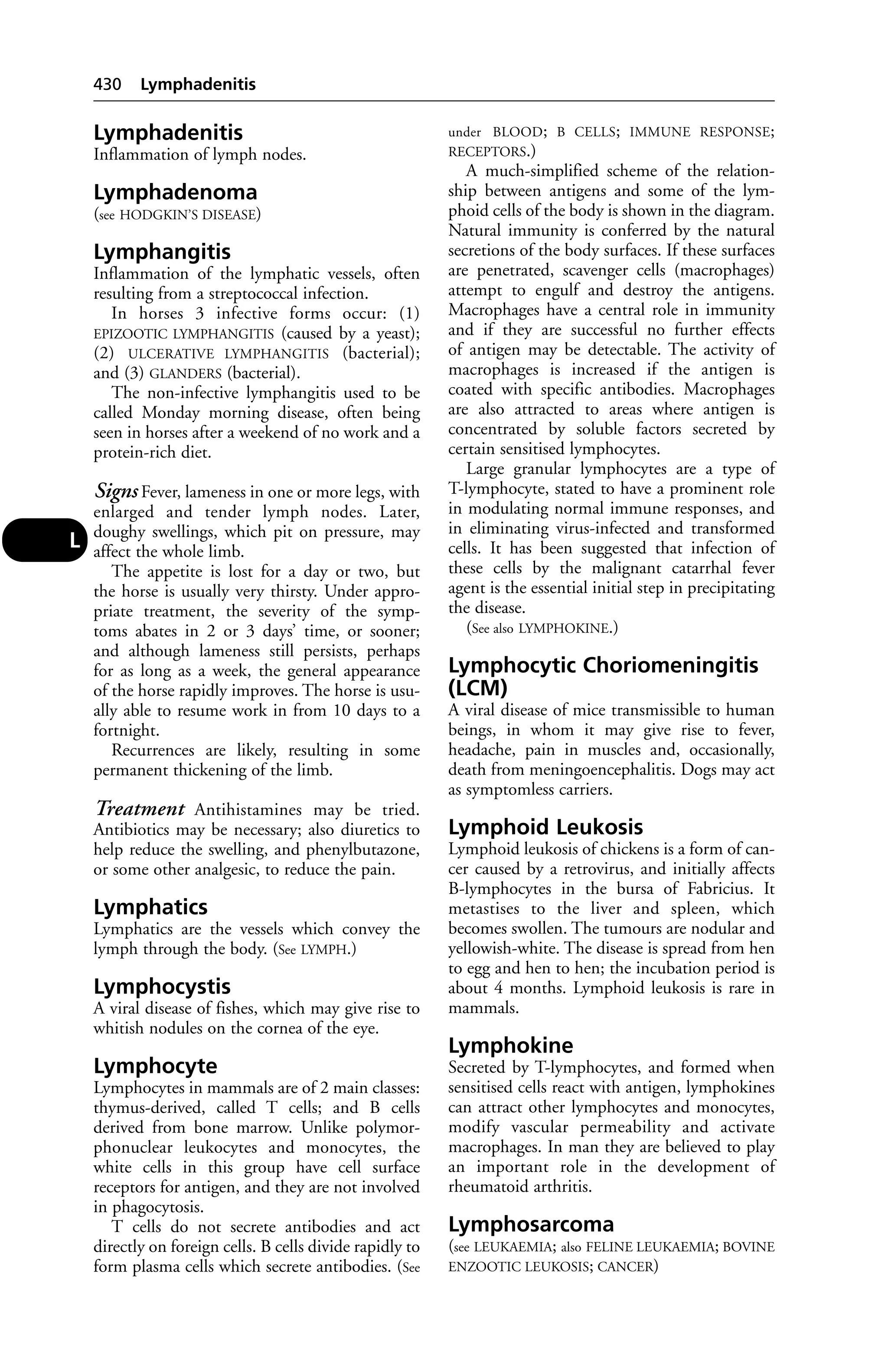
![blood. It is used as a measure of liver damage in
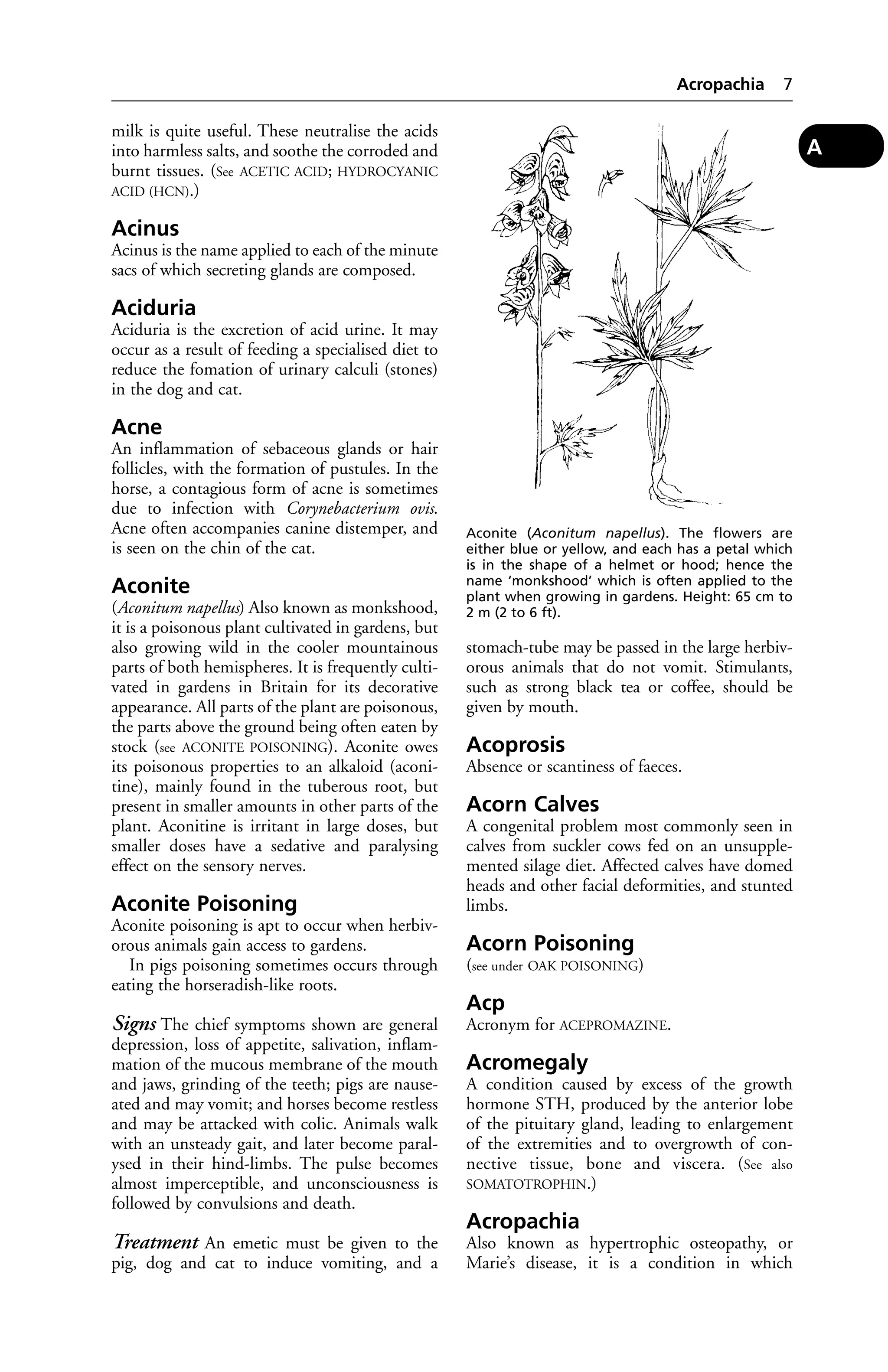
dogs and cats.
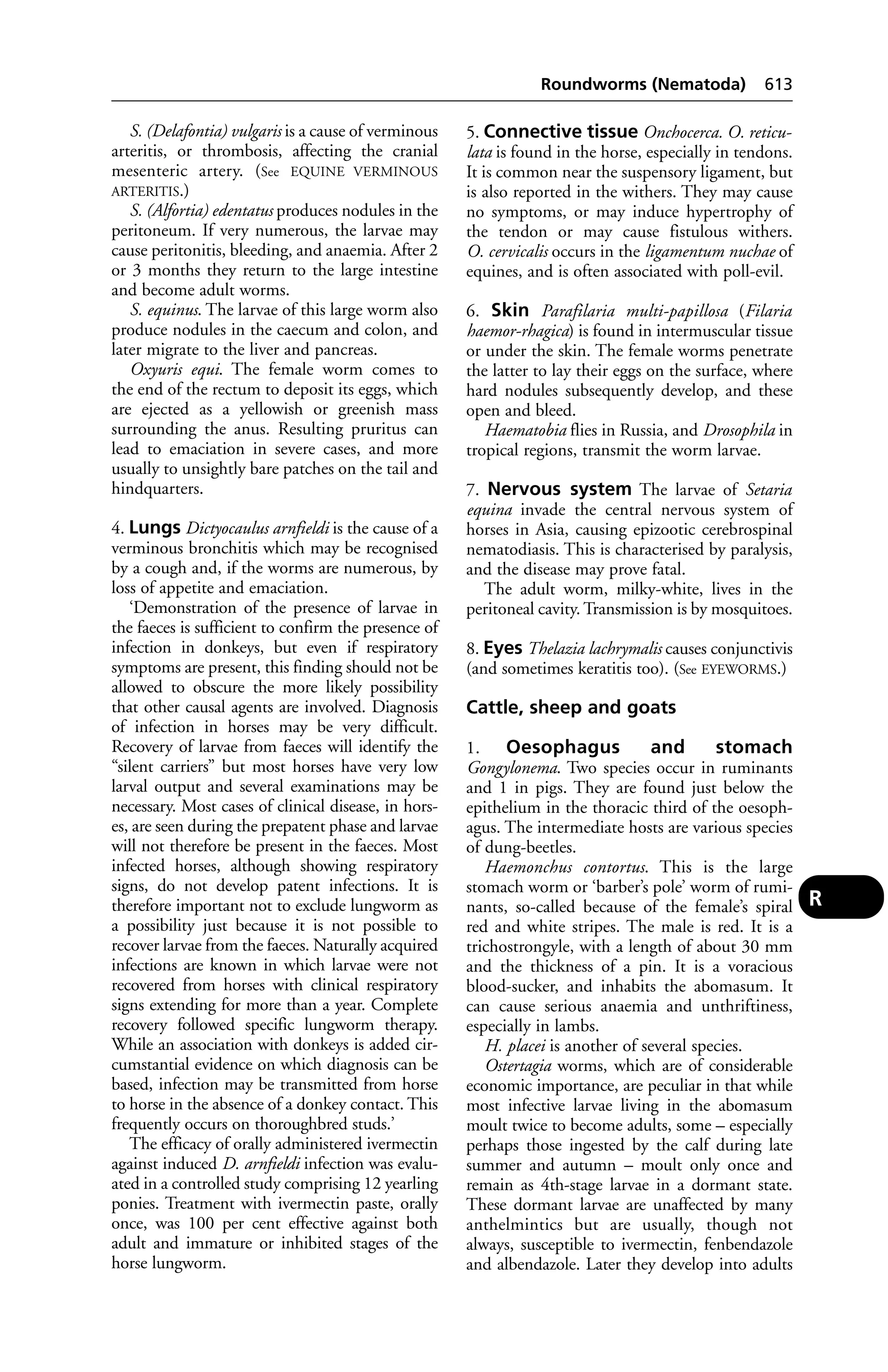
Alaskan Malamute
A breed of dog developed from the husky.
Dwarfism (chondrodysplasia) is inherited in
some litters. Day blindness may also be inherited
and congenital haemolytic anaemia occurs.
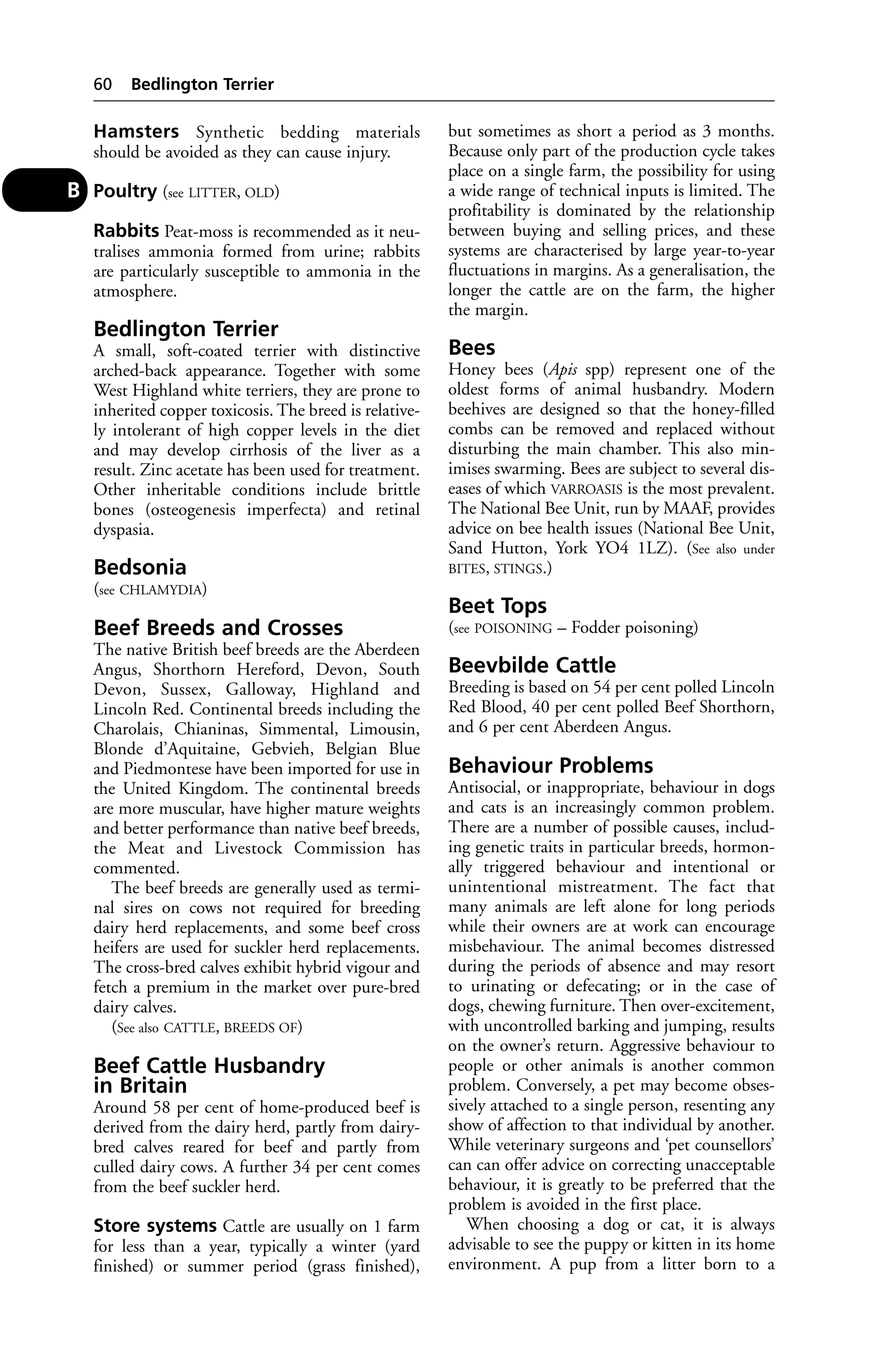
Albinism
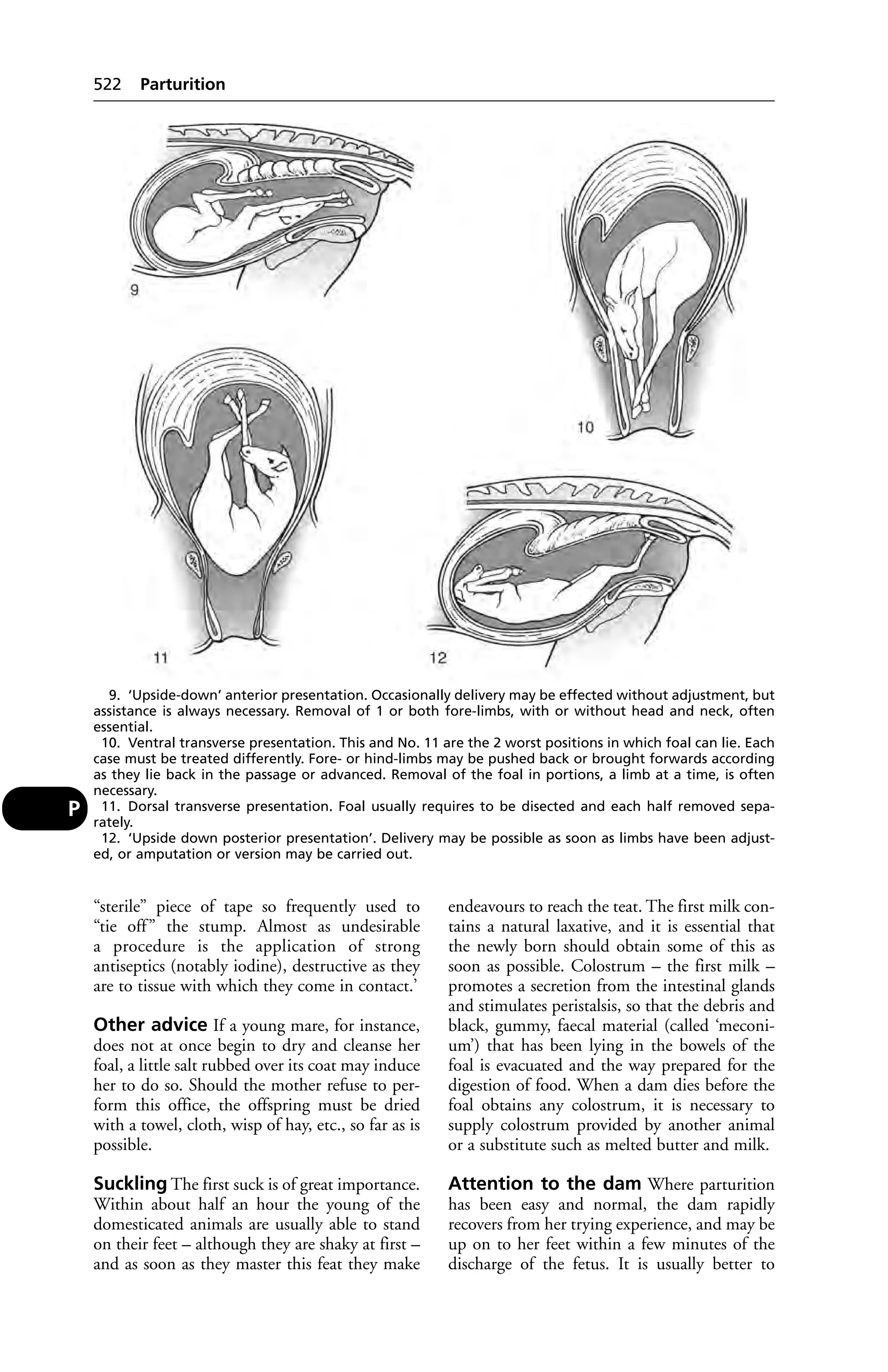
Albinism is a lack of the pigment melanin in
the skin – an inherited condition.
Albumins
(see PROTEINS; CONALBUMIN; ALBUMINURIA)
Albuminuria
The presence of albumin in the urine: one
of the earliest signs of NEPHRITIS and cystitis
(see URINARY BLADDER, DISEASES OF).
Alcohol Poisoning
Acute alcoholism is usually the result of too
large doses given bona fide, but occasionally the
larger herbivora and pigs eat fermenting wind-falls
in apple orchards; or are given or obtain,
fresh distillers’ grains, or other residue permeat-ed
with spirit, in such quantities that the ani-mals
become virtually drunk. In more serious
cases they may become comatose.
Aldosterone
This is a hormone secreted by the adrenal
gland. Aldosterone regulates the electrolyte
balance by increasing sodium retention and
potassium excretion. (See CORTICOSTEROIDS.)
Aldrin
A persistent insecticide; a chlorinated hydrocar-bon
used in agriculture and formerly in farm
animals. Its persistence has prevented its veteri-nary
use. Signs of toxicity include blindness,
salivation, convulsions, rapid breathing. (See
GAME BIRDS.)
Aleutian Disease
First described in 1956 in the USA, this disease
of mink also occurs in the UK, Denmark,
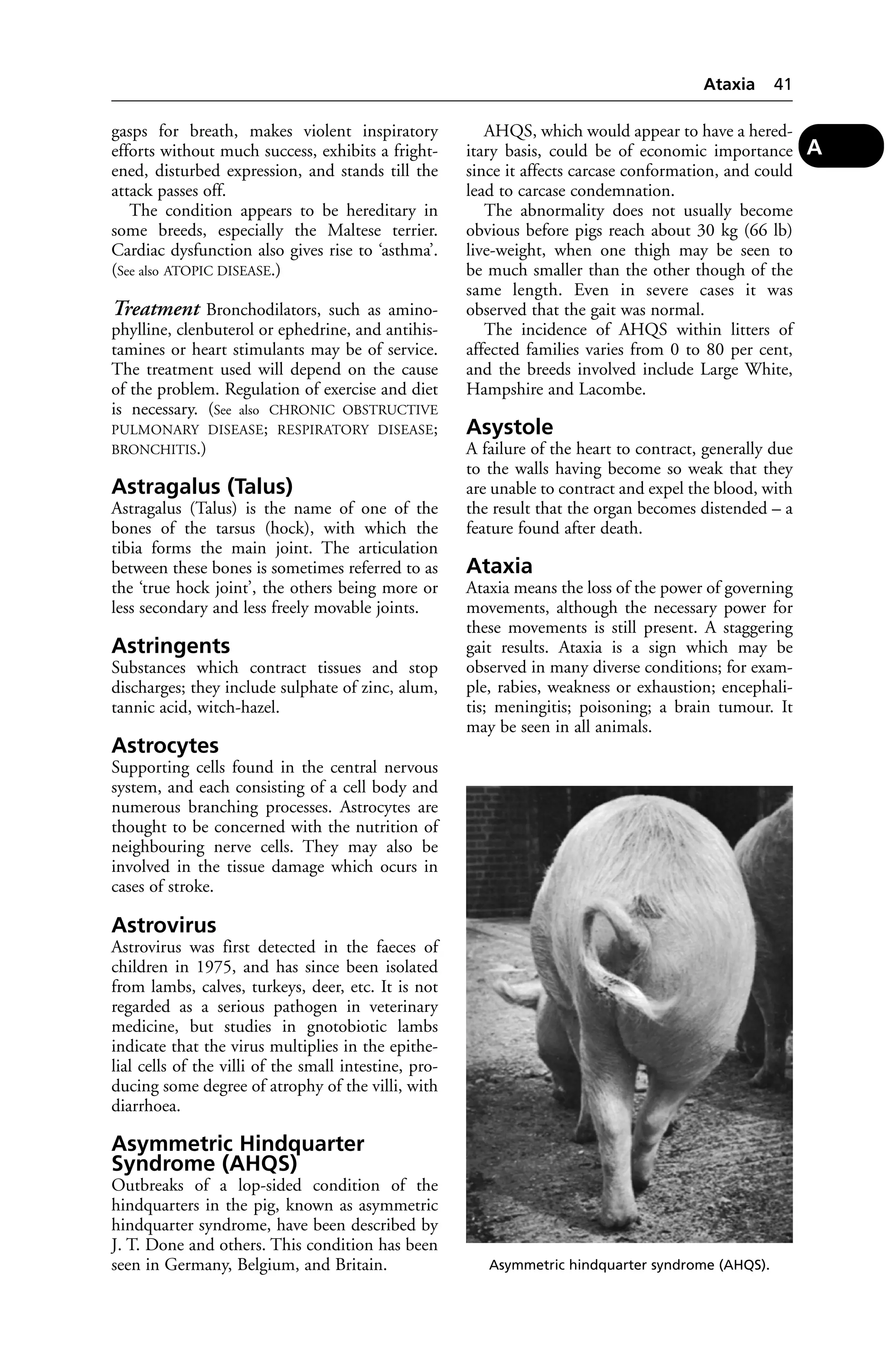
Sweden, New Zealand and Canada.
Mink
Signs include: failure to put on weight or even
loss of weight; thirst; the presence of undigested
food in the faeces – which may be tarry. Bleeding
from the mouth and anaemia may also be
observed. Death usually follows within a month.
Ferrets In these animals the disease is charac-terised
by a persistent viraemia.
Algae Poisoning 15
Signs include: loss of weight; malaise; chronic
respiratory infection; and paresis or paraplegia.
Bleeding from the mouth and anaemia may
also be observed. Death usually follows within
a month. The disease can be confused with the
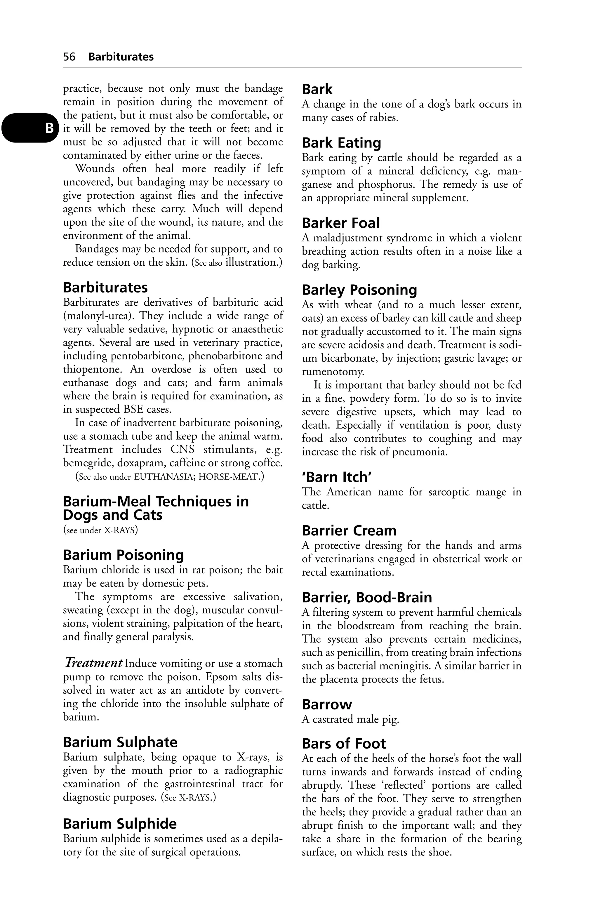
later stages of rabies.
Diagnosis In ferrets the counter-current
electrophoresis test has been used.
Alexin
(see COMPLEMENT)
Alfadalone
(see ALFAXALONE)
Alfaxalone
Used in combination with alfadalone (in Saffan
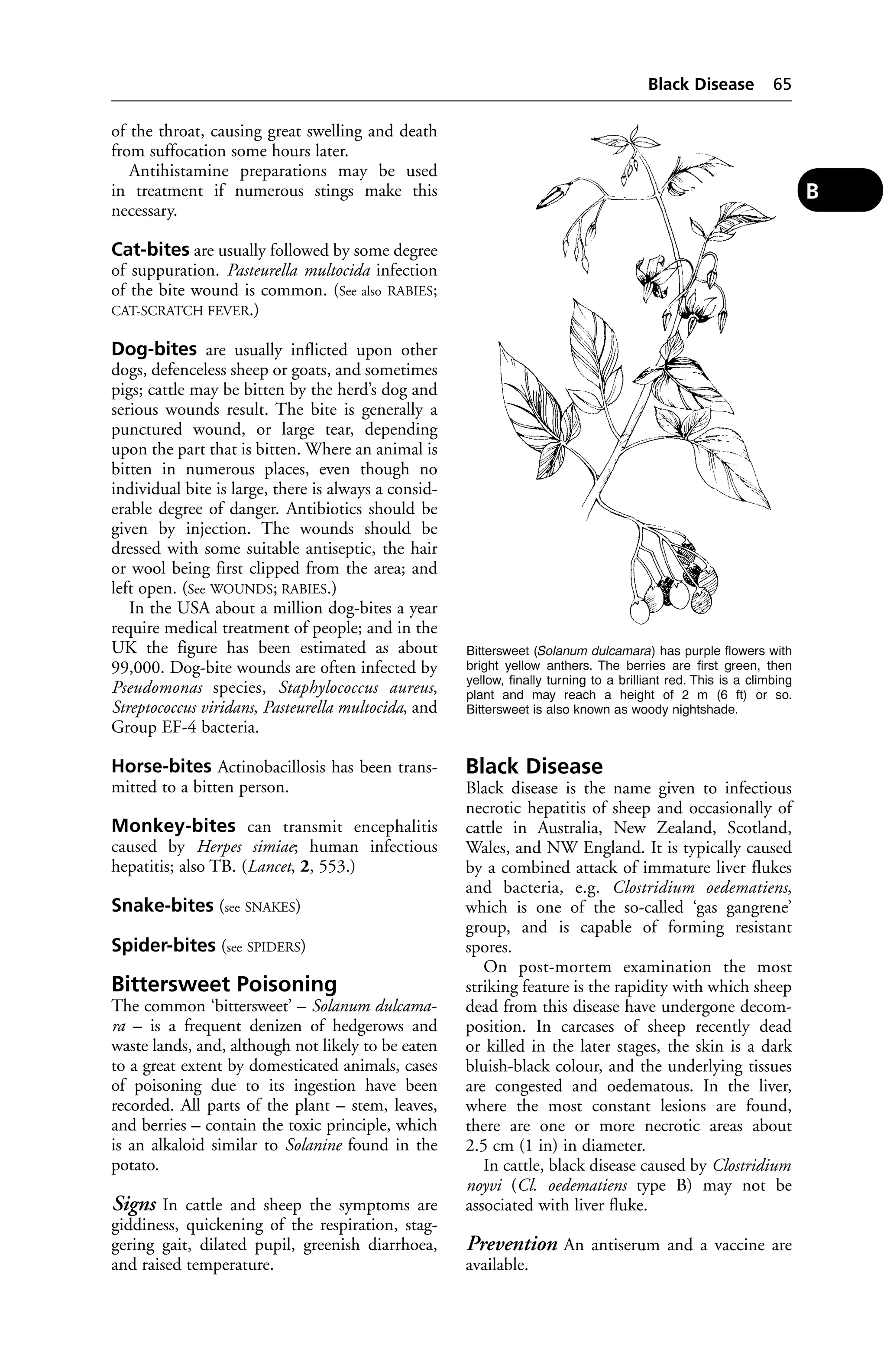
[Schering-Plough]) as a general anaesthetic in
cats; it must not be used in dogs. Given by
intravenous injection, It produces sedation in
9 seconds and anaesthesia after 25 seconds. It is
also given by deep intramuscular injection as an
induction for general anasthesia for long opera-tions.
It must not be given with other injectable
anaesthetics.
Algae
Simple plant life of very varied form and size,
ranging from single-cell organisms upwards to
large seaweed structures. Algae can be a nui-sance
on farms when they block pipes or clog
nipple drinkers. This happens especially in
warm buildings, where either an antibiotic
or sugar is being administered to poultry via
the drinking water. Filters may also become
blocked by algae.
The colourless Prototheca species are patho-genic
for both animals (cattle, deer, dogs,
pigs) and man. (See MASTITIS IN COWS – Algal
mastitis.)
The non-toxic algae of the Spirulina group
are used in the feed of some ornamental fish.
Algae Poisoning
Toxic freshwater algae, characteristically blue-green
in colour, are found in summer on lakes
in numerous locations, particularly where water
has a high phosphate and nitrate content
derived from farm land. Formed by the summer
blooms of cyanobacteria, they can form an oily,
paint-like layer several cm thick. Deaths have
occurred in cattle and sheep drinking from
affected water; photosensitivity is a common
sign among survivors. Dogs have also been
affected.
The main toxic freshwater cyanobacteria are
strains of the unicellular Microcystis aeruginosa,
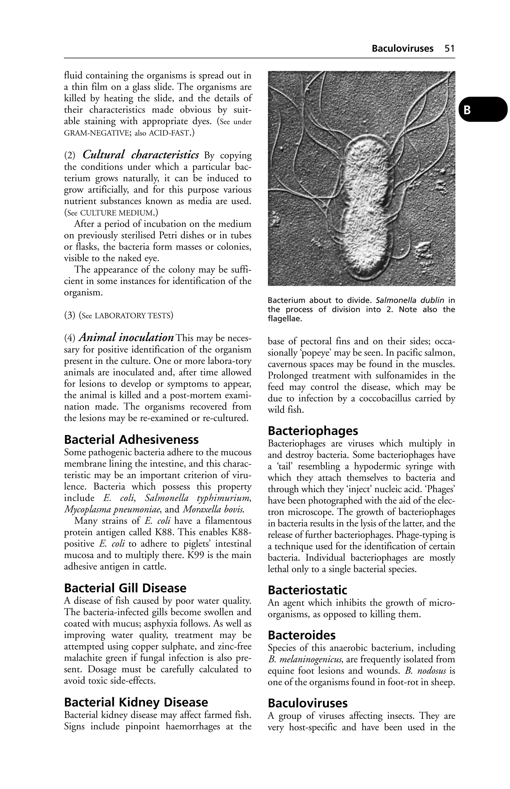
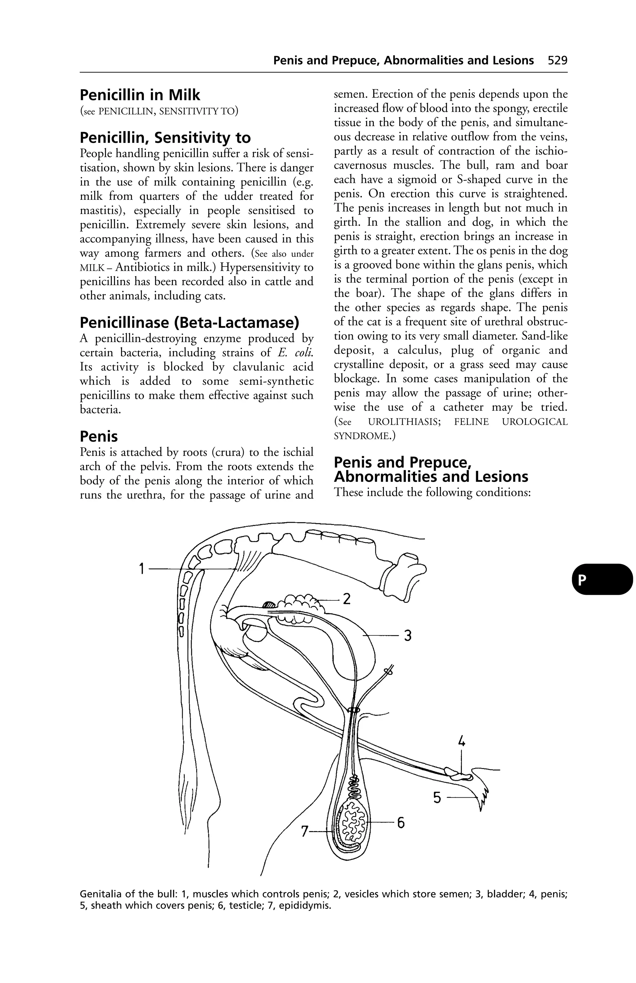
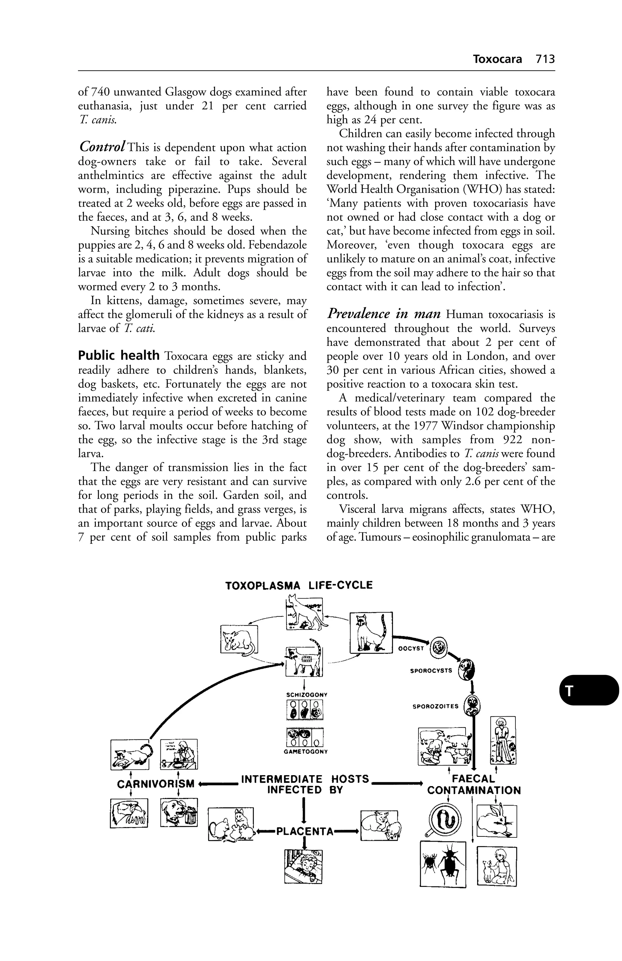
A](https://image.slidesharecdn.com/veterinarydictionary21stedition-140828090128-phpapp02/75/Veterinary-dictionary-_21st_edition-24-2048.jpg)


![by harness or other tackle from keeping their
nostrils above the level of the water; or they may
become panic-stricken and swim away from
shore. Remarkable instances of the powers of
swimming that are naturally possessed by ani-mals
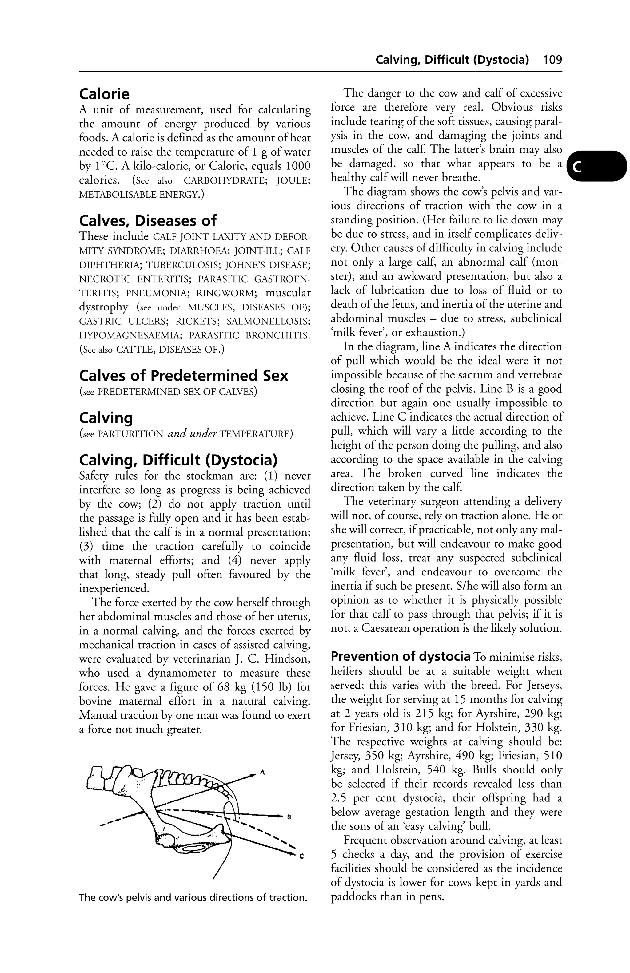
are on record; one example being that of a
heifer, which, becoming excited and frightened
on the southern banks of the Solway Firth,
entered the water and swam across to the
Scottish side, a distance of over 7 miles, and was
brought back the next day none the worse.
Recovery from drowning As soon as the
animal has been rescued from the water, it
should be placed in a position which will allow
water that has been taken into the lungs to run
out by the mouth and nostrils. Small animals
may be held up by the hind-legs and swung
from side to side. Larger ones should be laid on
their sides with the hindquarters elevated at a
higher level than their heads. If they can be
placed with their heads downhill, so much
the better. Pressure should be brought to bear
on the chest, by one person placing all their
weight on to the upper part of the chest wall, or
kneeling on this part. When no more fluid runs
from the mouth, the animal should be turned
over on to the opposite side and the process
repeated. No time should be lost in so doing,
especially if the animal has been in the water for
some time. (See ARTIFICIAL RESPIRATION.)
After-treatment As soon as possible the ani-mal
should be removed to warm surroundings
and dried by wiping or by vigorous rubbing
with a rough towel. Clothing should be applied,
and the smaller animals may be provided with
1 or more hot-water bottles. The danger that has
to be kept in mind is that of pneumonia, either
from the water in the lungs or from the general
chilling of the body, and the chest should be
especially well covered. Sometimes the ingestion
of salt water leads to salt poisoning in dogs, or to
a disturbance of the digestive functions, and
appropriate treatment is necessary.
Drug Interactions
For those in which one drug enhances the
action of another, see SYNERGISM.
Adverse drug interactions or reactions are
indicated by manufacturers in the product data
sheet. Unexpected adverse reactions should be
reported to the manufacturer or the Veterinary
Medicines Directorate.
Drug Residues in Food
Drug residues in food are regarded as very
important from the point of view of public
Dubbing 205
health. The permitted maximum level of drugs
remaining in meat, milk or eggs after medicines
have been administered (maximum residue limit
[MRL]) is specified by regulation for all EU
countries. The manufacturer’s recommended
withdrawal period between the last dose of
drug administered and the animal going for
slaughter, or the milk or eggs being sold for
human consumption, must be observed.
Carcases in abattoirs are monitored to ensure
that the residues are within allowable limits.
(See also HORMONES IN MEAT PRODUCTION;
MILK – Antibiotics in; SLAUGHTER.)
Drug Resistance
(see under ANTIBIOTIC RESISTANCE; DIPPING; FLY
CONTROL)
Drugs, Disease Caused by
(see IATROGENIC DISEASE)
Dry Eye
(see EYE, DISEASES OF)
Dry Feeding
Dry feeding of meal may give rise to PARAKER-ATOSIS
in pigs; to ‘CURLED TONGUE’ in turkey
poults; and to ‘SHOVEL BEAK’ in chicks.
Dry, Firm and Dark (DFD)
Dry, firm and dark (DFD) describes the meat
of animals that have undergone stress in trans-port
before slaughter. The condition is a result
of glycogen depletion in the body. The meat’s
acidity is reduced but it is safe for consumption.
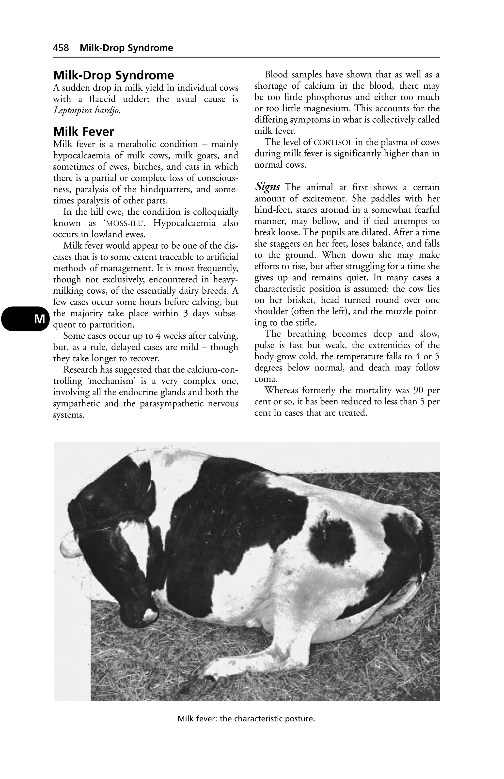
Dry Period
In cattle it is considered advisable on health
grounds that after a period of lactation, cows
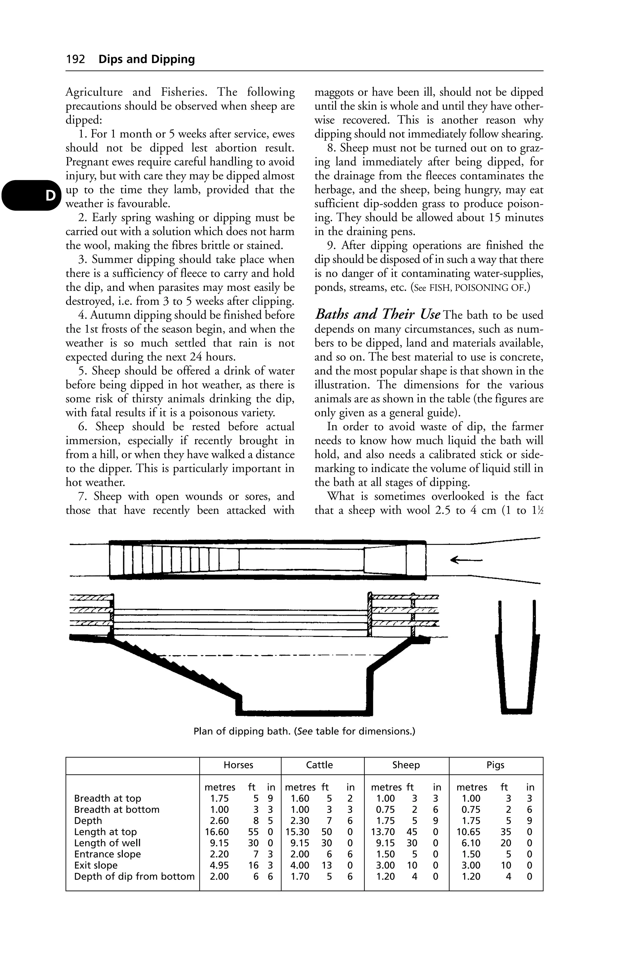
should not be milked for about 8 weeks – the dry
period. Cows are dry in the weeks before calving.
Drying-off Cows
After milking out completely, the teats should
be washed and a dry-cow intramammary prepa-ration
inserted in each teat. The cows should be
inspected daily.
If possible, keep the cows on dry food or very
short pasture for 3 days after drying off.
Drysdale
A sheep with a very good fleece bred in New
Zealand. A natural mutation of the Romney, it
was identified and developed by Dr F. W. Dry
of Massey University.
Dubbing
Trimming of the comb imay be performed,
with scissors, by poultry keepers, and involves
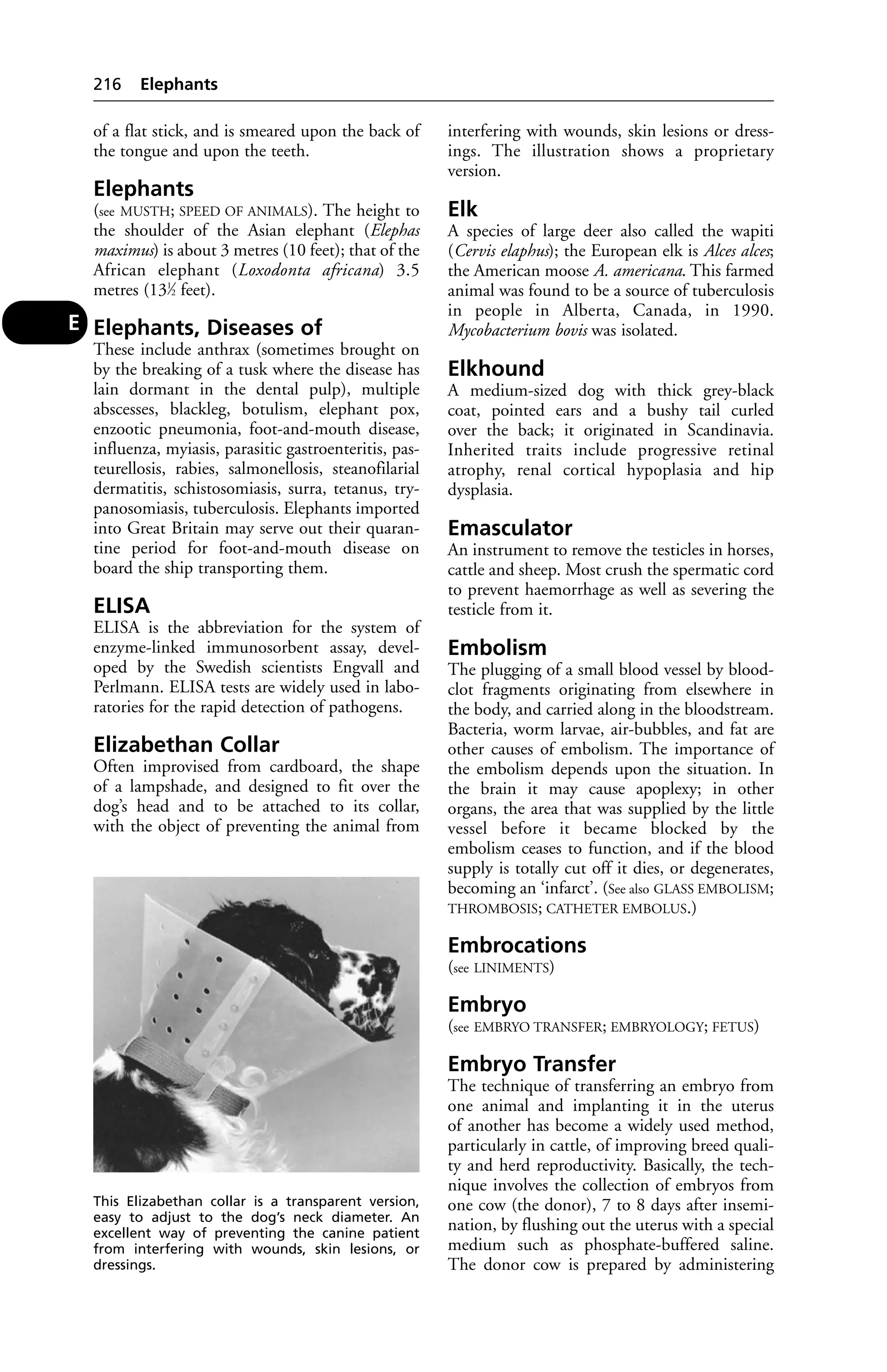
D](https://image.slidesharecdn.com/veterinarydictionary21stedition-140828090128-phpapp02/75/Veterinary-dictionary-_21st_edition-214-2048.jpg)

![may be affected, too. The infections cannot be
differentiated on clinical grounds; laboratory
tests are essential. (For signs, see under
ENCEPHALITIS.)
Control Mosquito control measures reduce
transmission of the disease; stabling horses dur-ing
outbreaks and applying insecticides can
help prevent mosquito attacks. Vaccines are
available for use in areas where the disease is
prevalent.
Public health In man, the disease takes the
form of an aseptic meningitis; outbreaks can be
very serious, and mortality can be high. In one
outbreak in Canada, 509 human cases were
reported, with 78 deaths; 12 of them among
children. Of 27 infants, many suffered brain
damage, resulting in convulsions, spasticity, and
hemiplegia.
Equine Filariasis
Infestation of horses with the filarid worm,
Seturia equina, the larvae being carried by mos-quitoes
and biting flies. It occurs in South and
Central Europe, and Asia.
Signs Malaise and anaemia, or fever, conjunc-tivitis,
and dropsical swellings.
Equine Gait Analysis
A combination of photographic recording and
computer analysis is used to study the motion
of the horse’s limbs as it trots or gallops on a
treadmill. The system was originally devised by
a Swiss, Bruno Kaegi. It helps to provide an
objective measurement of the degree of lame-ness
affecting a horse, and also a comparison
between the limbs.
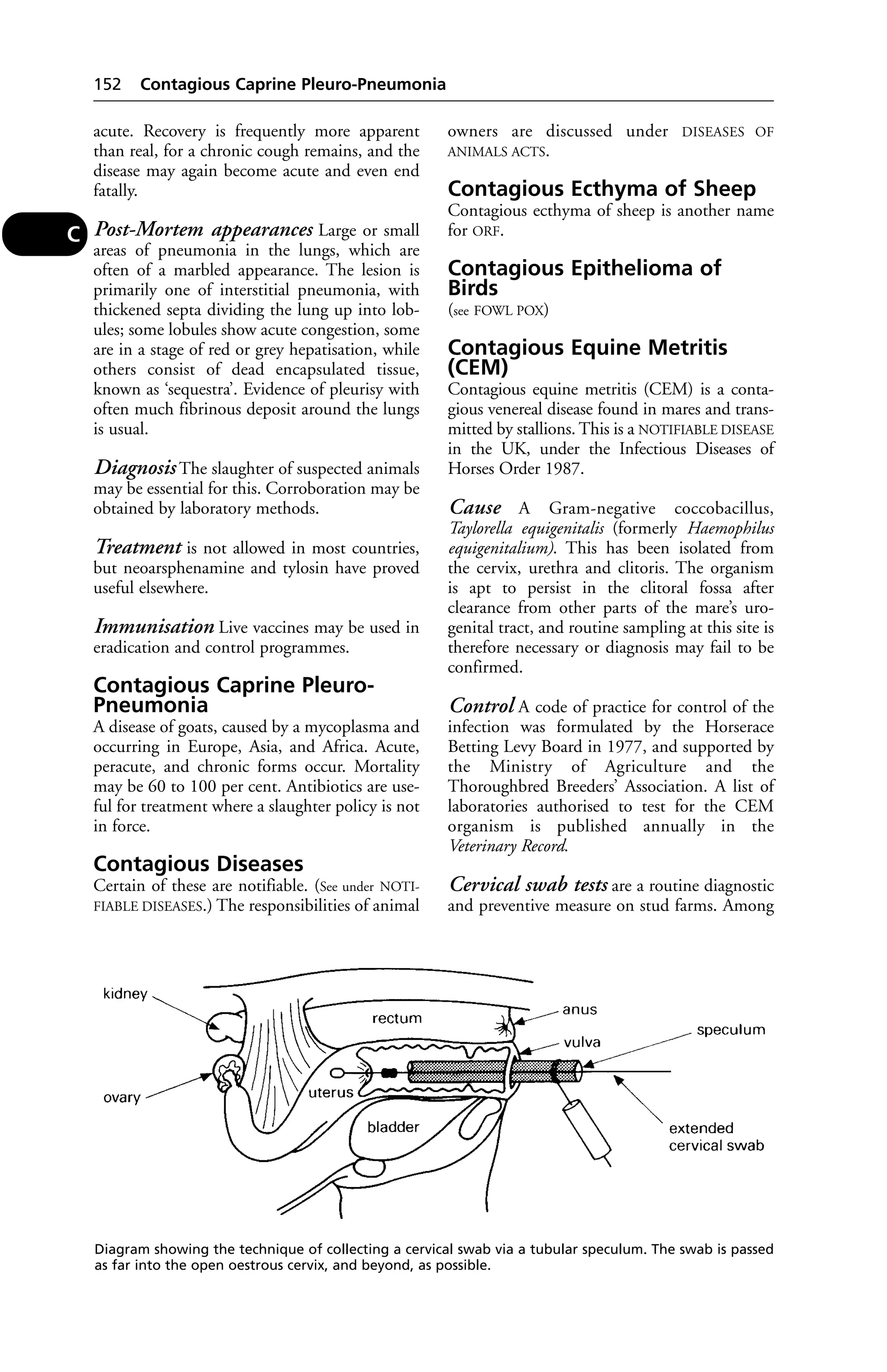
Equine Genital Infections in the
Mare
A wide range of organisms may be found on
taking cervical swabs. Some may be harmless,
but others may cause abortion or disease in the
mare or transmit infection to the stallion.
CONTAGIOUS EQUINE METRITIS (CEM), a
NOTIFIABLE DISEASE, is an important uterine
infection described in a separate entry. It is
caused by Taylorella equigenitalis.
Other infections include beta haemolytic
streptoccoci, Klebsiella aerogenes, Pseudomonas
species (see also LISTERIOSIS; LEPTOSPIROSIS; BRU-CELLOSIS).
Fungal infections have rarely been
reported, and include Aspergillus fumigatus and
Candida albicans.
Abortion caused by the virus of equine
rhinopneumonitis has also occurred in the UK
Equine Infectious Anaemia 227
for several years, most outbreaks being associated
with imported or visiting mares.
Equine Herpesviruses
These include EHV 1, the equine rhinopneu-monitis
or ‘equine abortion’ virus which has
also caused ataxia and paresis. Primarily affect-ing
the respiratory system, EHV1 is the cause
of much illness in young horses. EHV 3 causes
equine coital exanthema. (EHV 2 may be
non-pathogenic.)
Equine Hydatid Disease
(see HYDATID DISEASE)
Equine Hyperlipaemia
A disease of ponies, with an average age of
9 years, affecting the liver, kidneys, and pan-creas.
Mortality may reach 67 per cent.
Equine Infectious Anaemia
A NOTIFIABLE DISEASE. Synonyms include:
pernicious equine anaemia, swamp fever, horse
malaria.
A contagious disease of horses and mules
during the course of which changes occur in the
blood, and rapid emaciation with debility and
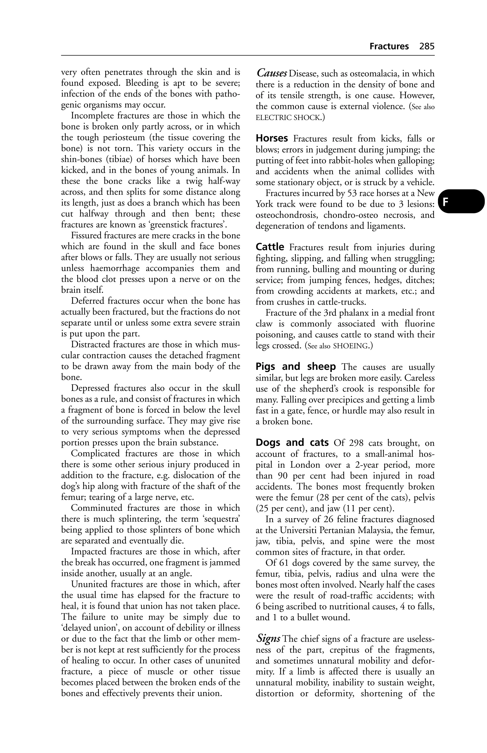
prostration are evident. It occurs chiefly in the
Western States of America and the North-
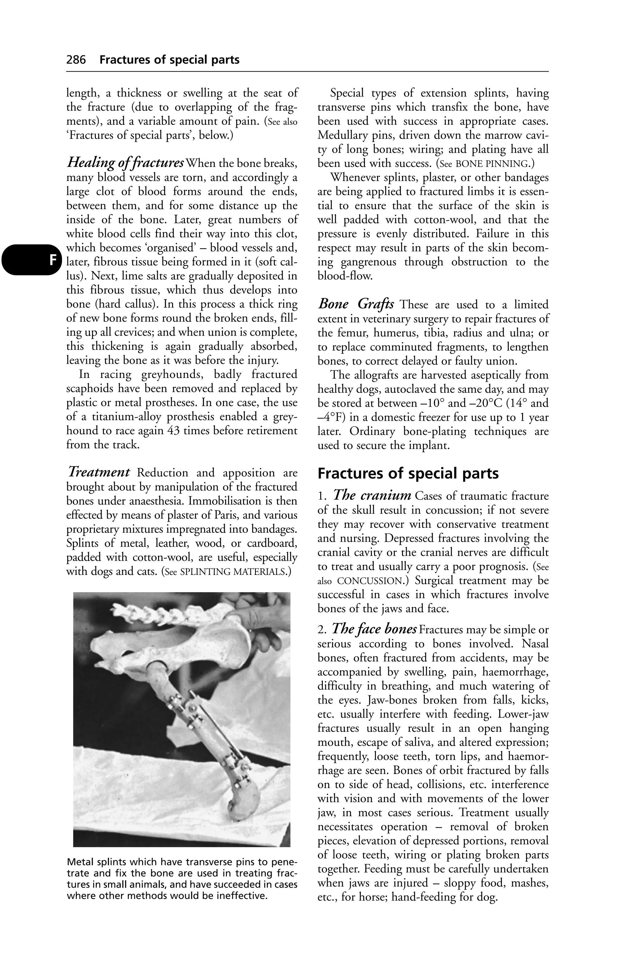
Western Provinces of Canada, as well as in most
countries of Europe, and in Asia, and Africa.
The first case in the UK was reported from
Newmarket in 1975.
Cause A virus. The horse is commonly infect-ed
by biting insects, e.g. horse flies, stable flies,
mosquitoes. Infected grooming tools if they
cause an abrasion, syringes, hypodermic needles
(or even contaminated vaccines) are other
means of transmission. The virus may be pre-sent
in urine, faeces, saliva, nasal secretions,
semen, and milk.
The disease is prevalent in low-lying,
swampy areas, especially during spring and
summer months.
The virus may cause illness in man (who may
infect a horse); also in pigs.
Signs After an incubation period of 2 to
4 weeks, equine infectious anaemia gives rise
to intermittent fever (with a temperature of up to
41°C [106°F]), depression and weakness. Often
there are tiny haemorrhages on the lining of the
eyelids and under the tongue. Jaundice, swelling
of the legs and lower part of the abdomen, and
anaemia may follow. In acute cases, death is
common. In chronic cases there may be a recur-rence
of fever, loss of appetite, and emaciation.
E](https://image.slidesharecdn.com/veterinarydictionary21stedition-140828090128-phpapp02/75/Veterinary-dictionary-_21st_edition-236-2048.jpg)

![Fatty Liver/Kidney Syndrome of Chickens (FLKS) 247
Intending farriers must undergo a 5-year
apprenticeship, including a period at an autho-rised
college, then take an examination for the
diploma of the Worshipful Company of Farriers
before they can practise independently. The
training is controlled by the Farriers Training
Council and a register of farriers kept by the
Farriers Registration Council, Sefton House,
Adam Court, Newark Road, Peterborough PE1
5PP. Its website is at www.farrier-reg.gov.uk.
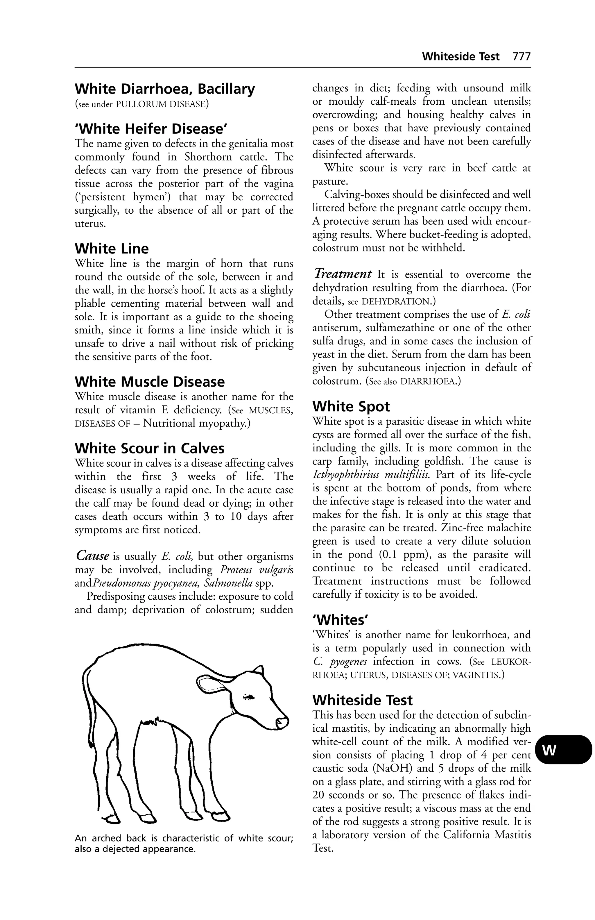
Farrowing
The act of parturition in the sow.
Farrowing Crates
A rectangular box in which the sow gives birth.
Their use is helpful in preventing overlying of
piglets by the sow, and so in obviating one cause
of piglet mortality; however, they are far from
ideal. Farrowing rails serve the same purpose
but perhaps the best arrangement is the circular
one which originated in New Zealand. (See
ROUNDHOUSE.)
Work at the University of Nebraska suggests
that a round stall is better, because the conven-tional
rectangular one does not allow the sow to
obey her natural nesting instincts, and may give
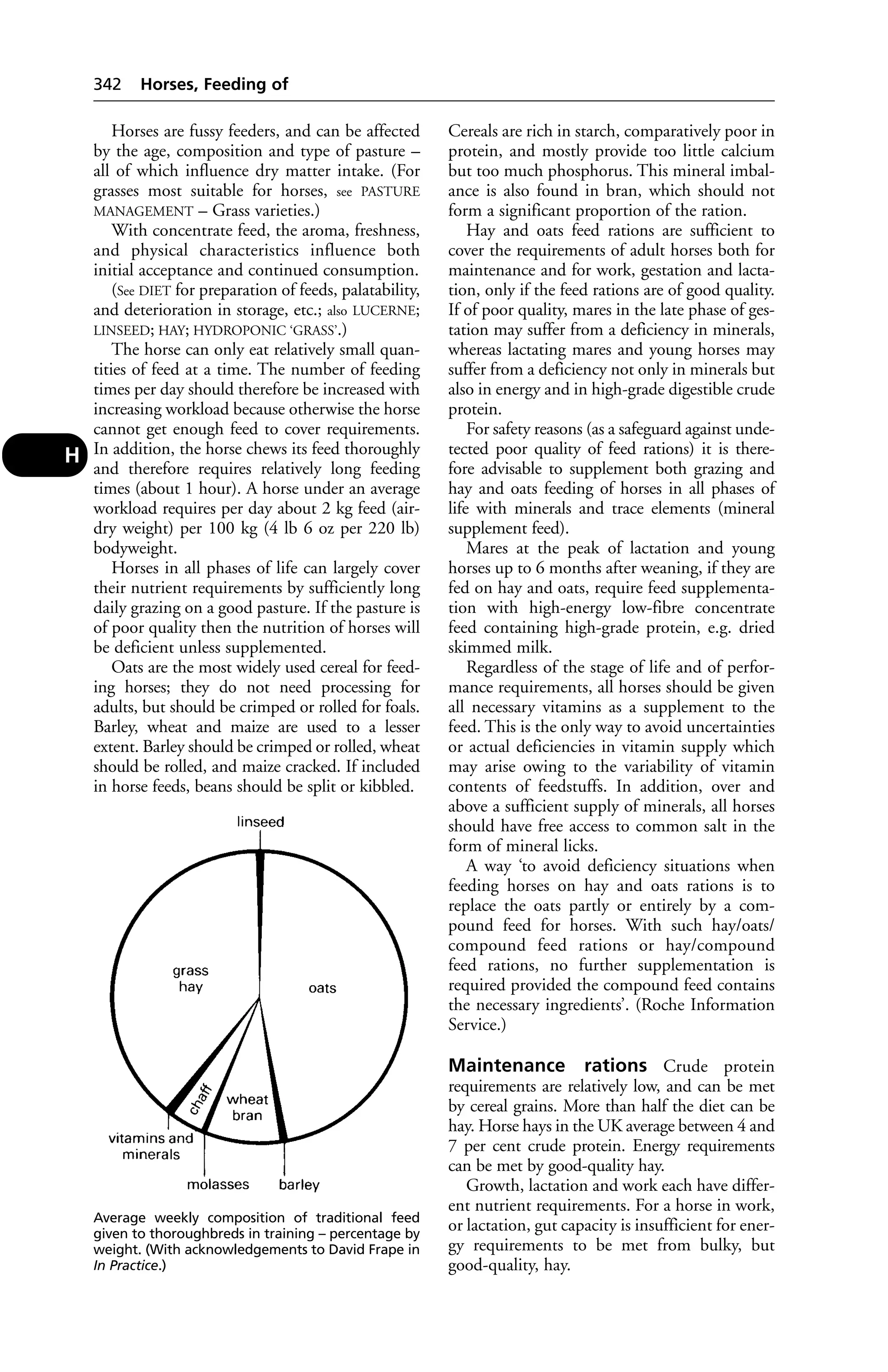
rise to stress, more stillbirths and agalactia.
Farrowing Rates
In the sow, the farrowing rate after 1 natural
service appears to be in the region of 86 per
cent. Following a 1st artificial insemination,
the farrowing rate appears to be appreciably
lower, but at the Lyndhurst, Hants AI Centre,
a farrowing rate of about 83 per cent was
obtained when only females which stood firm-ly
to be mounted at insemination time were
used. The national (British) average farrowing
rate has been estimated at 65 per cent for a
1st insemination.
Fascia
Sheets or bands of fibrous tissue which enclose
and connect the muscles.
Fascioliasis
Infestation with liver flukes.
Fat
Normal body fat is, chemically, an ester of 3
molecules of 1, 2, or 3 fatty acids, with 1 mol-ecule
of glycerol. Such fats are known as glyc-erides,
to distinguish them from other fats and
waxes in which an alcohol other than glycerol
has formed the ester. (See also LIPIDS [which
include fat]; FATTY ACIDS. For fat as a tissue, see ADI-POSE
TISSUE. A LIPOMA is a benign fatty tumour.
For other diseases associated with fat, see STEATI-TIS;
FATTY LIVER SYNDROME; OBESITY, DIET.)
Fat Supplements
In poultry rations these can lead to TOXIC FAT
DISEASE. (See LIPIDS for cattle supplement; also
ECZEMA in cats.)
Fatigue
(see EXERCISE; MUSCLE; NERVES)
Fatty Acids
These, with an alcohol, form FAT. Saturated
fatty acids have twice as many hydrogen atoms
as carbon atoms, and each molecule of fatty
acid contains 2 atoms of oxygen. Unsaturated
fatty acids contain less than twice as many
hydrogen atoms as carbon items, and 1 or more
pairs of adjacent atoms are connected by double
bonds. Polyunsaturated fatty acids are those in
which several pairs of adjacent carbon atoms
contain double bonds.
Fatty Degeneration
A condition in which there is an excess of fat in
the parenchyma cells of organs such as the liver,
heart, and kidneys.
Fatty Liver Haemorrhagic
Syndrome (FLHS)
This is a condition in laying hens which has
to be differentiated from FLKS (see next entry)
of high-carbohydrate broiler-chicks. Factors
involved include high carbohydrate diets, high
environmental temperatures, high oestrogen
levels, and the particular strain of bird. FLHS
in hens is improved by diets based on wheat as
compared with maize; whereas FLKS is aggra-vated
by diets based on wheat. Death is due to
haemorrhage from the enlarged liver.
Fatty Liver/Kidney Syndrome
of Chickens (FLKS)
A condition in which excessive amounts of fat are
present in the liver, kidneys, and myocardium.
The liver is pale and swollen, with haemorrhages
sometimes present, and the kidneys vary from
being slightly swollen and pale pink to being
excessively enlarged and white. Morbidity is
usually between 5 and 30 per cent.
Cause FLKS has been shown to respond to
biotin (see VITAMINS), and accordingly can be
prevented by suitable modification of the diet.
Signs A number of the more forward birds (usu-ally
2 to 3 weeks old) suddenly show symptoms
of paralysis. They lie down on their breasts with
F](https://image.slidesharecdn.com/veterinarydictionary21stedition-140828090128-phpapp02/75/Veterinary-dictionary-_21st_edition-256-2048.jpg)


This document is the 21st edition of Black's Veterinary Dictionary. It has been comprehensively updated from the 20th edition, with many new and amended entries to reflect developments in fields such as medication, newly identified conditions, and emerging diseases. A major addition is entries describing popular dog and cat breeds and their inheritable conditions. The dictionary has been a primary reference for veterinary practitioners, students, farmers and pet owners since its first publication in 1928.


