A stroke occurs when the blood supply to part of your brain is interrupted or severely reduced, depriving brain tissue of oxygen and food.
Within minutes, brain cells begin to die.
Stroke can be either ischemic or hemorrhagic.
A stroke occurs when the blood supply to part of your brain is interrupted or severely reduced, depriving brain tissue of oxygen and food.
Within minutes, brain cells begin to die.
Stroke can be either ischemic or hemorrhagic.
New Drug Discovery and Development .....NEHA GUPTA
The "New Drug Discovery and Development" process involves the identification, design, testing, and manufacturing of novel pharmaceutical compounds with the aim of introducing new and improved treatments for various medical conditions. This comprehensive endeavor encompasses various stages, including target identification, preclinical studies, clinical trials, regulatory approval, and post-market surveillance. It involves multidisciplinary collaboration among scientists, researchers, clinicians, regulatory experts, and pharmaceutical companies to bring innovative therapies to market and address unmet medical needs.
Title: Sense of Smell
Presenter: Dr. Faiza, Assistant Professor of Physiology
Qualifications:
MBBS (Best Graduate, AIMC Lahore)
FCPS Physiology
ICMT, CHPE, DHPE (STMU)
MPH (GC University, Faisalabad)
MBA (Virtual University of Pakistan)
Learning Objectives:
Describe the primary categories of smells and the concept of odor blindness.
Explain the structure and location of the olfactory membrane and mucosa, including the types and roles of cells involved in olfaction.
Describe the pathway and mechanisms of olfactory signal transmission from the olfactory receptors to the brain.
Illustrate the biochemical cascade triggered by odorant binding to olfactory receptors, including the role of G-proteins and second messengers in generating an action potential.
Identify different types of olfactory disorders such as anosmia, hyposmia, hyperosmia, and dysosmia, including their potential causes.
Key Topics:
Olfactory Genes:
3% of the human genome accounts for olfactory genes.
400 genes for odorant receptors.
Olfactory Membrane:
Located in the superior part of the nasal cavity.
Medially: Folds downward along the superior septum.
Laterally: Folds over the superior turbinate and upper surface of the middle turbinate.
Total surface area: 5-10 square centimeters.
Olfactory Mucosa:
Olfactory Cells: Bipolar nerve cells derived from the CNS (100 million), with 4-25 olfactory cilia per cell.
Sustentacular Cells: Produce mucus and maintain ionic and molecular environment.
Basal Cells: Replace worn-out olfactory cells with an average lifespan of 1-2 months.
Bowman’s Gland: Secretes mucus.
Stimulation of Olfactory Cells:
Odorant dissolves in mucus and attaches to receptors on olfactory cilia.
Involves a cascade effect through G-proteins and second messengers, leading to depolarization and action potential generation in the olfactory nerve.
Quality of a Good Odorant:
Small (3-20 Carbon atoms), volatile, water-soluble, and lipid-soluble.
Facilitated by odorant-binding proteins in mucus.
Membrane Potential and Action Potential:
Resting membrane potential: -55mV.
Action potential frequency in the olfactory nerve increases with odorant strength.
Adaptation Towards the Sense of Smell:
Rapid adaptation within the first second, with further slow adaptation.
Psychological adaptation greater than receptor adaptation, involving feedback inhibition from the central nervous system.
Primary Sensations of Smell:
Camphoraceous, Musky, Floral, Pepperminty, Ethereal, Pungent, Putrid.
Odor Detection Threshold:
Examples: Hydrogen sulfide (0.0005 ppm), Methyl-mercaptan (0.002 ppm).
Some toxic substances are odorless at lethal concentrations.
Characteristics of Smell:
Odor blindness for single substances due to lack of appropriate receptor protein.
Behavioral and emotional influences of smell.
Transmission of Olfactory Signals:
From olfactory cells to glomeruli in the olfactory bulb, involving lateral inhibition.
Primitive, less old, and new olfactory systems with different path
Best Ayurvedic medicine for Gas and IndigestionSwastikAyurveda
Here is the updated list of Top Best Ayurvedic medicine for Gas and Indigestion and those are Gas-O-Go Syp for Dyspepsia | Lavizyme Syrup for Acidity | Yumzyme Hepatoprotective Capsules etc
Recomendações da OMS sobre cuidados maternos e neonatais para uma experiência pós-natal positiva.
Em consonância com os ODS – Objetivos do Desenvolvimento Sustentável e a Estratégia Global para a Saúde das Mulheres, Crianças e Adolescentes, e aplicando uma abordagem baseada nos direitos humanos, os esforços de cuidados pós-natais devem expandir-se para além da cobertura e da simples sobrevivência, de modo a incluir cuidados de qualidade.
Estas diretrizes visam melhorar a qualidade dos cuidados pós-natais essenciais e de rotina prestados às mulheres e aos recém-nascidos, com o objetivo final de melhorar a saúde e o bem-estar materno e neonatal.
Uma “experiência pós-natal positiva” é um resultado importante para todas as mulheres que dão à luz e para os seus recém-nascidos, estabelecendo as bases para a melhoria da saúde e do bem-estar a curto e longo prazo. Uma experiência pós-natal positiva é definida como aquela em que as mulheres, pessoas que gestam, os recém-nascidos, os casais, os pais, os cuidadores e as famílias recebem informação consistente, garantia e apoio de profissionais de saúde motivados; e onde um sistema de saúde flexível e com recursos reconheça as necessidades das mulheres e dos bebês e respeite o seu contexto cultural.
Estas diretrizes consolidadas apresentam algumas recomendações novas e já bem fundamentadas sobre cuidados pós-natais de rotina para mulheres e neonatos que recebem cuidados no pós-parto em unidades de saúde ou na comunidade, independentemente dos recursos disponíveis.
É fornecido um conjunto abrangente de recomendações para cuidados durante o período puerperal, com ênfase nos cuidados essenciais que todas as mulheres e recém-nascidos devem receber, e com a devida atenção à qualidade dos cuidados; isto é, a entrega e a experiência do cuidado recebido. Estas diretrizes atualizam e ampliam as recomendações da OMS de 2014 sobre cuidados pós-natais da mãe e do recém-nascido e complementam as atuais diretrizes da OMS sobre a gestão de complicações pós-natais.
O estabelecimento da amamentação e o manejo das principais intercorrências é contemplada.
Recomendamos muito.
Vamos discutir essas recomendações no nosso curso de pós-graduação em Aleitamento no Instituto Ciclos.
Esta publicação só está disponível em inglês até o momento.
Prof. Marcus Renato de Carvalho
www.agostodourado.com
Muktapishti is a traditional Ayurvedic preparation made from Shoditha Mukta (Purified Pearl), is believed to help regulate thyroid function and reduce symptoms of hyperthyroidism due to its cooling and balancing properties. Clinical evidence on its efficacy remains limited, necessitating further research to validate its therapeutic benefits.
Tom Selleck Health: A Comprehensive Look at the Iconic Actor’s Wellness Journeygreendigital
Tom Selleck, an enduring figure in Hollywood. has captivated audiences for decades with his rugged charm, iconic moustache. and memorable roles in television and film. From his breakout role as Thomas Magnum in Magnum P.I. to his current portrayal of Frank Reagan in Blue Bloods. Selleck's career has spanned over 50 years. But beyond his professional achievements. fans have often been curious about Tom Selleck Health. especially as he has aged in the public eye.
Follow us on: Pinterest
Introduction
Many have been interested in Tom Selleck health. not only because of his enduring presence on screen but also because of the challenges. and lifestyle choices he has faced and made over the years. This article delves into the various aspects of Tom Selleck health. exploring his fitness regimen, diet, mental health. and the challenges he has encountered as he ages. We'll look at how he maintains his well-being. the health issues he has faced, and his approach to ageing .
Early Life and Career
Childhood and Athletic Beginnings
Tom Selleck was born on January 29, 1945, in Detroit, Michigan, and grew up in Sherman Oaks, California. From an early age, he was involved in sports, particularly basketball. which played a significant role in his physical development. His athletic pursuits continued into college. where he attended the University of Southern California (USC) on a basketball scholarship. This early involvement in sports laid a strong foundation for his physical health and disciplined lifestyle.
Transition to Acting
Selleck's transition from an athlete to an actor came with its physical demands. His first significant role in "Magnum P.I." required him to perform various stunts and maintain a fit appearance. This role, which he played from 1980 to 1988. necessitated a rigorous fitness routine to meet the show's demands. setting the stage for his long-term commitment to health and wellness.
Fitness Regimen
Workout Routine
Tom Selleck health and fitness regimen has evolved. adapting to his changing roles and age. During his "Magnum, P.I." days. Selleck's workouts were intense and focused on building and maintaining muscle mass. His routine included weightlifting, cardiovascular exercises. and specific training for the stunts he performed on the show.
Selleck adjusted his fitness routine as he aged to suit his body's needs. Today, his workouts focus on maintaining flexibility, strength, and cardiovascular health. He incorporates low-impact exercises such as swimming, walking, and light weightlifting. This balanced approach helps him stay fit without putting undue strain on his joints and muscles.
Importance of Flexibility and Mobility
In recent years, Selleck has emphasized the importance of flexibility and mobility in his fitness regimen. Understanding the natural decline in muscle mass and joint flexibility with age. he includes stretching and yoga in his routine. These practices help prevent injuries, improve posture, and maintain mobilit
The Gram stain is a fundamental technique in microbiology used to classify bacteria based on their cell wall structure. It provides a quick and simple method to distinguish between Gram-positive and Gram-negative bacteria, which have different susceptibilities to antibiotics
Maxilla, Mandible & Hyoid Bone & Clinical Correlations by Dr. RIG.pptx
Stroke.pptx
1.
2. Introduction
• Include ;
– TIA
– Stroke
– cerebrovascular anomalies such as intracranial
aneurysms
– arteriovenous malformations (AVMs).
8/7/2023 Dr.Tamiru S. 2
3. – Stroke; rapidly developed clinical sign of focal
disturbance of cerebral function of presumed
vascular origin and of more than 24 hours
– Cerebral ischemia is caused by a reduction in
blood flow that lasts longer than several seconds.
– Infarction – absence of blood flow more than a
few minutes
8/7/2023 3
Dr.Tamiru S.
4. – TIA; all neurologic signs and symptoms resolve
within 24 hours regardless of whether there is
imaging evidence of new permanent brain injury
– TIA recovery is complete within 24 hours.
• 10% of patients will go on to have a stroke.
8/7/2023 4
Dr.Tamiru S.
5. – Neurologic symptoms are manifest within seconds
because neurons lack glycogen, so energy failure is
rapid
– When blood flow is quickly restored, brain tissue
can recover fully and the patient's symptoms are
only transient
8/7/2023 5
Dr.Tamiru S.
6. – Focal ischemia or infarction is caused by
thrombosis of the cerebral vessels or by emboli
from a proximal arterial source or the heart.
– Intracranial hemorrhage is caused by bleeding
directly into or around the brain; it produces
neurologic symptoms;
• by producing a mass effect on neural structures
• from the toxic effects of blood itself
• increasing intracranial pressure.
8/7/2023 6
Dr.Tamiru S.
7. Stroke
– is the third largest killer in the Western World.
– is one of the major causes of disability, particularly in the
elderly.
– patients may present with a variety of physical, cognitive
and psychosocial problems.
– Most patients show signs of recovery over time.
8/7/2023 7
Dr.Tamiru S.
8. 8
Epidemiology
Ethiopia
– Important cause of hospital morbidity & mortality
– 6.5% of admissions were due to stroke
– Study in Tikur Anbessa hospital
( Sept. 2000 - Aug.2001)
• Hemorrhagic stroke – the most common
57% (of all patients)
59.2% (of those with CT)
9. • Peak incidence 55-64yrs
• HTN the most frequent risk factor (65.6%)
followed by cardiac disease (22.7%)
• The overall mortality 44.5%
Ethiop Med J. 2005 Oct;43(4):251-9
8/7/2023 Dr.Tamiru S. 9
12. Embolic
– The occlusion is caused by an embolus (solid,
liquid, or gaseous mass) carried to a blood vessel
from another area
– Most common emboli are blood clots
– Risk factors for blood clots include Atrial
Fibrillation and diseased or damaged carotid or
vertebral arteries
8/7/2023 12
Dr.Tamiru S.
13. – Rare causes of emboli include air, tumor tissue, and
fat
– Occurs suddenly & may rarely be accompanied by
headache
– Sudden, maximum deficit at the onset
– Common sites
»MCA or one of the branches
»PCA
»ACA( infrequently)
8/7/2023 Dr.Tamiru S. 13
14. Thrombotic
– The occlusion is caused by a cerebral thrombus; a
blood clot which develops gradually in a
previously diseased artery and obstructs it
– Caused by atherosclerosis:
• platelets adhere to the roughened surface of the
plaque deposit and a blood clot is created
• atheromatous plaque results in narrowing and
reduction of blood flow
8/7/2023 14
Dr.Tamiru S.
15. Ischemic Stroke
CBF & Ischemic Thresholds
– Normal CBF 50-60 ml/100 g/minute
• Varies in different regions of the brain
– CBF 20-30ml/100g/min Loss of electrical
activity
– CBF 10 ml/100g/min Neuronal death
8/7/2023 15
Dr.Tamiru S.
16. Brain can’t tolerate decrease in blood flow for a
long time
- 0ml/100 g tissue per minute ---4-10min
- 16-18ml/100 g tissue per minute---hours
- <20ml/100 g tissue per minute---ischemia unless
prolonged for hours or days
8/7/2023 16
Dr.Tamiru S.
17. – Acute occlusion of an intracranial vessel causes
reduction in blood flow to the brain region it
supplies.
– The magnitude of flow reduction is a function of
collateral blood flow and this depends on
individual vascular anatomy, the site of occlusion,
and likely systemic blood pressure.
8/7/2023 17
Dr.Tamiru S.
18. – Tissue surrounding the core region of infarction is
ischemic but reversibly dysfunctional and is
referred to as the ischemic penumbra.
– The ischemic penumbra will eventually infarct if
no change in flow occurs, and hence saving the
ischemic penumbra is the goal of revascularization
therapies.
8/7/2023 18
Dr.Tamiru S.
19. Focal cerebral infarction occurs via two distinct
pathways
(1)a necrotic pathway; cellular cytoskeletal
breakdown due to energy failure of the cell
(2)apoptotic pathway; cells become programmed to
die.
8/7/2023 19
Dr.Tamiru S.
20. – Ischemia produces necrosis by starving neurons of
glucose and oxygen, which in turn results in failure
of mitochondria to produce ATP.
– Without ATP, membrane ion pumps stop
functioning and neurons depolarize, allowing
intracellular calcium to rise.
8/7/2023 20
Dr.Tamiru S.
21. – Cellular depolarization also causes glutamate
release from synaptic terminals; excess
extracellular glutamate produces neurotoxicity by
activating postsynaptic glutamate receptors that
increase neuronal calcium influx.
8/7/2023 21
Dr.Tamiru S.
22. – Free radicals are produced by membrane lipid
degradation and mitochondrial dysfunction.
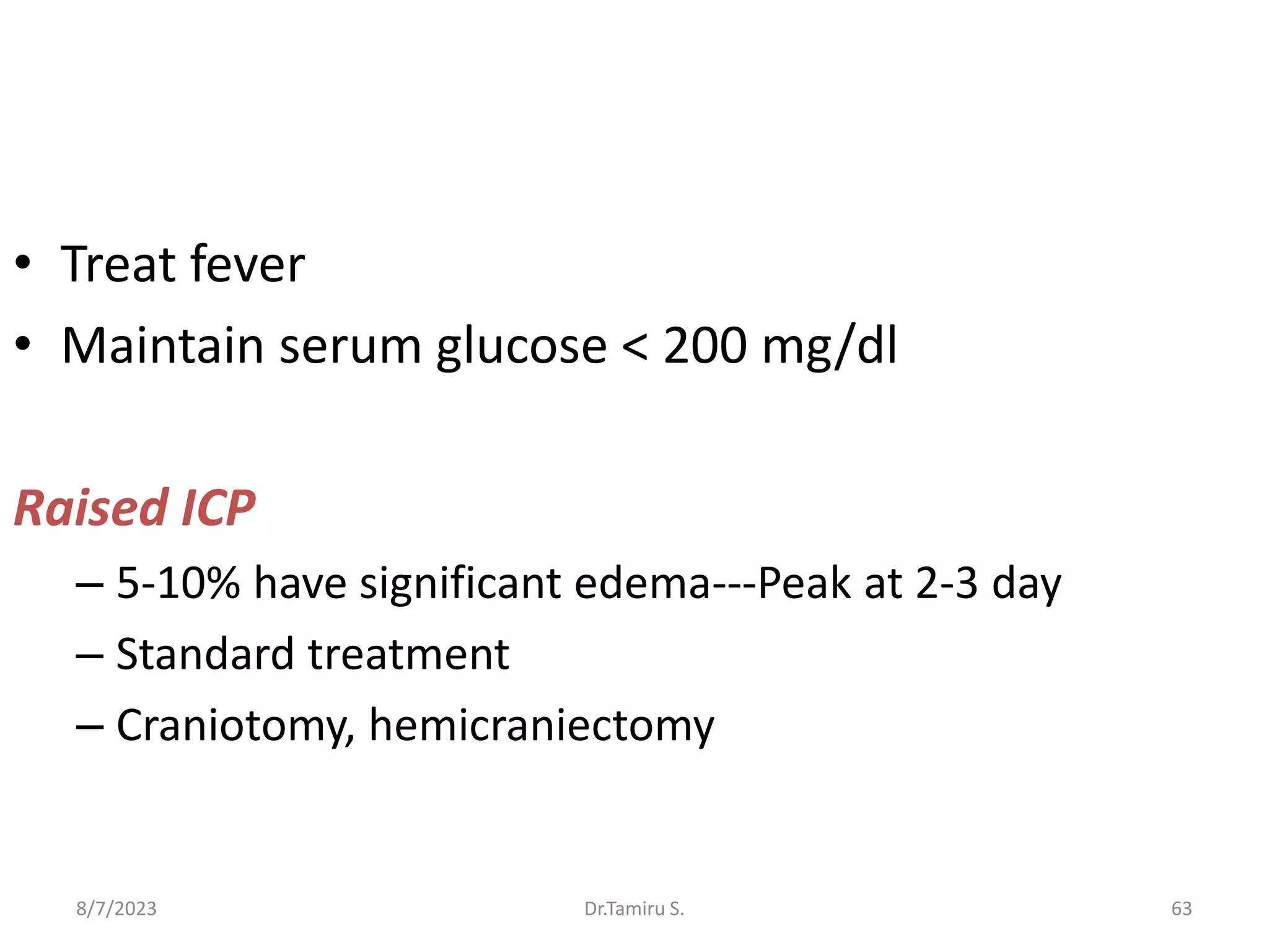
– Fever dramatically worsens brain injury during
ischemia, as does hyperglycemia [ 200 mg/dl].
8/7/2023 Dr.Tamiru S. 22
23. Causes of Ischemic Stroke
Common Causes
• Thrombosis
Lacunar stroke (small
vessel)
Large vessel thrombosis
Dehydration
• Embolic occlusion
1. Artery-to-artery
Carotid bifurcation
Aortic arch
Arterial dissection
8/7/2023 23
Dr.Tamiru S.
27. Risk Factors
Ethiopia
Established hypertension- 65.6%
Cardiac disease - 22.7%
Established diabetes -11.7%
TIA
Previous stroke
Atrial fibrillation
*most patient were not on treatment for hypertension
*majority of cardiac pts have VHD
*AF was found in most of the cardiac patients but treatment
was not started for most of them
8/7/2023 27
Dr.Tamiru S.
29. • Nonmodifiable
– Age
– Gender
– Race/ethnicity
– Family history
– Genetics
8/7/2023 29
Dr.Tamiru S.
30. • Patients at risk should be brought to ER if they
develop
–Loss of sensory/motor function
–Change in vision, gait, ability to speak
or understand
–Severe headache
8/7/2023 30
Dr.Tamiru S.
31. Clinical Manifestations
Internal Carotid Artery
– often clinically silent if the circle of Willis is
complete.
– transient monocular blindness (also called
amaurosis fugax).
8/7/2023 31
Dr.Tamiru S.
32. – Severe stenosis if bilateral, can cause
hypoperfusion of the cerebral hemispheres and
symptoms in border zones between the MCA and
other major vascular territories (watershed areas),
especially if superimposed on generalized
hypoperfusion secondary to severe hypotension.
8/7/2023 32
Dr.Tamiru S.
33. Anterior Cerebral Artery
– is relatively rare in comparison to strokes in other
major branches of the circle of Willis
– account for 2% of all cerebral infarcts.
– upper motor neuron weakness and cortical
sensory deficits (neglect) in contralateral leg.
– leg weakness > arm weakness
8/7/2023 33
Dr.Tamiru S.
34. – urinary incontinence, generalized depression of
psychomotor activity (abulia), and transcortical
motor aphasia manifested as loss of verbal fluency
with preserved ability to repeat.
– bilateral damage usually causes a patient to be
mute, with severe mood disturbances and long-
lasting incontinence.
8/7/2023 34
Dr.Tamiru S.
35. Middle Cerebral Artery
– are the most common site of stroke 2/3 of all
infarcts.
– edema during the first 3 to 4 days may lead to
severely increased intracranial pressure and
herniation.
8/7/2023 35
Dr.Tamiru S.
36. – Contralateral weakness
– Face & arm > leg
– Aphasia – dominant hemisphere
– Neglect – non-dominant hemisphere
– Neglect may be motor, verbal or visual
– Head & eye deviation toward side of infarct.
– hemianopia common in large MCA infarcts
8/7/2023 36
Dr.Tamiru S.
37. – Strokes of the inferior division in the dominant
hemisphere characteristically produce receptive
aphasia of the Wernicke type (severe loss of
speech comprehension with preserved spoken
and written language).
– Occlusion of more distal branches causes less
clinical damage.
8/7/2023 37
Dr.Tamiru S.
38. Posterior Cerebral Artery
– In about 3/4, both posterior cerebral arteries
(PCAs) arise from the basilar artery; in most
others, one PCA arises from the basilar artery and
the other arises from the ICA.
– In a few individuals, both PCAs originate from the
ICAs
– As a consequence, the syndromes associated with
occlusion of the PCA are highly variable.
8/7/2023 38
Dr.Tamiru S.
39. Posterior Cerebral Artery Infarction
Thalamic lesions:
– Sensory symptoms: loss of tactile, temperature & pain
sensation
– Typically complain of numbness & tingling on one side of
the body
– Hypoesthesia and dysesthesia common
8/7/2023 39
Dr.Tamiru S.
40. Occipital Lesions:
– Homonymous visual field defect
– May cause midbrain lesions leading to oculomotor
palsy or internuclear ophthalmoplegia
8/7/2023 40
Dr.Tamiru S.
41. Cerebellar Infarction
– May cause a pure vestibular syndrome leading to
misdiagnosis of labyrinthitis
– Increased ICP 6-12 h secondary to edema may require
emergent surgical decompression
– Vertigo, headache, Nystagmus
8/7/2023 41
Dr.Tamiru S.
42. – Strokes of the perforating branches cause
complete contralateral hemianesthesia with loss
of all sensation because of collateral blood supply
from the MCA.
8/7/2023 42
Dr.Tamiru S.
43. – Difficulty reading (dyslexia) and performing
calculations (dyscalculia) may occur.
– Recovery is often good, but the initial numbness
may be replaced by paresthesias or excruciating
pain; this Dejerine-Roussy syndrome is caused by
damage to the thalamus.
8/7/2023 43
Dr.Tamiru S.
44. – Involvement of the subthalamic nucleus may
produce hemiballismus, with wild flinging
movements of the limbs on one side of the body.
– Distal branch occlusions of the PCA cause partial
syndromes; occlusion of the terminal branch can
produce a variety of incomplete visual field
deficits.
8/7/2023 44
Dr.Tamiru S.
45. Vertebral and Basilar Arteries
– Characteristic of occlusion of the blood supply to
the brain stem are “crossed syndromes” (i.e.,
contralateral loss of strength and selected
contralateral and ipsilateral sensory symptoms
below the level of the lesion, in addition to
ipsilateral motor and sensory deficits localized to
the level of the lesion).
8/7/2023 45
Dr.Tamiru S.
46. – Severe vertigo, nausea, vomiting, nystagmus,
ipsilateral ataxia (of the cerebellar type), and
ipsilateral Horner's syndrome (ptosis, myosis, and
decreased sweating).
8/7/2023 46
Dr.Tamiru S.
47. Diagnosis
• History
• Time of onset of symptoms
• Did the patient wake up with symptoms?
8/7/2023 47
Dr.Tamiru S.
48. Physical Examination
– Helps localize the site of the lesion
– Arterial blood pressure, including measurement in
both arms to evaluate the possibility of aortic
dissection or vascular abnormalities that result in
reduced blood flow to the brain when the arms
are exercised (subclavian steal).
– The pulse may reveal arrhythmias
8/7/2023 48
Dr.Tamiru S.
49. – Cardiac murmurs may suggest valvular lesions
– Bruits of the carotid arteries
– Ophthalmoscopic visualization of retinal
cholesterol or platelet-fibrin emboli suggests more
proximal atherosclerotic disease
8/7/2023 49
Dr.Tamiru S.
50. Laboratory Examination
– CBC
– ESR
– RBS/FBS
– A prothrombin time and partial thromboplastin
time
– Lipid profile
– VDRL
8/7/2023 50
Dr.Tamiru S.
51. – Tests for homocysteinuria, collagen vascular
diseases, amyloidosis
– Antiphospholipid antibodies are elevated in some
patients with immune-related diseases.
– Measurement of protein C, protein S,
antithrombin III, blood viscosity, and platelet
function
8/7/2023 51
Dr.Tamiru S.
53. Noninvasive Brain Imaging
– Which is essential to verify causes of focal
neurologic dysfunction, can generally distinguish
ischemic stroke from other diseases.
8/7/2023 53
Dr.Tamiru S.
54. Imaging Studies in Stroke
CT SCAN
– Preferred standard initial modality in all acute strokes
– Better than MRI for detection of Acute blood
– Can identify other ICSOL mimickers of stroke
8/7/2023 54
Dr.Tamiru S.
55. • Pitfalls
–Infarcts missed / not reliably seen in the first
24-48 hrs
–Misses small cortical infarcts
–Misses posterior fossa infarcts b/c of bone
artifact
8/7/2023 Dr.Tamiru S. 55
56. MRI
– Shows location and extent of infarction in all areas
– Less sensitive for acute blood
– More expensive, time consuming, not readily
available
– cannot be used in patients who have
ferromagnetic materials within their bodies, is
often impossible to use in critically ill patients
– Outside the acute period – more useful
– MRI perfusion studies, Diffusion weighted image
– MR angiography
8/7/2023 56
Dr.Tamiru S.
57. Cerebral Angiography
– is reserved for patients who are suspected to have
a surgically correctable lesion
8/7/2023 57
Dr.Tamiru S.
58. Cerebral Angiography
• “Gold- Standard” for stenosis, aneurysm,
vasospasm, thrombi,vasculitis,FMD,AVF
• Therapeutic
–Stents
–Balloon angioplasty
–Embolization
–Intra- Arterial thrombolytics
• Invasive, not widely available, risky
8/7/2023 58
Dr.Tamiru S.
59. Ultrasound
• “Duplex U/Sound”– can identify stenosis at the
origin of internal carotid artery
• TCD of MCA, ACA, PCA ,Vertebro-basilar flow
Perfusion Techniques
• CT , MRI perfusion scans
• SPECT
8/7/2023 59
Dr.Tamiru S.
60. Treatment
• After the clinical diagnosis of stroke is made,
an orderly process of evaluation and
treatment should follow
• The first goal is to prevent or reverse brain
injury.
8/7/2023 60
Dr.Tamiru S.
61. • ABC
• Immediate Imaging
• Ischemic Stroke diagnosed…………..
1. Medical Support
2. Thrombolysis
3. Anticoagulation
4. Antiplatelet agents
5. Neuroprotection
6. Stroke centers and rehabilitation.
8/7/2023 61
Dr.Tamiru S.
62. Medical Support
• Optimize cerebral perfusion to the penumbra
• Prevent/Treat complications of bedridden pts
• Infections, DVT, PE
Blood Pressure Mx
• BP Should be lowered only if
»Malignant HTN
»>185/110
»Thrombolytic planned
• B blockers ( esmolol, labetolol)
8/7/2023 62
Dr.Tamiru S.
63. • Treat fever
• Maintain serum glucose < 200 mg/dl
Raised ICP
– 5-10% have significant edema---Peak at 2-3 day
– Standard treatment
– Craniotomy, hemicraniectomy
8/7/2023 63
Dr.Tamiru S.
66. HEMORRHAGIC CEREBROVASCULAR DISEASE
– Can be diffuse (i.e., bleeding into the
subarachnoid or intraventricular spaces) or focal
(intraparenchymal hemorrhage).
– About 2/3 of intracranial bleeding are
predominantly subarachnoid hemorrhages,
whereas about a third are intracerebral
hemorrhages.
8/7/2023 66
Dr.Tamiru S.
68. Saccular (“Berry”) Aneurysm
– About 2% of adults harbor intracranial aneurysms
– For patients who arrive alive at hospital, the
mortality rate over the next month is about 45%.
– From survivors, >50% left with major neurologic
deficits as a result of the initial hemorrhage,
cerebral vasospasm with infarction, or
hydrocephalus.
8/7/2023 68
Dr.Tamiru S.
69. – If the patient survives but the aneurysm is not
obliterated, the rate of rebleeding is about 20% in the
first 2 weeks and about 3% per year afterwards.
– Unruptured, asymptomatic aneurysms are much less
dangerous than a recently ruptured aneurysm.
– The annual risk of rupture for aneurysms <10 mm in
size is ~0.1%, and for aneurysms ≥10 mm in size is
~0.5 to 1%;
8/7/2023 69
Dr.Tamiru S.
70. – The three most common locations are the
terminal internal carotid artery, MCA bifurcation,
and top of the basilar artery.
– They often cause symptoms by compressing the
adjacent brain or cranial nerves
8/7/2023 70
Dr.Tamiru S.
71. – Approximately 85% of aneurysms occur in the
anterior circulation, mostly on the circle of Willis.
– About 20% of patients have multiple aneurysms,
many at mirror sites bilaterally.
8/7/2023 71
Dr.Tamiru S.
72. Clinical Manifestations
– Most unruptured are completely asymptomatic.
– The classic symptom of a subarachnoid
hemorrhage is a very rapidly developing, severe
headache, typically called the “worst headache of
my life,” that is sometimes accompanied by a stiff
neck.
– The headache is usually generalized, often with
neck stiffness, and vomiting is common.
8/7/2023 72
Dr.Tamiru S.
73. – sudden transient loss of consciousness that occurs
in nearly half of patients- 10 % prolonged
– most patients first complain of headache upon
regaining consciousness.
8/7/2023 73
Dr.Tamiru S.
74. Delayed Complications
Rerupture
• At 2wks-20%
• At 4 wks-30%
• Peak 7 days
• Mortality – 60%
• Early treatment eliminates this risk.
8/7/2023 74
Dr.Tamiru S.
75. Hydrocephalus
– Acute hydrocephalus can cause stupor and coma.
– Subacute hydrocephalus develops over a few days
or weeks and causes progressive drowsiness or
slowed mentation (abulia) with incontinence.
– Chronic hydrocephalus may develop weeks to
months after SAH and manifest as gait difficulty,
incontinence, or impaired mentation.
8/7/2023 75
Dr.Tamiru S.
76. Vasospasm
– Narrowing of the arteries at the base of the brain
following SAH causes symptomatic ischemia and
infarction in ~30%
– Major cause of delayed morbidity and death.
– Appear 4 to 14 days after the hemorrhage, most
often at 7 days.
– The severity and distribution of vasospasm
determine whether infarction will occur.
8/7/2023 76
Dr.Tamiru S.
77. Hyponatremia
– In the first 2 weeks following SAH
– It usually results from inappropriate secretion of
vasopressin and secretion of atrial and brain
natriuretic factors, which produce a natriuresis.
– clears over the course of 1 to 2 wks
– should not be treated with free-water restriction
as this may increase the risk of stroke.
8/7/2023 77
Dr.Tamiru S.
78. Laboratory Findings
– CBC
– Clotting times should be determined to assess
whether the patient has an infection or clotting
abnormalities.
– Blood should also be sent for electrolyte analysis
to serve as a baseline for detecting later
complications.
8/7/2023 78
Dr.Tamiru S.
79. Imaging
95 % - detected by CT Scan within 72 hrs
LP
spinal fluid yellow (xanthochromic) within 6 to 12
h of SAH.
Peaks at 48 h
lasts for 1 to 4 weeks, depending on the amount
of subarachnoid blood.
8/7/2023 79
Dr.Tamiru S.
80. – Conventional x-ray angiography (both carotids and
both vertebrals) to localize and define the
anatomic details of the aneurysm and to
determine if other unruptured aneurysms exist
– CT angiography is an alternative method for
locating the aneurysm
8/7/2023 80
Dr.Tamiru S.
81. • ECG
– ST-segment and T-wave changes
– Prolonged QRS complex, increased QT interval,
and prominent “peaked” or deeply inverted
symmetric T waves are usually secondary to the
intracranial hemorrhage
8/7/2023 81
Dr.Tamiru S.
82. TREATMENT
• Medical
– ABC
– BP control, maintain adequate cerebral perfusion
– Raised ICP (e.g., mild hyperventilation, mannitol,
and sedation)
– bed rest in a quiet room and stool softeners
– mild sedation and analgesia
– hydration is necessary to avoid a decrease in
blood volume predisposing to brain ischemia.
8/7/2023 82
Dr.Tamiru S.
83. – phenytoin is often given as prophylactic therapy
since a seizure may promote rebleeding.
– Glucocorticoids routine use is not recommended.
8/7/2023 83
Dr.Tamiru S.
84. • Antifibrinolytic agents are not routinely
prescribed but may be considered in patients
in whom aneurysm treatment cannot proceed
immediately.
– associated with a reduced incidence of
aneurysmal rerupture but associated with an
increased incidence of delayed cerebral infarction
and DVT.
8/7/2023 84
Dr.Tamiru S.
85. • Vasospasm
– remains the leading cause of morbidity and
mortality
– Treatment with the calcium channel antagonist
nimodipine (60 mg orally every 4 h) improves
outcome
8/7/2023 85
Dr.Tamiru S.
86. – Free-water restriction is contraindicated
– supplemental oral salt coupled with normal saline
will mitigate hyponatremia, but often patients also
require hypertonic saline.
8/7/2023 86
Dr.Tamiru S.
87. – Pneumatic compression stockings applied to
prevent pulmonary embolism
– Systemic heparin is contraindicated in patients
with ruptured and untreated aneurysms
8/7/2023 Dr.Tamiru S. 87
88. – ventricular drainage- raised ICP
– An aneurysm can be “clipped” by a neurosurgeon
or “coiled” by a neurointerventional radiologist.
– Surgical repair involves placing a metal clip across
the aneurysm neck, thereby immediately
eliminating the risk of rebleeding.
8/7/2023 88
Dr.Tamiru S.
89. INTRAPARENCHYMAL HEMORRHAGE
– is the most common type of intracranial
hemorrhage.
– It accounts for about 10% of all strokes and is
associated with a 50% case fatality rate.
8/7/2023 89
Dr.Tamiru S.
90. – Hypertension, trauma, and cerebral amyloid
angiopathy cause the majority of these
hemorrhages.
– Advanced age and heavy alcohol consumption
increase the risk
– cocaine use is one of the most important causes in
the young.
8/7/2023 90
Dr.Tamiru S.
91. Hypertensive Intraparenchymal Hemorrhage
– Usually results from spontaneous rupture of a
small penetrating artery deep in the brain.
– The most common sites are the basal ganglia
(especially the putamen), thalamus, cerebellum,
and pons.
8/7/2023 91
Dr.Tamiru S.
92. – When occur in other brain areas or in
nonhypertensive patients, greater consideration
should be given to hemorrhagic disorders,
neoplasms, vascular malformations, and other
causes.
8/7/2023 92
Dr.Tamiru S.
93. – Most develop over 30–90 minutes, whereas those
associated with anticoagulant therapy may evolve
for as long as 24–48 hours.
– Within 48 hours macrophages begin to
phagocytize the hemorrhage at its outer surface.
– After 1–6 months, the hemorrhage is generally
resolved to a slitlike orange cavity lined with glial
scar and hemosiderin-laden macrophages.
8/7/2023 93
Dr.Tamiru S.
94. Clinical Manifestations
– almost always occur while the patient is awake
and sometimes when stressed.
– presents as the abrupt onset of focal neurologic
deficit- worsens steadily over 30–90 minutes .
– Seizures are uncommon.
– associated with a diminishing level of
consciousness and signs of increased ICP such as
headache and vomiting.
8/7/2023 94
Dr.Tamiru S.
95. Putamen
– Most common site
– Adjacent internal capsule is invariably involved
– hemiparesis
– Facial palsy
– Slurred speech
– Eyes- away from hemiparesis
– If large—raised ICP and Brainstem compression
8/7/2023 95
Dr.Tamiru S.
97. Pons
– Deep coma, quadriplegia
– Decerebrate rigidity
– Pin-point pupils, reacting to light
– Absent Doll’s eye, occulovestibular reflex
– Hyperpnea, hypertension, hyperhidrosis
– Death in few hrs , small bleed can survive
8/7/2023 97
Dr.Tamiru S.
98. Cerebellum
– Develop over several hrs
– Occipital headache, repeated vomiting, ataxia
– Dizziness, vertigo
– Dysarthria and dysphagia may occur.
– Brainstem compression, hydrocephalus
– High index of suspicion
8/7/2023 98
Dr.Tamiru S.
99. Treatment
– ABC
– Nearly 50% of patients with a hypertensive ICH die
– coagulopathy should be reversed as soon as
possible
– Treat severe hypertension to prevent hematoma
progression
8/7/2023 99
Dr.Tamiru S.
100. Evacuation of hematoma
– Evacuation of supratentorial hematomas does not
appear to improve outcome.
– Beneficial for Cerebellar
• >3 cm –evacuate
• 1-3 cm –monitor for raised ICP
• <1 cm – if pt is alert and without brainstem
signs, evacuation is unnecessary
8/7/2023 100
Dr.Tamiru S.