Vegf @ ant vegf
•Download as PPTX, PDF•
22 likes•2,549 views

Anti VEGF cause regression of neovascularization. There is no effect on the basic pathology responsible for neovascularization (hypoxia). It’s the disease that is to be cured to prevent hypoxia and its effects. They are a valuable ammunition in our armamentarium but alone are not curative of the condition. They give us time & VALUABLE breathing space during which we can plan our course of further action.
![HISTORY OF VEGF
Michaelson 1940:
“Under condition of Hypoxia a diffusible factor
[Factor X] is released by ischemic tissue [retina]
that leads to neovascularization of retina and
anterior segment”](data:image/gif;base64,R0lGODlhAQABAIAAAAAAAP///yH5BAEAAAAALAAAAAABAAEAAAIBRAA7)
More Related Content
What's hot
What's hot (20)
Viewers also liked
Viewers also liked (20)
Similar to Vegf @ ant vegf
Similar to Vegf @ ant vegf (20)
Recently uploaded
Recently uploaded (20)
Vegf @ ant vegf
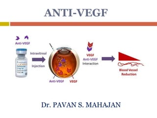
- 1. ANTI-VEGF Dr. PAVAN S. MAHAJAN
- 2. HISTORY OF VEGF Michaelson 1940: “Under condition of Hypoxia a diffusible factor [Factor X] is released by ischemic tissue [retina] that leads to neovascularization of retina and anterior segment”
- 3. Napoleone Ferrare and Colleagues: 1989 “A molecule in conditioned media from bovine pituitary follicular cells promotes proliferation of Endothelial cells” This molecule went on to be known as VASCULAR ENDOTHELIAL GROWTH FACTOR (VEGF) 1992: It was hypothesized that the Michaelson’s Factor X could be VEGF.
- 4. Angiogenesis •Angiogenesis – Growth of blood vessels
- 5. Angiogenesis – A Natural Process Physiological angiogenesis – Embryonic development – Wound healing – Endometrium, ovary
- 6. Angiogenesis – A Pathologic Problem Pathological angiogenesis – Cancer – Eye disease ie. ARMD, DME etc.
- 8. Role of VEGF-A in angiogenesis Stimulates angiogenesis Increase permeability Chemotactic factor for inflammatory cells – Promotes inflammation
- 9. VEGF-A is present in the healthy eye VEGF and its receptors naturally expressed in healthy eye High concentrations of VEGF in RPE Receptors primarily located on vascular endothelial cells In healthy eye, VEGF may play a protective role in maintaining adequate blood flow (choroidal) to RPE and photoreceptors
- 10. Pathologic VEGF-A secreted by RPE • Hypoxia • Accumulation of lipid metabolic byproducts • Oxidative stress to retina & RPE • Alterations in Bruch’s membrane • Drusen (Reduction in the choriocapillaries blood flow and block diffusion of oxygen and nutrients to RPE and photoreceptors) Initiating stimuli for VEGF release Witmer et al, Prog Retin Eye Res, 2003; Ferrara et al, Nat Med, 2003. 10
- 11. Source Stimuli Endothelium Muller cells Astrocytes RPE cells Neurons pericytes Hypoxia Cytokines ROS Age High glucose oncogenes
- 12. 12 Neovascular AMD and DME primarily affect different vascular systems • Primarily associated with breakdown of the inner BRB2 • Primarily associated with breakdown of the outer BRB1 1. Cummings M, Cunha-Vaz J. Clin Ophthalmol 2008;2:369–375 2. Bhagat N et al. Surv Ophthalmol 2009;54:1–32 Neovascular AMD DME RPE layer Retinal capillaryMicroaneurysm Fovea Fovea Choroid Druse n PR L ON L INL IPL OP L IPL, inner plexiform layer; INL, inner nuclear layer; OPL, outer plexiform layer; ONL, outer nuclear layer; PRL, photoreceptor layer Choroidal neovascularization (CNV) Edema Retina Hard exudate
- 13. 13 Structural changes observed1,2 • Retinal thickening • Subretinal fluid accumulation • Cystoid spaces • Pigment epithelial detachment • CNV OCT of neovascular AMD Structural changes observed3,4 • Retinal swelling (thickening) • Cystoid macular edema • Serous retinal detachment • Vitreomacular traction • Hard exudates OCT of DME Differences in neovascular AMD and DME are evident from OCT images 1. Liakopoulos S et al. Invest Ophthalmol Vis Sci 2008;49:5048–5054 2. The Royal College of Ophthalmologists. AMD: guidelines for management. 2009. http://www.rcophth.ac.uk/docs/publications/AMD_GUIDELINES_FINAL_VERSION_Feb_09.pdf [accessed Sep 2009] 3. Bhagat N et al. Surv Ophthalmol 2009;54:1–32 4. Lang GE. In Developments in ophthalmology. 2007. p31–47 Retina RPE layer Choroid
- 14. 14 Common rationale for targeting VEGF Augustin AJ, Kirchhoff J. Expert Opin Ther Targets 2009;13:641–651 Kijlstra A et al. In Uveitis and immunological disorders. 2009. p73–85 Bhagat N et al. Surv Ophthalmol 2009;54:1–32 Upregulation in expression of VEGF Changes in the ageing eye Sustained hyperglycaemia Neovascularization Neovascular AMD Hyperpermeability Macular edema Phosphorylation of tight junction proteins Disorganization of BRB
- 15. The Angiogenic Cascade Hypoxia • Hypoxia stimulates production of VEGF and other angiogenic growth factors in the subretinal space
- 16. The Angiogenic Cascade (cont) VEGF FGF Other Angiogenic Growth Factors Vascular Endothelial Cell • VEGF and other angiogenic factors bind to endothelial cells of nearby capillaries and activate them Hypoxia
- 17. The Angiogenic Cascade (cont) Proliferation Migration Proteolysis VEGF FGF Other Angiogenic Growth Factors Vascular Endothelial Cell • Activated endothelial cells proliferate, migrate, and release proteases Hypoxia
- 18. The Angiogenic Cascade (cont) Proliferation Migration Proteolysis VEGF FGF Other Angiogenic Growth Factors Vascular Endothelial Cell • Enzymes permeabilize the basement membrane Hypoxia Basement Membrane
- 19. The Angiogenic Cascade (cont) Proliferation Migration Proteolysis VEGF FGF Other Angiogenic Growth Factors Vascular Endothelial Cell • Migrating endothelial cells form new blood vessels in formerly avascular space Hypoxia Basement Membrane
- 20. The angiogenic cascade in AMD
- 21. Characteristics of new vessels
- 22. VEGF-A isoforms VEGF-A is a single gene that codes for distinct protein isoforms Human VEGF-A isoforms include: 121, 165, 189 and 206 Isoform number refers to number of amino acids contained in the mature, secreted proteins Murine (rodent) isoforms contain 1 less amino acid than human isoforms Thus, murine equivalent of VEGF165 is VEGF164 Neufeld et al, FASEB J, 1999; Robinson and Stringer, J Cell Sci, 2001; Ferrara et al, Endocr Rev, 1992; Adamis and Shima, In press, 2004; Shima et al, J Biol Chem, 1996.
- 23. VEGF-A is the best studied, has been most strongly associated with angiogenesis, and is the target of most current anti-VEGF treatments. VEGF-A signals through two receptor tyrosine kinases, VEGFR1 and VEGFR2, and is the only member of the VEGF gene family found to be induced by hypoxia.
- 24. Studies have established that VEGF165 is the most abundantly expressed VEGF-A isoform and has a vital role in angiogenesis. one study has found that VEGF121, although less abundant, is more mitogenic than VEGF165 or VEGF189.
- 25. Ferrara et al, Nat Med. 2003; 9: 669 1651 - Most abundant isoform expressed in humans & largest contributor to angiogenesis - Sequestered in the extracellular matrix 1 189 - Highly diffusible and bioactive isoform VEGF-A121 86-89 1 121 1 206 - Highest molecular weight isoform bound to extracellular matrix VEGFR Binding Domain Heparin Binding Domain VEGF-A206 86-89 VEGF-A189 86-89 VEGF-A165 86-89 VEGF-A isoforms
- 26. All VEGF-A isoforms except VEGF121 contain a plasmin cleavage site and theoretically may be cleaved by plasmin to generate the smaller VEGF110 form.
- 27. VEGF-A110 Soluble & bioactive plasmin cleavage product Plasmin 1651 VEGF-A165 VEGF-A110 86-89 121110 1 11086-89 VEGF Receptor Binding Domain Keyt et al, J Biol Chem. 1996; 271: 7788 VEGF Receptor Binding Domain Heparin Binding Domain Targeted binding site
- 29. Anti-VEGFS
- 30. HYPOXIA VEGF Isoforms VEGF A Proteins Anti VEGF VEGF receptors Neovascularization Retina Anterior Segment
- 31. Indications - Wet ARMD CRVO/BRVO PDR DME Pre op (5-7 days) in surgery for PDR and vitreous hmg. ROP Eales disease Refractory post surgical CME Coats disease Neovascular glaucoma Iris neovascularisation Before keratoplasty to reduce corneal neovascularisation Pterygium Trabeculectomy ( to modulate wound healing ) Posterior segment Anterior segment
- 33. PEGAPTANIB (MACUGEN) pegaptanib sodium (MacugenTM; OSI- Eyetech, Inc. and Pfizer, Inc.) approved in December 2004. Pegaptanib is an RNA aptamer that inhibits the VEGF165 isoform while sparing VEGF121
- 34. PEGAPTANIB (MACUGEN) First Anti Angiogenic Agent 28 Base RNA Aptamer NON-IMMUNOGENIC NATURE Selectively binds extra cellular VEGF 165 DOES NOT EFFECT NORMAL VASCUALR GROWTH
- 35. V.I.S.I.O.N. (VEGF Inhibition Study in Ocular Neovascularization) trials A total of 1186 subjects with any angiographic subtypes of neovascular AMD were included. Patients received intravitreous injections of 0.3 mg, 1 mg or 3 mg pegaptanib or sham injections every six weeks for 48 weeks. Subjects with predominantly classic lesions could also have received photodynamic therapy with verteporfin (PDT; VisudyneTM, Novartis) at investigator discretion.
- 36. After one year, the 0.3 mg dose conferred a signifi cant clinical benefit compared to sham treatment as measured by proportions of patients losing <15 letters of visual acuity. In contrast to PDT, clinical benefi t was seen irrespective of angiographic AMD subtype, baseline vision or lesion size and led to the clinical approval of pegaptanib for the treatment of all angiographic subtypes of neovascular AMD.
- 37. The 1 mg and 3 mg doses showed no additional benefit beyond the 0.3 mg dose. In an extension of the V.I.S.I.O.N. study, patients in the pegaptanib arms were rerandomized to continue or discontinue therapy for 48 more weeks. Compared to patients discontinuing pegaptanib or receiving usual care, those remaining on 0.3 mg pegaptanib received additional significant clinical benefit in the second year. Further subgroup analyses suggested that pegaptanib treatment was especially effective in those patients who were treated early in the course of their disease.1
- 38. BEVACIZUMAB (AVASTIN) full-length, humanized monoclonal antibody directed against all the biologically active isoforms of vascular endothelial growth factor (VEGF-A). FDA approved drug for treatment of metastatic colorectal cancer. Off label drug…
- 39. It is a recombinant IgG1 antibody with a molecular weight of about 149kD that is produced in a Chinese Hamster Ovary mammalian cell expression system in a nutrient medium containing the antibiotic gentamicin. It is a clear to slightly opalescent, colorless to pale brown, sterile solution with pH 6.2. It was originally designed for intravenous (IV) infusion and is supplied in 100 mg and 400 mg preservative-free, single-use vials to deliver 4 mL or 16 mL of (25 mg/mL).
- 40. Inhibits vegf mediated angiogenesis and vascular leakage. Short term results are rewarding though long term efficacy and toxicity is not conclusively established. Many multicenter trials are going on to establish long term safety . Since 2006, there have been reports of over fifty one ocular entities being treated with bevacizumab, generally those associated with neovascularization or vascular leakage as a consequence of an underlying disease.
- 41. Intravitreal bevacizumab administration is now used as a first line therapy for several diseases by many retina specialists and accounts for more than 50% of anti- VEGF administrations in the US. Additionally, the markedly lower cost of bevacizumab as compared to similarly effective drugs such as ranibizumab has led it its adoption for treatment of exudative AMD throughout the world.
- 42. Ranibizumab Ranibizumab (Lucentis, Genentech, Inc., South San Francisco, CA) is a humanized, affinity-matured VEGF antibody fragment that binds to and neutralizes all isoforms of VEGF-A and their biologically active degradation products
- 43. RANIBIZUMAB (LUCENTIS) NON BINDING FRAGMENT Makes it Humanized Therefore Less antigenic Fab FRAGMENT Mouse Derived Active against all Isoforms of VEGF High affinity binding site
- 44. BEVACIZUMAB (AVASTIN) Full Sized Antibody. 148 kilodaltons. Half Life 20 days. Clearance is slow. Long action & less dosage. Cost’s less. RANIBIZUMAB (LUCENTIS) Antibody Fragment. 48 kilodaltons. Half Life of 3 days. Clearance 100 folds faster. 140 times higher affinity. Costly.
- 45. Aflibercept(Vegf Trap Eye/VTE) Fusion protein Binds to VEGF-A, VEGF-B, and PIGF creating inert complexes. These receptors play a role in angiogenesis
- 46. Route of Administration Intravitreal Bioavailability Mean Cmax of free aflibercept in the plasma was 0.02 mcg/mL (range: 0- 0.054 mcg/mL), attained in 1-3 days. Time to Peak 1-3 days Multiple Dosing 2 mg monthly for the first 3 months, followed by 2 mg every 2 months, Long duration,leser injection Clearance No drug metabolism studies have been conducted. Expected to undergo elimination through both target- mediated disposition via binding to free endogenous VEGF and metabolism via proteolysis.
- 47. Clinical Efficacy of Eylea (aflibercept) Eylea (aflibercept) Phase III Clinical Trials VIEW 1 and VIEW 2---Study indication for wet AMD Copernicus---Study indication for macular edema due to CRVO Galileo---Study indication for macular edema due to CRVO
- 48. VIEW 1 and VIEW 2 Phase III, Randomized, Double-Blind Head-to-head against ranibizumab 52 Week Total for each study Clinic visits every 4 weeks for 52 weeks Primary Endpoint Proportion of patients who maintained vision at Week 52 Maintenance of vision defined as loss of ≤ 15 letters in the ETDRS letter score as compared to baseline
- 49. Primary Endpoint Results Study Group VIEW 1 VIEW 2 Aflibercept 2mg every 4 weeks 95.1% 95.6% Aflibercept 0.5mg every 4 weeks 95.9% 96.3% Aflibercept 2mg every 8 weeks 95.1% 95.7% Ranibizumab 0.5mg every 4 weeks 94.4% 94.4% *All results are at Week 52
- 50. VIEW 1 and VIEW 2 Strengths Power set and met Power was set at 190, and +260 enrolled in each study group Randomized and Double-blind Limitations No clear reporting of withdraws from trial Exclusion criteria of: “No prior treatment with anti- VEGF.” Manufacturer funded study Delfini Evidence: Grade B VEGF=vascular endothelial growth factor
- 51. Copernicus Phase III, Randomized, Double-Blind 52 Weeks Either Aflibercept or sham injection every 4 weeks for 24 weeks, then XO PRN aflibercept injections Primary Endpoint Proportion of patients who gained vision at Week 24 and Week 52 Gain in vision defined as gain of ≥ 15 letters in the ETDRS letter score as compared to baseline
- 52. Primary Endpoint Results Study Group for Copernicus Week 24 Week 52 Aflibercept 2mg every 4 weeks 56.1% --- Sham Injections every 4 weeks 12.3% --- Aflibercept 2mg every 4 weeks to Aflibercept 2mg PRN --- 55.3% Sham Injections every 4 weeks to Aflibercept 2mg PRN --- 30.1%
- 53. Copernicus Strengths Power was set and met Power set at 169, and 189 patients enrolled in study Randomized and Double-blind Limitations Lack of head-to-head study design Manufacturer funded study Delfini Evidence: Grade B-U
- 54. Galileo Phase III, Randomized, Double-Blind 52 Weeks Either Aflibercept or sham injection every 4 weeks Primary Endpoint Proportion of patients who gained vision at Week 24 and Week 52 Gain in vision defined as gain of ≥ 15 letters in the ETDRS letter score as compared to baseline
- 55. Primary Endpoint Results Study Groups for Galileo Week 24 Week 52 Aflibercept 2mg every 4 weeks 63.1% 60.2% Sham injections every 4 weeks 22.1% 32.4%
- 56. Galileo Strengths Randomized and Double-blind Limitations Power was not set or met Lack of head-to-head study design Manufacturer funded study Delfini Evidence: Grade B-U
- 57. Bevasiranib(Cand5) siRNA-based anti-angiogenic agent proposed for the treatment of wet AMD. Administered as bevasiranib sodium, the chemical structure is a complex of two 21-nucleotide RNA eicosasodium molecules. Upon introduction into the cell, a siRNA binds to and activates the RNA-induced silencing complex (RISC).
- 58. A single activated RISC complex can bind to and destroy hundreds of mRNAs, thus preventing translation and protein synthesis. By silencing the synthesis of VEGF protein, bevasiranib is an ideal therapeutic strategy for the eye, which, because of its limited volume, requires potent molecules for effective local administration. No data on the metabolism or elimination of bevasiranib.
- 59. Preliminary results of Phase 1 and 2 clinical trials of bevasiranib have shown promising results for the treatment of wet AMD and diabetic macular edema. But the lack of available data from randomized placebo controlled or comparative studies makes it difficult to objectively evaluate the role of bevasiranib.
- 60. pegaptani b bevacizu mab ranibizu mab Vegf trap bevasiran ib Active against VEGF A 165 VEGF A VEGF A VEGF A , PLGF VEGF A FDA APROVAL YES, NO YES NO NO NATURE Pegylated aptamer Full length monoclona l ab. Fab fragment Extracellul ar VEGF blocker siRNA Dose in 0.05, Frequency o.3 mg, Every 6 week 1.25 mg, Every 4 week 0.5 mg, Every 4 week 0.5-2.0mg, Every 4 or 12 week 0.5-2.0 mg, Every 12 week
- 61. BENEFITS OF BEVACIZUMAB High efficacy. Longer half life up to 20 days and thus fewer injections. Lack of preservative. Higher safety dose: Normal i/v dose is 1.25mg Retinal toxicity occurs at dosage > 3.5mg. Insignificant systemic absorption and effect. No experimentally proven toxicity. Lower cost. Wide availability.
- 62. INTRAVITREAL BEVACIZUMAB 4 ml vial of Bevacizumab has 100 mg of drug. In a tuberculin Syringe we take 0.05ml. thus (1.25mg/0.05ml) With a 30 Gauge needle and this syringe. Drug is injected from the limbus - 3.5 mm in case of Pseudophakic. - 4.0 mm in case of a Phakic.
- 63. STEPS OF INTRAVITREAL ANTI-VEGF Under topical anesthesia. Betadine painting over lid, 1-2 drops of Betadine instilled in the conjunctival sac, then washed with normal saline. Drug is taken in tuberculin syringe with 30G needle. (1.25mg/0.05ml) Ask the patient to look up. From the inferior-temporal quadrant at 4 mm from limbus (Phakic) the needle is directed towards the centre of the globe and drug is injected. Injection site pressed with a cotton bud. IOP and CRA perfusion is assessed. Topical Antibiotic is administered for one week. (TDS to QID)
- 64. Ocular A/E ---- Ocular inflammation – 2.1-2.9% TRD in eyes with PDR – 5.2% RPE tears – 0.8-2.2% RPE tears with vascularised PED – 22% Vitreous Hemorrhage. Accidental injury to Lens capsule. Raised IOP. Central Retinal Artery Occlusion. Endophthalmitis – 0.019-0.029%
- 65. Systemic A/E Non ocular hemorrhage – Patients with history of stroke are at incresed risk. No increase in risk for MI/Death.
- 66. REPORTS REGARDING ANTERIOR SEGMENT REACH OF INTRAVITREAL Anti VEGFS “Intravitreal injection of Bevacizumab penetrates quickly into the ciliary body, iris and anterior chamber angle”. “Penetration into Iris appears to be faster than that into ciliary body and anterior chamber angle.” “The highest concentration is seen in the anterior chamber from day 1 to 4 after an Intra-Vitreal injection and it regresses by day 14.” European Ophthalmic Review 2009; 3(1): 36-38
- 67. ADVANTAGE OF Anti VEGF IN NVI & NVA Causes regression of iris and angle neovascularization within 48 hours. IOP lowering effect seen in some cases. A window period is gained during which an effective P.R.P can be done. Effective in reducing the chances of failure of and intra operative bleed during filtering surgery, when given 48-72 hours prior to surgery.
- 68. Co-Relation between VEGF concentration and NVI / NVA RETINAL HYPOXIA VEGF Conc. > 890 pg/ml of Aqueous Iris and Angle Neovascularization Intravitreal injection of Anti VEGF(AVASTIN) VEGF Conc. < 550 pg/ml of Aqueous NEOVASCULARIZATION REGRESSES
- 69. CAUSES OF NEOVASCULARIZATION OF IRIS & ANGLE Ocular vascular Diseases: Ischemic CRVO Diabetic Retinopathy BRVO CRAO Choroidal Hemangioma Sickle Cell retinopathy Extra Ocular Diseases: Carotid artery disease/ ligation Ocular Ischemia Carotid Cavernous Fistula
- 70. Other Ocular Diseases - Retinal Detachment - Eales Disease - Coats Disease - Retinopathy of prematurity - Persistent hyper plastic primary Vitreous. - Norrie’s Disease - Sticklers Syndrome Ocular Inflammatory Disease - Chronic Uveitis - Sympathetic Ophthalmia - Endopthalmitis - V.K.H. Syndrome
- 71. Post therapy - After Cataract Extraction in diabetic retinopathy. - After Vitrectomy in Diabetic retinopathy - Retinal Detachment Surgery - Post Radiation therapy - Laser Coreoplasty Ocular Neoplasm Malignant Melanoma Retinoblastoma
- 72. ANTERIOR SEGMENT USES OF Anti - VEGF Glaucoma - Iris and Angle Neovascularization - Neovascular Glaucoma - During Trabeculectomy for NVG Corneal Vascularization Pterygium
- 73. BEVACIZUMAB (AVASTIN) FOR CORNEAL VASCULARISATION Causes of corneal Vascularization: - Trauma: (physical, chemical, thermal or radiation) - Post infective: ( viral, bacterial, fungal, protozoal) - Corneal dystrophies and degeneration. - Endothelial malfunction: (PBK, ABK, uveitis) - Inflammatory corneal disorders.
- 74. PROPOSED MECHANISM OF Anti VEGF ACTION Various pathologies Corneal inflammation Expression of VEGF & other factors Vascular growth S/C injection of Anti VEGF Superficial Vascularization Deep Vascularization PARTIAL REGRESSION
- 75. SUBCONJUNCTIVAL INJECTION OF Anti VEGF Dosage: 2.25 mg in 0.1ml. (exact dosage ??) Preferred site is to inject near corneal vascularization. Injection site could be more than one. Regression seen is usually partial and may require repeat injections. There is a possibility that the drug might not work in old vascularized cornea. The effect are less pronounced for centrally located vessels. (?? intrastromal injection of Anti-VEGF)
- 76. ADVANTAGES OF SUBCONJUNCTIVAL Anti VEGF • Reduced vascularity of cornea. • Better outcomes of penetrating keratoplasty. • Visual improvement in few patients. • Can be used in adjunct to steroid therapy or immunosuppressive drugs.
- 77. TOPICAL BEVACIZUMAB (AVASTIN) WITH ANTIBIOTICS Post operative penetrating keratoplasty cases, specially in vascularized cornea. Avastin is given in drop form with antibiotics drops. Dosage: ??? 0.01ml in 3ml antibiotic twice a week for 3 weeks. However topical steroids, antibiotics & lubricants are continued.
- 78. SUBCONJUNCTIVAL Anti VEGF IN PTERYGIUM PRIMARY PTERYGIUM : Subconjunctival Bevacizumab (Avastin) 1.25mg/0.05ml causes regression of vascularity, symptoms (irritation, redness) up to 7 wks. post injection only. Teng CC, et al. Cornea. 2009 May; 28(4):468-70
- 79. TOPICAL Anti VEGF IN PTERYGIUM RECURRENT PTRYGIUM: Topical Bevacizumab (Avastin) 25mg/ml QID dosing for 3 weeks, in a case of recurrent impending pterygium prevented recurrence up 6 mths follow up. Wu PC, et al. Cornea.2009 Jan;28(1):103-4
- 80. STUDIES
- 81. The CRUISE Study To assess the efficacy and safety of intraocular injections of 0.3 mg or 0.5 mg ranibizumab in patients with macular edema after central retinal vein occlusion (CRVO). The CRUISE was a 6-month Phase III Multicenter Randomized Injection-controlled study Additional 6 months of followup (total 12 months)
- 83. Interpretation Monthly ranibizumab therapy improved mean BCVA and increased the proportion of patients gaining 15 ETDRS letters Patients treated with ranibizumab were twice as likely to have BCVA of 20/40 compared with the sham group at month 6 The rapid and significant resolution of macular edema by day 7 in both ranibizumab groups suggests that the majority of retinal edema in CRVO is VEGF mediated.
- 84. Gaps and Unanswered Questions Does not address whether ranibizumab treatment is beneficial to patients who present with VA 20/40 or better The duration of ranibizumab treatment required for patients with macular edema following CRVO What percentage of patients will require treatment beyond the mandated 6 monthly treatments require further exploration?
- 85. DRCR.net study 2011 Ranibizumab plus Prompt or Deferred Macular Laser Photocoagulation versus Triamcinolone plus Macular Laser Photocoagulation Multicenter, randomized clinical trial which included 854 eyes of 691 patients M. J. Elman, N. M. Bressler, H. Qin et al., “Expanded 2-year follow-up of ranibizumab plus prompt or deferred laser or triamcinolone plus prompt laser for diabetic macular edema,” Ophthalmology, vol. 118, no. 4, pp. 609–614, 2011.
- 86. DRCR.net study 2011 Four treatment groups: Prompt laser with sham injection, 0.5 mg of ranibizumab with prompt laser 0.5mg of ranibizumab with laser deferred for at least 24 weeks 4mg of triamcinolone with prompt laser.
- 87. DRCR.net study 2011 At one year, the two groups treated with ranibizumab had a significant change in mean VA from the baseline. The triamcinolone and laser alone groups did not show a significant change in VA. a subgroup analysis of pseudophakic eyes in the triamcinolone group showed similar results as for those in the ranibizumab groups.
- 88. BOLT study The most meaningful study concerning Bevacizumab for DME Compare the efficacy of anti-VEGF therapy to focal laser photocoagulation in 80 patients with CSME . A prospective, randomized phase III clinical trial
- 89. BOLT study Bevacizumab injections given every 6 weeks or laser treatment performed every 4 months. Injected eyes received 3-9 injections, whereas the focal laser eyes received 1-4 treatments in the 12-month study period.
- 90. BOLT study Patients in the bevacizumab group were 5.1 times as likely to gain at least 10 letters. BOLT 2012 : long term effect of Bevacizumab is maintained at 24 months The BOLT study suggested that intravitreal bevacizumab therapy should be considered as a first choice in the management of center- involving DME.
- 91. PACORES study The Pan-American Collaborative Retina Study Group Retrospective interventional multicenter study evaluated the retinal thickness and visual acuity data of 80 consecutive patients (139 eyes) receiving intravitreal Avastin of 1.25 or 2.5mg with a minimum followup of 24 months Arevalo JF, et al. Primary intravitreal bevacizumab (Avastin) for diabetic macular edema: results from the Pan-American Collaborative Retina Study Group at 6-month follow-up. Ophthalmology 2007;114(4):743-750.
- 92. PACORES study Results showed that at 24 months 44.6% eyes remained stable, 51.8% improved 2 or more lines, and 3.6 % decreased 2 or more lines. Patients who received on average 5.8 injections of single or double dose Avastin demonstrated a maintained partial resolution of macular edema
- 93. RIDE/RISE study Double-blinded, sham-controlled randomized studies with a followup of 36 months. Patients received monthly injections of 0.3 mg ranibizumab, 0.5 mg ranibizumab, or sham. As twice as many patients in the ranibizumab groups gained ≥15 letters compared to the sham group D. S. Boyer, J. Sy, A. C. Rundle et al., Ranibizumab for Vision Loss due to Diabetic Macular Edema—Results of two Phase III Randomized trials, American Diabetes Association 71st Scientific Sessions, San Diego, Calif, USA, 2011.
- 94. RESOLVE Study The Safety and Efficacy of Ranibizumab in Diabetic Macular Edema A multicenter, randomized, and double masked evaluated the efficacy and safety of intravitreal ranibizumab (0.3 or 0.5mg) compared with sham treatment in 151eyes with DME over 12 months
- 95. RESOLVE Study Results showed a significant and continuous improvement in BCVA and central retinal thickness for ranibizumab versus sham. P. Massin, F. Bandello, J. G. Garweg et al., “Safety and efficacy of ranibizumab in diabetic macular edema (RESOLVE study): a 12-month, randomized, controlled, double-masked, multicenter phase II study,” Diabetes Care, vol. 33, no. 11, pp. 2399–2405, 2010.
- 96. RESTORE study A randomized, double-masked, multicenter phase III study over 12 months compared ranibizumab + sham laser and ranibizumab + laser with laser + sham injection for DM in 345 patients P. Mitchell, F. Bandello, U. Schmidt-Erfurth et al., “The RESTORE study: ranibizumab monotherapy or combined with laser versus laser monotherapy for diabetic macular edema,” Ophthalmology, vol. 118, no. 4, pp. 615–625, 2011.
- 97. RESTORE study Ranibizumab or sham injections were given monthly for three months and then PRN; laser or sham laser was given at baseline and then PRN after an interval of at least three months.
- 98. RESTORE study In the ranibizumab and ranibizumab + laser groups a rapid improvement of VA was observed after one month which continued up to three months and was sustained until month 12 Likewise, the percentage of patients reaching VA ≥ 20/40 was greater in the two ranibizumab groups Ranibizumab monotherapy and combination with laser treatment are superior to laser treatment alone for DME.
- 99. Ranibizumab for wet-AMD ANCHOR Study Randomized 1:1:1 Verteporfin PDT Sham PDT Sham PDT Sham injection (n = 143) Ranibizumab 0.3 mg (n = 140) Ranibizumab 0.5 mg (n = 140) Predominantly classic lesions secondary to AMD (N = 423) Brown et al. N Engl J Med 2006; 355: 1432- Phase III, multicenter, double-masked, 24-month study AMD, age-related macular degeneration PDT, photodynamic therapy
- 100. Conclusions ANCHOR study Ranibizumab demonstrated efficacy in patients with subfoveal, predominantly classic CNV associated with neovascular AMD over a 2-year period treatment with monthly intravitreal Ranibizumab prevented central vision loss and improved mean VA VA benefit from Ranibizumab was both rapid (within one month) and sustained (over the 2-year study period) Ranibizumab was superior to treatment with verteporfin PDT for patients losing <15 letters from baseline and mean change in VA over time Brown et al. Ophthalmology 2009; 116: 57-65
- 101. Ranibizumab for wet-AMD Marina Study )0for wet-AMD Marina Study Randomized 1:1:1 Sham (n = 238) Ranibizumab 0.3 mg (n = 238) Ranibizumab 0.5 mg (n = 240) Minimally classic or occult with no classic lesions secondary to AMD (N = 716) Rosenfeld et al. N Engl J Med 2006; 355: 1419-1431 Phase IInPhase III, multicenter, double-masked, 24-month study AMD, age-related macular degeneration
- 102. Conclusions MARINA study The results from MARINA demonstrate that intravitreal Ranibizumab is associated with clinically and statistically significant benefits with respect to VA in patients with minimally classic or occult lesions with no classic CNV associated with neovascular AMD over a 2-year period In patients treated with Ranibizumab, efficacy was maintained throughout the 2-year period whereas patients in the sham group continued to experience a decline in vision Rosenfeld et al. N Engl J Med 2006; 355: 1419-1431
- 103. Conclusions Ranibizumab was well tolerated over a 2-year period Efficacy outcomes were achieved with a low rate of serious ocular AEs and no clear difference from the sham-treated group in the rate of non-ocular AEs Subsequent to the results of the ANCHOR and MARINA trials, Ranibizumab was licensed for the treatment of neovascular AMD by the US Food and Drug Administration in 2006 and in the European Union in 2007 AE, adverse event Rosenfeld et al. N Engl J Med 2006; 355: 1419-1431
- 104. Conclusions Improvements in VA from baseline seen (2-year) are greater in ANCHOR than MARINA ANCHOR patients had predominantly classic CNV lesions; MARINA patients had minimally classic or occult with no classic CNV lesions average CNV lesion size was smaller in ANCHOR; however, predominantly classic lesions are typically more aggressive and lead to more rapid loss of VA than minimally classic lesions, therefore the potential for improvement is greater predominantly classic lesions are typically diagnosed early and therefore treated earlier than occult lesions which may account for the greater improved VA outcomes observed recent VA loss associated with rapidly progressing predominantly classic CNV may be partially reversible whereas earlier VA loss due to slowly progressing occult CNV may be irreversible, providing little opportunity for VA improvement with treatment Brown et al. Ophthalmology 2009; 116: 57-65 Rosenfeld et al. N Engl J Med 2006; 355: 1419-1431
- 105. SUMMING UP Anti VEGF’s have a definitive role in suppressing neovascularization and to some extent the severity of the disease in Choroidal and Retinal neovascularization. Neovascular glaucoma. Corneal vascularization. (still in nascent stage) Vascularization of primary & recurrent Pterygium. (still in nascent stage) Ocular tumors & neoplasms.????
- 106. LIMITATIONS Anti VEGF cause regression of neovascularization. There is no effect on the basic pathology responsible for neovascularization (hypoxia). It’s the disease that is to be cured to prevent hypoxia and its effects. They are a valuable ammunition in our armamentarium but alone are not curative of the condition. They give us time & VALUABLE breathing space during which we can plan our course of further action.
- 107. Thank you