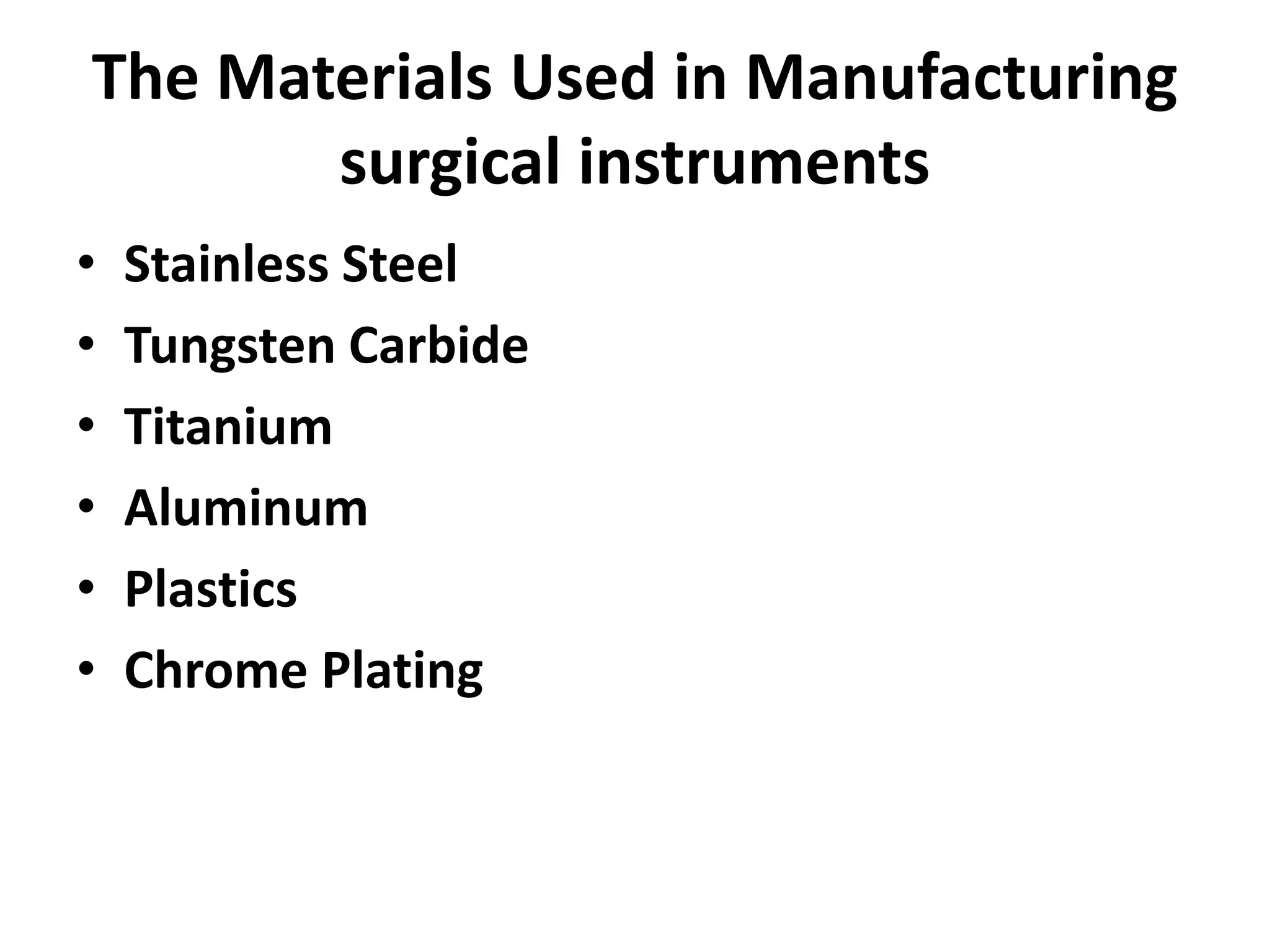
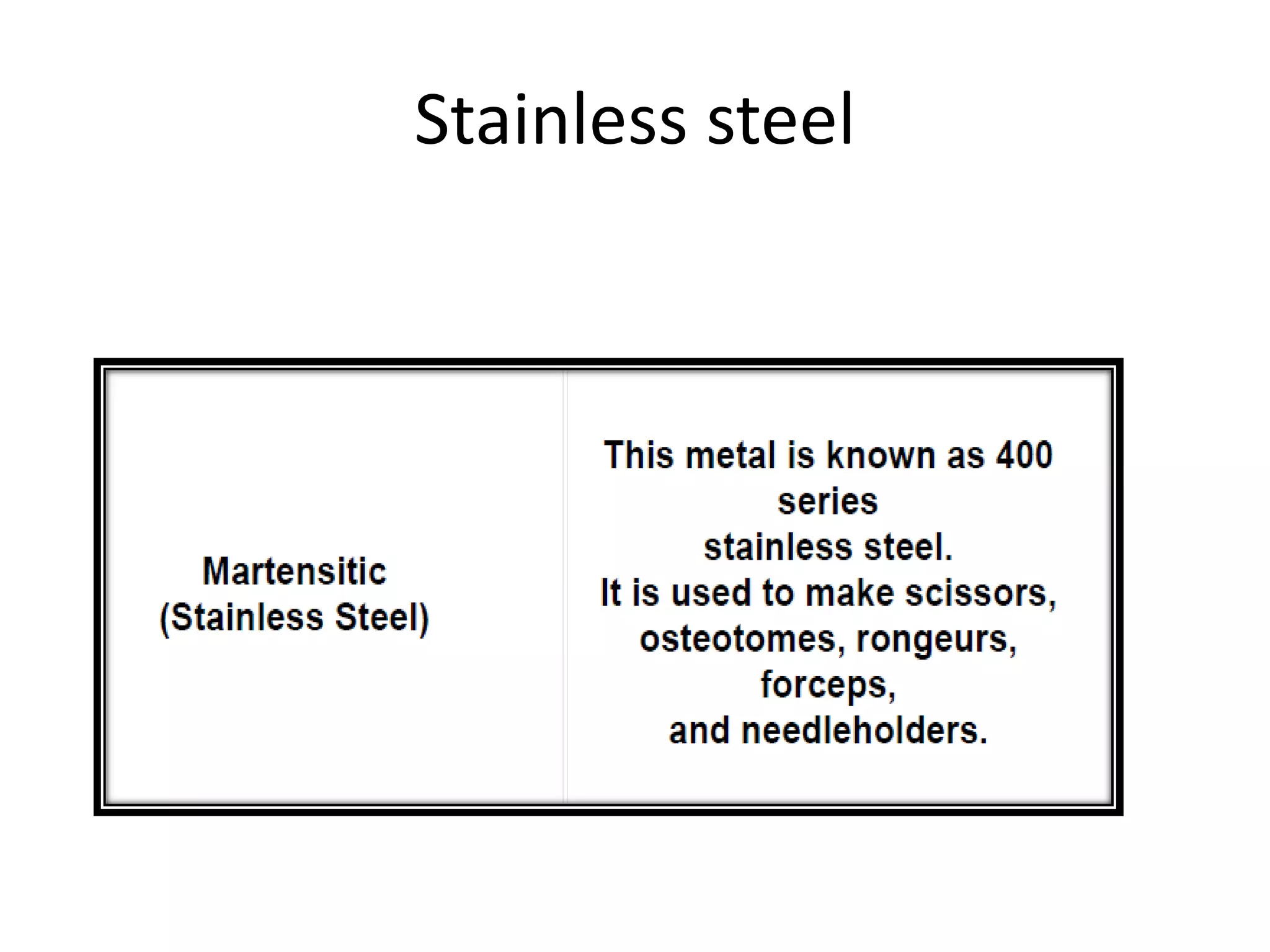
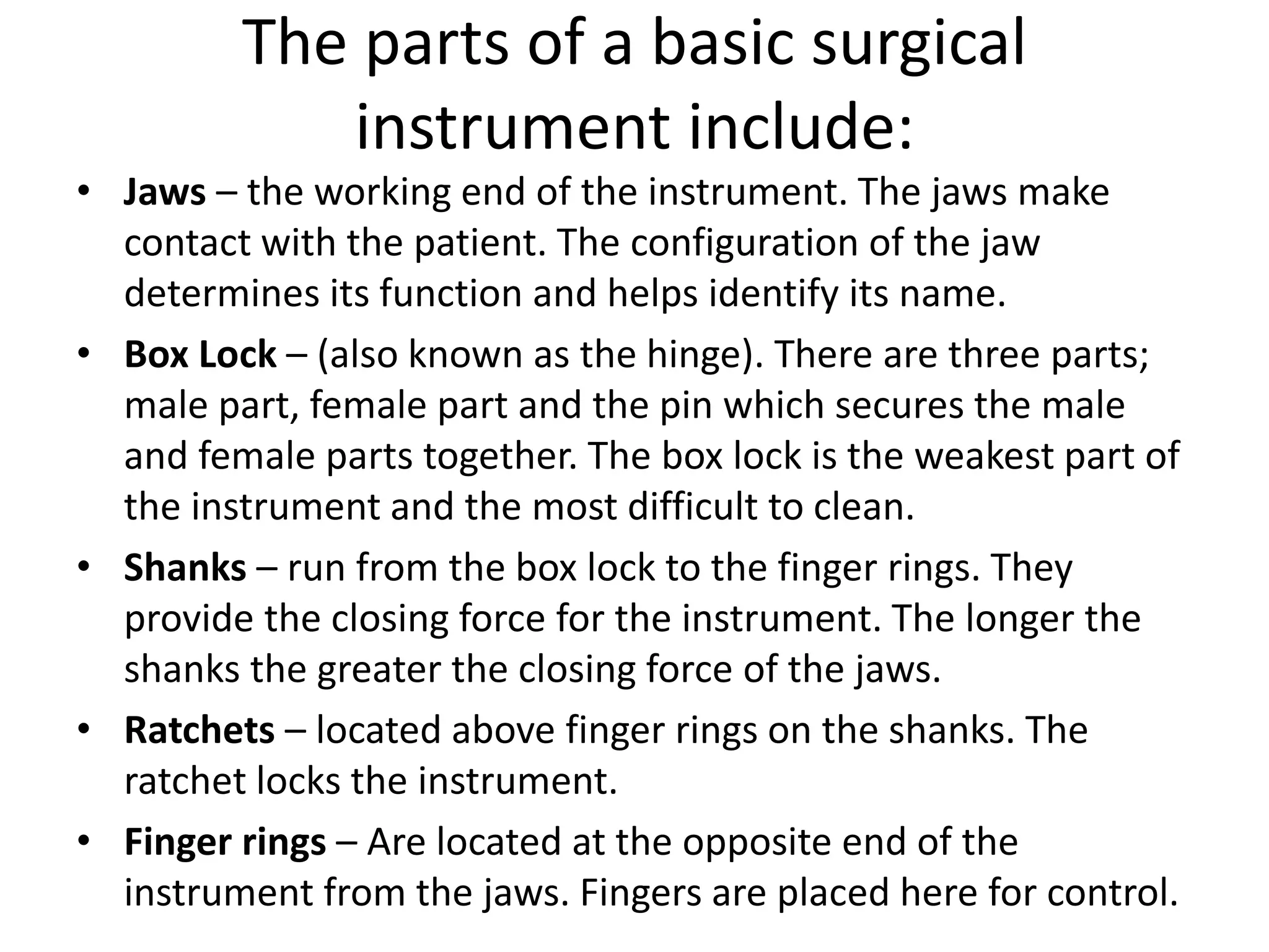
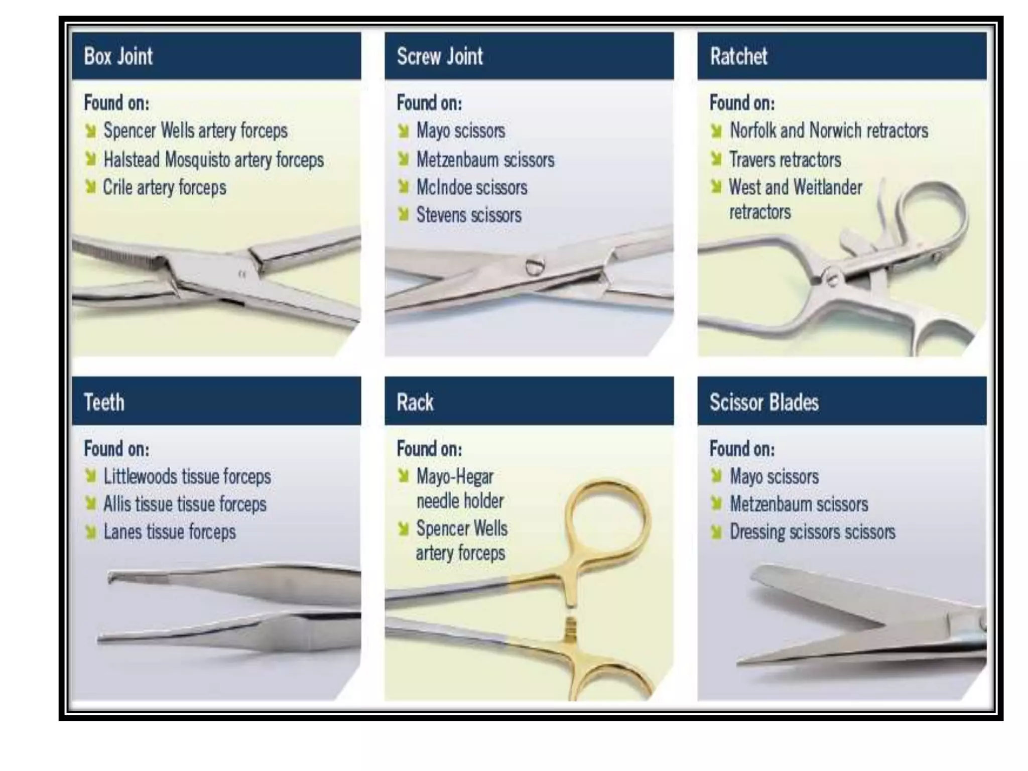
This document provides information on the manufacturing process and types of surgical instruments. It discusses the materials used, key parts of instruments like jaws and handles. Different categories of instruments are outlined including cutting instruments, grasping instruments, hemostatic instruments, and retractors. Specific instruments are described in detail like scalpels, scissors, forceps, needle holders. Fine vascular instruments for microsurgery are also covered. The document aims to educate on the variety of instruments used in surgery and their functions.
Surgical retractors are instruments used to separate the edges of an incision and hold back underlying tissues, allowing access to body parts. There are two main types - hand retractors which must be held manually, and self-retaining retractors which use mechanisms like screws or clamps to hold tissues independently. Common hand retractors include Senn, Army-Navy, Ribbon, Hohmann, Farabeuf, Meyerding, Deaver, and Richardson retractors used in various procedures. Examples of self-retaining retractors are Weitlaner, Balfour Abdominal, Finochietto Rib, Hip, Gelpi, Joll, and Omni-Tract Wishbone retract
1. Surgical instruments are divided into four main groups: cutting/dissecting, clamping/occluding, grasping/holding, and retracting/exposing. Common instruments include scalpels, scissors, forceps, and retractors.
2. Instruments must be properly cleaned and maintained to function correctly. Stainless steel instruments are stronger but some non-ferrous alternatives are needed for MRI guided surgery.
3. Specific instruments have distinct designs and uses - for example, metzenbaum scissors for delicate tissue, kelly clamps for larger vessels, and gelpi retractors for shallow incisions. Proper technique is required for safe handling of each instrument.
Debridement is an important component of the wound bed preparation (WBP) management Model.
Cause of the wound and patient-centered concerns, debridement is a necessary step in local wound care.
Debridement is the removal of necrotic tissue, exudate, bacteria, and metabolic waste from a wound in order to improve or facilitate the healing process
Common surgical instruments for medical students.
The the PPT contains: introduction, history, handling & care, classification and description of individual surgical instrument with good pictures, synonyms, uses, design description & insight.
Hope this is worth sharing.
Surgical drains have several purposes and types. They help evacuate fluids from surgical sites to prevent infection and allow wounds to heal. Common types include closed suction drains like Jackson-Pratt drains and open drains like Penrose drains. Placement, securing, and care of drains is important to avoid complications like infection, displacement, or blockage. Drains are removed once drainage decreases significantly or they are no longer needed.
Suprapubic cystostomy is a procedure where a catheter is placed through the bladder wall in the suprapubic region to drain urine. It is indicated when urethral catheterization fails or is not possible due to conditions like urethral injury. There are two main types - open or percutaneous, and temporary or permanent procedures. The document describes the pre-operative preparation, surgical steps including incision and catheter placement, closure techniques, post-operative management, and potential complications of suprapubic cystostomy.
The passage discusses the importance of teaching children about money at a young age through age-appropriate activities like allowances, savings jars, and helping with family shopping. Learning good financial habits early can set kids up for financial success as adults by developing an understanding of earning, spending, saving and the value of a dollar. Hands-on learning about money through real world activities can instill lifelong skills for managing personal finances responsibly.
The document provides an outline for performing an open appendectomy surgery. It discusses the relevant anatomy of the appendix, causes of appendicitis, pre-operative care including investigations and antibiotic treatment. It describes the surgical technique including common incisions used, identifying and ligating the appendix and closing the wound. Post-operative care involves monitoring for complications and managing patients depending on whether the case was complicated or uncomplicated.
Burst abdomen refers to the disruption of an abdominal wound between the 6th and 8th days after surgery, usually caused by sutures in the deep layers tearing under strain. A patient may experience a sudden giving way of the wound, pinkish fluid discharge, and protrusion of intestines or omentum. Risk factors include the type of sutures used, midline or vertical incisions, peritonitis or major abdominal surgeries, and the patient's general health status. Treatment requires emergency surgery to cleanse and return all protruding organs, followed by reapproximation of the abdominal wall in layers and wound support.
Preoperative preparation of patients for surgeryErum Khateeb
The document discusses preoperative preparation and optimization of patients for surgery. It covers preoperative care, investigations, assessing surgical risk, and preparing specific patient groups. The goals of preoperative preparation are to anticipate difficulties, enhance patient safety, minimize complications, and optimize high-risk patients. Key aspects include obtaining medical history, conducting physical exams and tests, discussing risks and obtaining consent, and providing prophylaxis for issues like thrombosis. Careful preoperative preparation helps improve surgical outcomes.
An exploratory laparotomy is a surgical procedure where the abdomen is opened to examine the internal organs. It is performed under general anesthesia through an upper midline incision. Indications include trauma, infections, malignancy, complications of other procedures, and removal of foreign bodies. During the procedure, the surgeon examines the abdominal organs and treats any issues found. Potential complications include ileus, infection, hernia, and adhesive obstruction.
This topic is been added in the new edition ( 26th ) of Bailey & Love. This topic covers the types, uses and also the principles of removal of a drain. Every MBBS student should be aware of drains & its uses in surgery.
The document discusses sutures and suture materials used in surgery. It covers the history of sutures, classifications, materials, ideal properties, principles of selection and wound closure techniques. Sutures are used to hold tissues together after injury and come in absorbable and non-absorbable varieties made from natural or synthetic materials like catgut, silk, nylon and polyglycolic acid. Selection depends on tissue type and healing properties. The goal of wound closure is to bring edges together without gaps or tension.
A tourniquet is a device that controls blood flow to an extremity by applying circumferential pressure. It is used medically to create a bloodless surgical field or stop traumatic bleeding. The earliest known usage was by Romans in 199 BC during amputations. Modern tourniquets are used widely in orthopedic and plastic surgery. Complications can include post-tourniquet syndrome if applied for too long or at insufficient pressure, especially in patients with vascular issues. Proper application and monitoring can prevent complications.
This document describes various surgical instruments used in procedures. It discusses forceps, needles, scalpel handles and blades, scissors, suture materials, and retractors. Forceps are used for grasping or compressing tissue and include thumb forceps, hemostats, and needle holders. Scalpel blades come in various sizes like size 10, 11, and 15. Scissors can be blunt-blunt, blunt-sharp, or sharp-sharp and may be straight or curved. Suture materials range in thickness from size 6 to 12-0 and can be synthetic like nylon or natural like silk. Retractors are used to pull tissue away from the surgical site and may be hand-held or self-retaining.
Surgical drains are devices that drain fluids, blood, or air that can accumulate after surgery. There are different types of drains classified as open or closed, and active or passive. Drains are indicated for therapeutic, diagnostic, prophylactic, monitoring, or palliative purposes. Common types include Jackson-Pratt, hemovac, pigtail, and penrose drains. Drains must be properly assessed, maintained if needed, and removed once drainage decreases to prevent complications like infection, blockage, or tissue damage.
This document provides an overview of wound healing and vacuum assisted closure (VAC) therapy. It discusses the standard process of wound healing, novel wound dressing concepts, and how VAC therapy works by applying negative pressure to wounds to promote granulation tissue formation, blood flow, and wound contraction. The document outlines the methodology for VAC application and reviews its uses for different wound types as well as advantages like reduced dressing changes and bacteria. It also discusses future developments and concludes that VAC is a new and improved tool to help convert complicated wounds into simpler wounds.
The principles of vascular repair with sutures were established in the first decade of the 20th century by Alexis Carrel, who in 1912 was awarded the Nobel Prize for medicine for his work .Since then, technical refinements of suture materials have made possible surgical reconstruction of most arteries from the root of the aorta to microvascular anastomosis or repair of the smallest vessels, e.g., digital arteries or those on the surface of the brain.
A laparotomy is a surgical procedure involving an incision through the abdominal wall to access the abdominal cavity. It can be performed for both diagnostic and therapeutic purposes when there is a need for surgery but no definitive pre-operative diagnosis. The key steps include pre-operative preparation, gaining surgical access, systematically exploring the abdominal cavity including solid organs and intestines, performing any necessary procedures, and closing the abdominal wall. Complications can include infection, adhesions, hernias and more, so thorough exploration and drainage if needed is important.
This document discusses suture materials, including threads and needles. It describes the ideal properties of suture threads and classifies threads based on absorbability, raw material, and structure. Common absorbable and non-absorbable suture materials are listed. Needle types such as cutting, taper, and blunt point needles are also defined. Proper suture size and packaging are additionally covered.
Sutures are materials used in surgery for a variety of reason ranging from surgical repair of wounds, ligature, etc. There are a wide variety of sutures with different characteristics that must be born in mind while choosing a suture
THYROIDECTOMY- Operative Surgery
Dear viewers,
Greetings from “Surgical Educator”
Because of the popular demand by viewers of the YouTube channel “Surgical Educator”, I have decided to create and upload videos on common surgeries. I have already uploaded two videos on open and Laparoscopic Appendicectomy. In this video today, I have discussed Thyroidectomy Surgery. However, these videos are not real surgeries but the theoretical aspect of operative surgery like going through an atlas of operative surgery. Along with these videos, I recommend you to watch real operative surgery videos as well and I will give a link for each surgery in the end of the video as end-cards, which I think will be very useful. This will give a very good opportunity for the surgical trainees to mentally rehearse various surgical steps in a sequential manner prior to actual surgery. You can watch the video in the following links:
Surgicaleducator.blogspot.com
Youtube.com/c/surgicaleducator
Thank you for watching the videos.
This document provides information on surgical drains, including:
- Surgical drains are tubes used to remove fluids like pus, blood or serum from surgical sites or wounds.
- Drains are classified as open or closed systems and can be active, using suction, or passive, relying on gravity. Common types include Jackson-Pratt, chest, and nasogastric tubes.
- Drains must be properly inserted, secured, and monitored for drainage volume and signs of infection. They are usually removed once drainage decreases to less than 25ml/day to avoid complications like infection or blockage.
Surgical staplers are medical devices that can be used instead of sutures to close wounds and incisions. They close large wounds or incisions more quickly than sutures and are less painful for patients. Surgical staplers come in reusable and disposable models and resemble construction staplers. They are used internally to seal tissue during surgery and are useful for minimally invasive procedures. Surgical staples are typically made of stainless steel or titanium and are designed to insert and close multiple staples at once to quickly seal wounds and prevent bleeding.
This document describes various types of surgical instruments used for clamping and occluding, specifically focusing on clamping instruments. It defines crushing clamps that are designed to crush tissue versus non-crushing clamps used to occlude tissue temporarily. The document outlines different types of haemostatic clamps including mosquito haemostats, Kelly haemostats, and right angle haemostats. It also discusses atraumatic non-crushing clamps and bulldog clamps. The anatomy of instruments is defined including joints, shanks, ratchets, finger rings, and jaws. Methods for passing instruments to surgeons are provided.
The document describes various surgical instruments including forceps, scissors, needle holders, and scalpels. It provides details on their uses, parts, and sizes. Some key instruments summarized are:
1. Sponge holding forceps which are long and straight with round fenestrated ends used to hold antiseptic materials.
2. Cheatle forceps which are large and heavy with serrated blades used to safely transport sterile items between trays.
3. Needle holders which have serrated tips and a box lock close to the tip for grasping needles during suturing.
4. Scalpel handles which come in different sizes to attach interchangeable surgical blades for cutting tasks.
This document provides an overview of basic orthopedic surgical instruments. It describes the main categories of instruments used in orthopedic surgeries, including forceps, scissors, retractors, periosteal elevators, rongeurs, nerve hooks, curettes, and dissectors/probes. For each category, it provides details on specific instrument types, including their names, parts, uses, and images. The summary focuses on the instrument categories and does not include details on specific instrument names or uses.
Surgical instruments are specially designed tools used to modify tissue or provide access during surgery. Common instruments include scalpels, forceps, scissors, needles, and retractors. Proper handling is important for safety, efficiency of movement, and to prevent damage to tissues and instruments. Scalpels should be held like a pen for precise cuts. Needles should be held two-thirds from the tip. Scissors and forceps are held with fingers in the rings. Hemostats are used gently to clamp vessels. Electrocautery uses a pen grip with the index finger as a trigger. Proper handling allows surgery to be facilitated with minimal tissue damage.
This document describes various types of instruments used in minor oral surgery. It discusses different joints, handles, retaining systems and blade tips for surgical instruments. It also categorizes instruments based on their usage, such as for cutting, retracting tissue, grasping or dilating. Specific instruments are described, including scalpels, periosteal elevators, tissue retractors, scissors, forceps, hemostats, needle holders, bone curettes, rongeurs, chisels and osteotomes. Clinical tips are provided for using many of these instruments properly and effectively.
The document describes various surgical instruments used in oral surgery. It discusses diagnostic instruments like mouth mirrors and probes. It also covers instruments for gaining surgical access such as scalpels and blades, and instruments used for retraction like skin hooks and retractors. Additionally, it mentions instruments for reflecting flaps like periosteal elevators and stripers as well as instruments for holding tissues like artery forceps and Adson's tissue holding forceps. The objectives of the armamentarium are to provide an optimal surgical field, optimal visibility, and decrease surgeon strain.
Surgical instruments have evolved significantly over time and are now made from materials like stainless steel, titanium, and polymers. They are categorized based on their function, such as cutting, grasping, retracting, or viewing. The document provides details on the typical components of instruments like handles, blades, and tips. It also describes common instruments from each category like forceps, retractors, and suction tips. Surgical instrument sets are assembled for specific procedures and sterilized for later use in surgery.
Instruments in major oral and maxillofacial surgeryDrKamini Dadsena
A surgical instrument is a specially designed tool or device for performing specific actions and carrying out desired effects during surgery or operations.
This document discusses the classification and basic terminology of surgical instruments. It provides descriptions and images of various common instruments used for cutting, dissecting, grasping, clamping, retracting, suturing, dilating, suctioning, viewing and measuring tissues in surgery. Instruments covered include blades, scissors, forceps, retractors, clamps, catheters and staplers. The document explains how instruments are designed for specific surgical tasks and how their shapes and features facilitate those tasks.
Instrumentation for Basic Oral Surgery Dr.Ali Mohammed AbuTrabAli Mohammed AbuTrab
This document describes various instruments used in oral surgery. It discusses scalpels and blades for incising tissue, periosteal elevators for reflecting tissue, retractors for providing access and visibility, forceps for grasping and removing tissue, rongeurs and burs for removing bone, curettes for removing pathological tissue, suture needles and materials for closing wounds. The key instruments and their uses are outlined for each surgical step and tissue management task.
This document lists and describes various instruments used in implant dentistry for incising and reflecting soft tissues, removing bone and tissue, grafting, retracting tissues, and drilling osteotomies. Key instruments include scalpels and blades for incisions, periosteal elevators and forceps for tissue reflection and grasping, rongeurs and burs for bone removal, bone scrapers and grafting tools for harvesting and placement of grafts, and osteotomes and piezosurgery units for bone modification and cutting. Proper instrumentation is essential for achieving optimal surgical outcomes in dental implant procedures.
This document lists surgical instruments and their uses. It describes various forceps including artery forceps, mosquito forceps, Kocher's forceps and sponge holding forceps. Scissors such as Mayo's scissors and suture cutting scissors are also outlined. Other instruments mentioned include surgical knives, needle holders, retractors such as Langenbeck and Morris retractors, and skin hooks. For each item, the document provides details on the design features and common surgical applications.
This document describes various types of instruments used in oral surgery. It discusses different joints, handles, retaining systems and blade tips that are part of surgical instruments. It then categorizes instruments based on their usage, such as for cutting, retracting tissue, grasping or suturing. Specific soft tissue instruments are explained like scalpels, periosteal elevators, tissue retractors, scissors and forceps. Bone instruments include curettes, files, rongeurs, chisels and gouges. Tips are provided for proper use of different instruments.
The document provides an overview of common surgical instruments used in the operating room, including:
1) Cutting instruments like scalpel blades, scissors, and knives used to cut tissue.
2) Forceps used to grasp tissues, including smooth, toothed, and locking forceps.
3) Hemostatic forceps and clamps used to clamp blood vessels.
4) Retractors used to expose the surgical site by pulling tissues aside, including handheld, malleable, and self-retaining retractors.
general surgery Instruments-Revision copy.pptxm7sd4y47jv
This document describes various common surgical instruments and their uses. It discusses the parts of instruments like finger bows and shafts. It provides details on specific instruments like sponge holding forceps, towel clips, Bard Parker handles, surgical blades, Allis forceps, dissection forceps, artery forceps, needle holders, scissors, retractors, and Desjardins forceps. The key functions and applications of each instrument are outlined.
This document provides an overview of common basic surgical instruments used in various procedures. It describes the purpose and key features of different types of forceps, scissors, needle holders, retractors, and other instruments. Examples given include cheatles forceps, 1x2 toothed tissue forceps, curved mosquito hemostat, mayo-hegar needle holders, metzenbaum scissors, deaver retractor, balfour abdominal retractor, and lane twin clamp. The document serves as a reference for the essential surgical tools and their applications.
This document defines various terms related to maintaining surgical instruments and apparatus. It describes the different types of instruments used for tasks like cutting, dissecting, grasping tissues, clamping blood vessels, retracting tissues, and improving visualization. It provides examples of specific instruments that fall into each category, such as scalpels, scissors, forceps, retractors, and hemostats. It also outlines the key parts of instruments like blades, handles, jaws, and locks. Maintaining efficiency and preventing failure of these tools is important for ensuring safe and effective surgical procedures.
Classification of surgical instruments.pptxahsanashfaq19
This document summarizes different types of surgical instruments used in procedures. It describes retracting instruments like Deaver and Richardson retractors which are used to hold back tissues. Cutting instruments include scalpels with different blade and handle combinations for various tissues. Scissors like Mayo and Metzenbaum are used for cutting sutures, heavy tissues, and delicate tissues. Bone cutters come in unpowered, reciprocating, sonic, and specialized forms like costotomes and are used for cutting and reshaping bones. Grasping instruments are also discussed.
Robot-controlled
instrument that mimics
surgeon's hand motions at
the surgical site.
Arthroscope: Endoscope
used to visualize joint
spaces for diagnosis or
procedures.
Console: Surgeon sits here
to control robotic arms and
instruments via hand
controls and foot pedals.
Power Tools: Drills, saws,
reamers, etc. used to
prepare bone surfaces and
implant prostheses.
Robot Cart: Mobile unit
containing robotic arms and
instruments. Maintains
sterility at surgical site.
Implants: Joint replacement
components made of
metals, plastics, or
ceramics.
Endow
1) The document discusses aortoiliac aneurysms, including definitions, epidemiology, risk factors, rupture risk, associated aneurysms, pathophysiology, diagnosis, imaging, decision making for treatment, medical management, and indications for intervention.
2) Key risk factors for aneurysm rupture include diameter greater than 5.5 cm, female sex, smoking, and saccular aneurysm morphology. Imaging recommendations include ultrasound screening and CT or MRI for diagnosis.
3) Treatment is generally recommended for aneurysms greater than 5.5 cm in men or 5 cm in women, or those showing rapid growth. Immediate repair is indicated for ruptured aneurysms.
Aneurysms of upper and lower extremities + aneurysmsTapish Sahu
The document discusses aneurysms of the upper and lower extremity arteries. It defines an aneurysm as a permanent localized dilation of an artery with at least a 50% increase in diameter compared to normal. The most commonly affected vessel is the abdominal aorta. The document discusses the classification, presentation, diagnosis and treatment options for various types of peripheral artery aneurysms including femoral, popliteal, profunda femoris and persistent sciatic artery aneurysms. Treatment involves open or endovascular surgical repair depending on the location and size of the aneurysm.
Aneurysms of splanchnic and visceral arteriesTapish Sahu
This document provides information on aneurysms of splanchnic and visceral arteries. It discusses their definition, epidemiology, etiology, clinical presentation, treatment principles, and management approaches for different types of aneurysms including splenic artery aneurysms. The key points are that splanchnic artery aneurysms are rare but lethal, various treatment modalities exist including open surgery, endovascular techniques, and observation depending on the specific aneurysm characteristics and patient factors.
1. Acute limb ischemia (ALI) occurs due to sudden deterioration of arterial blood supply to the limb. The main causes are thrombosis from atherosclerosis or embolism from cardiac sources.
2. ALI is clinically evaluated and classified into 3 classes based on severity of symptoms. Treatment depends on the class and involves early heparinization, catheter-directed thrombolysis, percutaneous thrombectomy, or surgical thrombectomy/bypass.
3. For less severe class I and IIA ALI, endovascular options like catheter-directed thrombolysis or pharmacomechanical thrombectomy are preferred to rapidly restore blood flow while minimizing risks. More severe class III ALI often requires emergency surgical intervention or
This document discusses penetrating aortic ulcers (PAUs), which are focal ulcerative lesions in the aortic wall. PAUs have unknown causes but are often associated with atherosclerosis. They can progress to pseudoaneurysms or ruptured aneurysms. Symptomatic PAUs usually require intervention to prevent rupture, while asymptomatic cases may be monitored. Computed tomography is the primary diagnostic tool and endovascular stent grafting is now usually the preferred treatment approach over open surgery due to lower risks, though complications can still occur.
1. Arterial aneurysms most commonly occur in the abdominal aorta and can be caused by degenerative processes, infections, trauma, or genetic conditions.
2. Abdominal aortic aneurysms are the most prevalent type of aneurysm in the United States and rupture of aneurysms is a serious complication.
3. Aneurysms can be classified by their morphology (fusiform vs. saccular), etiology (degenerative, inflammatory, infectious), and location (aortic, iliac, femoral, etc.). Management depends on the type and severity of the individual aneurysm.
Acute mesenteric arterial disease can result from occlusion of the mesenteric arteries or veins, reducing or stopping blood flow to the intestines. It has high morbidity and mortality rates of 60-70% despite aggressive treatment. Diagnosis involves clinical evaluation, lab tests, CT angiography and mesenteric angiography. Treatment depends on the severity and includes resuscitation, antibiotics, surgery to remove dead bowel and restore blood flow, and endovascular procedures in some cases. Prompt diagnosis and treatment is needed to prevent intestinal infarction and improve outcomes.
Upper extremity arterial disease can be caused by large vessel occlusive diseases like atherosclerosis or embolism, or small vessel diseases like autoimmune disorders. Symptoms range from Raynaud's phenomenon to acute ischemia with pain and pallor. Evaluation involves vascular exams, imaging like ultrasound and angiography. Treatment depends on severity and includes medications for vasospasm, endovascular interventions for stenoses, or open surgery for severe occlusions.
1. Autogenous vein grafts have histological properties that make them less than ideal as vascular conduits compared to arteries, such as a lack of vasa vasorum and compliance mismatch.
2. Prosthetic grafts are more standardized but have higher risk of thrombosis and intimal hyperplasia. Various modifications aim to improve patency such as heparin bonding and venous adjuncts.
3. Surveillance of vascular grafts with duplex ultrasound is important to detect failing grafts which can be treated with thrombectomy or new bypass to preserve limb.
1. Vascular surgery patients present unique challenges due to multiple comorbidities. Anesthesia aims to provide analgesia, amnesia, and muscle relaxation while minimizing physiologic stress.
2. Regional techniques like epidurals combined with light general anesthesia result in better outcomes for aortic and infrainguinal surgery compared to general anesthesia alone by reducing sympathetic activation.
3. Strict intraoperative monitoring of vital signs, oxygenation, ventilation, and neurologic function is important due to the physiologic perturbations of vascular surgery. Tight glycemic control and maintenance of normothermia and hemostasis also impact outcomes.
Graft thrombosis is a major cause of failed arterial bypass surgery. Several techniques can be used to assess grafts intraoperatively, including inspection, palpation, arteriography, ultrasonography, angioscopy, and intravascular ultrasonography. Early graft failure within 30 days is often due to technical errors, while late failure over 30 days is usually caused by atherosclerosis or intimal hyperplasia. Treatment depends on the timing and cause of failure. Early failures may be treated with thrombectomy or thrombolysis, while late failures respond better to thrombolysis or open revision. The optimal conduit depends on patient and graft factors. Close surveillance after treatment is important to monitor for recurrent stenosis.
This document discusses antiplatelet agents used for cardiovascular disease. It describes the mechanisms of action, indications, dosing, side effects, and perioperative management of various antiplatelet drugs including aspirin, clopidogrel, ticlopidore, ticagrelor, prasugrel, cangrelor, abciximab, eptifibatide, tirofiban, dipyridamole, vorapaxar, and atopaxar. It also discusses the use of antiplatelet therapy for primary and secondary prevention of cardiovascular events such as cardiovascular death, stroke, and myocardial infarction, as well as for peripheral artery disease.
Preoperative evaluation and management of vascular surgery patients is important to minimize complications. It includes assessing cardiac, pulmonary, renal, and diabetic status through history, exams, labs, and testing. Patients found to be high-risk may require optimization like smoking cessation or glucose control prior to elective surgery. During surgery, prophylaxis against DVT is recommended according to patient risk factors. Postoperative care focuses on glycemic control and resuming medications appropriately.
The document discusses normal coagulation, coagulopathies, and hemorrhage. It begins by introducing coagulation as a defense mechanism to maintain circulatory system integrity during vascular injury. Coagulation involves thrombin generation, fibrin clot formation, and fibrin clot dissolution through a balance of procoagulant, anticoagulant, and fibrinolytic factors. The mechanisms of hemostasis include vasoconstriction, platelet activation and aggregation, coagulation, and fibrinolysis. The major components and processes of coagulation are then described, including vitamin K-dependent proteins, fibrinolysis proteins, the roles of endothelium and platelets, the coagulation cascade, and tests to monitor blood coagulation.
The document summarizes several cases of hypercoagulable states and deep vein thrombosis (DVT). It describes three cases: 1) A 36-year-old with recurrent DVT who presented with abdominal pain and bleeding, 2) A 33-year-old man with sudden dyspnea and chest pain along with leg edema, and 3) A 21-year-old woman referred for contraceptive evaluation given her family history of thrombosis. It then reviews hypercoagulable states, including definitions and classifications of congenital and acquired causes. Specific conditions discussed in detail include deficiencies of antithrombin, protein C, and protein S; factor V Leiden; prothrombin gene mutation; antiphosph
Vascular surgery patients are at high risk for postoperative complications due to complex surgeries and preexisting health issues. The safest environment is determined by medical comorbidities, surgery risks, and ability to maintain homeostasis. High-risk patients are admitted to the ICU or step-down unit. Hemodynamic monitoring guides resuscitation through indicators like blood pressure, CVP, urine output, and lactate levels. Central lines and arterial lines are used for monitoring and access but carry risks if improperly placed. Postoperative complications include hypertension, hypotension, arrhythmias, myocardial infarction, and pulmonary issues treated through medication, pacing, or ventilation.
CT, MRI, and angiography are important imaging modalities in vascular surgery. CT was developed in the 1970s and provides detailed cross-sectional images using x-rays. It is now widely used to evaluate vascular conditions like aneurysms, arterial blockages, and venous diseases. CT angiography with intravenous contrast allows visualization of blood vessels. While very useful, CT does expose patients to radiation. Other limitations include beam hardening artifacts from dense tissues and partial volume effects.
Molecular imaging techniques such as positron emission tomography (PET) and single photon emission computed tomography (SPECT) can help diagnose and monitor various vascular diseases. PET provides better resolution than SPECT but is more expensive. Tracers like 18-FDG are used to detect vascular inflammation. Molecular imaging helps assess atherosclerosis, aortic diseases, vasculitis, and vascular graft infections. Intravascular ultrasound (IVUS) provides high resolution imaging of blood vessels and plaque morphology. It helps with vascular interventions, aneurysm treatment, and diagnosing various aortic and venous conditions. Both molecular imaging and IVUS provide additional information to angiography with benefits for treatment planning and monitoring.
This document discusses radiation safety in vascular and endovascular procedures. It defines key terms like absorbed dose, equivalent dose, and effective dose. It describes the biological effects of radiation as either deterministic or stochastic. It provides guidance on minimizing radiation exposure for patients and operators during diagnostic imaging and endovascular interventions through techniques like collimation, distance, barriers, and monitoring dose. The goal is to justify and optimize radiation use to provide benefit while limiting harm.
This document provides an overview of vascular laboratory assessments for peripheral arterial disease (PAD). It discusses the importance of noninvasive tests like ankle-brachial pressure index (ABPI) in evaluating PAD and outlines the history, indications, modalities, and clinical applications of various physiologic tests. These include segmental limb pressure monitoring, exercise testing, reactive hyperemia testing, toe-brachial indexing, and plethysmography for evaluating the severity and location of PAD.
This particular slides consist of- what is hypotension,what are it's causes and it's effect on body, risk factors, symptoms,complications, diagnosis and role of physiotherapy in it.
This slide is very helpful for physiotherapy students and also for other medical and healthcare students.
Here is the summary of hypotension:
Hypotension, or low blood pressure, is when the pressure of blood circulating in the body is lower than normal or expected. It's only a problem if it negatively impacts the body and causes symptoms. Normal blood pressure is usually between 90/60 mmHg and 120/80 mmHg, but pressures below 90/60 are generally considered hypotensive.
TEST BANK For Accounting Information Systems, 3rd Edition by Vernon Richardso...rightmanforbloodline
TEST BANK For Accounting Information Systems, 3rd Edition by Vernon Richardson, Verified Chapters 1 - 18, Complete Newest Version
2024 HIPAA Compliance Training Guide to the Compliance OfficersConference Panel
Join us for a comprehensive 90-minute lesson designed specifically for Compliance Officers and Practice/Business Managers. This 2024 HIPAA Training session will guide you through the critical steps needed to ensure your practice is fully prepared for upcoming audits. Key updates and significant changes under the Omnibus Rule will be covered, along with the latest applicable updates for 2024.
Key Areas Covered:
Texting and Email Communication: Understand the compliance requirements for electronic communication.
Encryption Standards: Learn what is necessary and what is overhyped.
Medical Messaging and Voice Data: Ensure secure handling of sensitive information.
IT Risk Factors: Identify and mitigate risks related to your IT infrastructure.
Why Attend:
Expert Instructor: Brian Tuttle, with over 20 years in Health IT and Compliance Consulting, brings invaluable experience and knowledge, including insights from over 1000 risk assessments and direct dealings with Office of Civil Rights HIPAA auditors.
Actionable Insights: Receive practical advice on preparing for audits and avoiding common mistakes.
Clarity on Compliance: Clear up misconceptions and understand the reality of HIPAA regulations.
Ensure your compliance strategy is up-to-date and effective. Enroll now and be prepared for the 2024 HIPAA audits.
Enroll Now to secure your spot in this crucial training session and ensure your HIPAA compliance is robust and audit-ready.
https://conferencepanel.com/conference/hipaa-training-for-the-compliance-officer-2024-updates
About this webinar: This talk will introduce what cancer rehabilitation is, where it fits into the cancer trajectory, and who can benefit from it. In addition, the current landscape of cancer rehabilitation in Canada will be discussed and the need for advocacy to increase access to this essential component of cancer care.
We are one of the top Massage Spa Ajman Our highly skilled, experienced, and certified massage therapists from different corners of the world are committed to serving you with a soothing and relaxing experience. Luxuriate yourself at our spas in Sharjah and Ajman, which are indeed enriched with an ambiance of relaxation and tranquility. We could confidently claim that we are one of the most affordable Spa Ajman and Sharjah as well, where you can book the massage session of your choice for just 99 AED at any time as we are open 24 hours a day, 7 days a week.
Visit : https://massagespaajman.com/
Call : 052 987 1315
Hypertension and it's role of physiotherapy in it.Vishal kr Thakur
This particular slides consist of- what is hypertension,what are it's causes and it's effect on body, risk factors, symptoms,complications, diagnosis and role of physiotherapy in it.
This slide is very helpful for physiotherapy students and also for other medical and healthcare students.
Here is summary of hypertension -
Hypertension, also known as high blood pressure, is a serious medical condition that occurs when blood pressure in the body's arteries is consistently too high. Blood pressure is the force of blood pushing against the walls of blood vessels as the heart pumps it. Hypertension can increase the risk of heart disease, brain disease, kidney disease, and premature death.
The best massage spa Ajman is Chandrima Spa Ajman, which was founded in 2023 and is exclusively for men 24 hours a day. As of right now, our parent firm has been providing massage services to over 50,000+ clients in Ajman for the past 10 years. It has about 8+ branches. This demonstrates that Chandrima Spa Ajman is among the most reasonably priced spas in Ajman and the ideal place to unwind and rejuvenate. We provide a wide range of Spa massage treatments, including Indian, Pakistani, Kerala, Malayali, and body-to-body massages. Numerous massage techniques are available, including deep tissue, Swedish, Thai, Russian, and hot stone massages. Our massage therapists produce genuinely unique treatments that generate a revitalized sense of inner serenely by fusing modern techniques, the cleanest natural substances, and traditional holistic therapists.
TEST BANK FOR Health Assessment in Nursing 7th Edition by Weber Chapters 1 - ...rightmanforbloodline
TEST BANK FOR Health Assessment in Nursing 7th Edition by Weber Chapters 1 - 34.
This particular slides consist of- what is Pneumothorax,what are it's causes and it's effect on body, risk factors, symptoms,complications, diagnosis and role of physiotherapy in it.
This slide is very helpful for physiotherapy students and also for other medical and healthcare students.
Here is a summary of Pneumothorax:
Pneumothorax, also known as a collapsed lung, is a condition that occurs when air leaks into the space between the lung and chest wall. This air buildup puts pressure on the lung, preventing it from expanding fully when you breathe. A pneumothorax can cause a complete or partial collapse of the lung.
Can Allopathy and Homeopathy Be Used Together in India.pdfDharma Homoeopathy
This article explores the potential for combining allopathy and homeopathy in India, examining the benefits, challenges, and the emerging field of integrative medicine.
Healthy Eating Habits:
Understanding Nutrition Labels: Teaches how to read and interpret food labels, focusing on serving sizes, calorie intake, and nutrients to limit or include.
Tips for Healthy Eating: Offers practical advice such as incorporating a variety of foods, practicing moderation, staying hydrated, and eating mindfully.
Benefits of Regular Exercise:
Physical Benefits: Discusses how exercise aids in weight management, muscle and bone health, cardiovascular health, and flexibility.
Mental Benefits: Explains the psychological advantages, including stress reduction, improved mood, and better sleep.
Tips for Staying Active:
Encourages consistency, variety in exercises, setting realistic goals, and finding enjoyable activities to maintain motivation.
Maintaining a Balanced Lifestyle:
Integrating Nutrition and Exercise: Suggests meal planning and incorporating physical activity into daily routines.
Monitoring Progress: Recommends tracking food intake and exercise, regular health check-ups, and provides tips for achieving balance, such as getting sufficient sleep, managing stress, and staying socially active.
34. The parts of a basic surgical
instrument include:
• Jaws – the working end of the instrument. The jaws make
contact with the patient. The configuration of the jaw
determines its function and helps identify its name.
• Box Lock – (also known as the hinge). There are three parts;
male part, female part and the pin which secures the male
and female parts together. The box lock is the weakest part of
the instrument and the most difficult to clean.
• Shanks – run from the box lock to the finger rings. They
provide the closing force for the instrument. The longer the
shanks the greater the closing force of the jaws.
• Ratchets – located above finger rings on the shanks. The
ratchet locks the instrument.
• Finger rings – Are located at the opposite end of the
instrument from the jaws. Fingers are placed here for control.
49. Types of surgical instruments
• 1. Cutting and dissecting instruments,
• 2. Grasping instruments,
• 3. Instruments used for hemostasis,
• 4. Retracting instruments
50. 1.Cutting and dissecting instruments
• Their function is to cut or dissect the tissue and
to remove the unnecessary tissues during the
surgery.
• Scalpels or scissors are most frequently used
instruments for these purposes.
• The following instruments also belong to this
category: hemostats used to prepare the tissues,
dissectors, diathermy pencil (mono- or bipolar
diathermy or electrocautry), amputation knife,
saws
51. Scalpels
• During the tissue dissection scalpels cause
minimum traumatization of the tissue.
• It is used for 1) making an incision on the skin,
2) dissecting the connective tissues, and 3)
preparation of a scarred tissue.
54. Holding of a scalpel
• In long, straight incisions, the scalpel is held
like a fiddle bow: the handle is gripped
horizontally between the thumb and middle
fingers while the index finger is staying above
the handle. The ring and little fingers are
holding the end of the handle.
• 2. In short or fine incisions, the scalpel is held
like a pencil, and the cutting is made mostly
with the tip
55. Figure 10. Holding of the scalpel
A. Fiddle bow holding, B. Pencil holding
71. Correct holding of the scissors and all
ring-ended instruments:
• The thumb and the fourth finger are inserted
into the rings which are located at the handle,
while the index finger is placed distally over
the handle to stabilize the scissors
(1st-4th rule of holding the instrument)
72. Uses of Scissors
• Scissors are also suitable for blunt dissection
and preparation of the tissues.
• In this case the scissors are introduced into
the tissues with their tips closed.
• Thereafter, we open the scissors and do the
dissection with the lateral blunt edges of the
blades
73. 2.Grasping instruments
• These instruments are used to grasp, pick up, and
hold the tissues or organs during the operation
for the purpose of having a better retraction, a
more precise incision and a more effective
movement.
• The minimum requirement for most of them is to
produce as little as possible injury to the tissue or
organ while grasping it.
• Forceps, towel clamps, vascular clamps, needle
holders, organ holders, and sponge holding
forceps belong to this category
74. Non-locking grasping instruments:
thumb forceps
• These are the simplest grasping tools. Forceps are
made of different sizes, with straight, curved or
angled blades. They can have blunt (smooth
forceps), sharp (splinter forceps), or ring tips .
• Forceps are used to hold the tissues during
cutting and suturing, to retract tissues for
exposure, to grasp vessels for electrocautery, to
pack sponges and gauze strips in the case of
bleeding, to soak up the blood, and to extract
foreign bodies.
79. • Forceps should be held like a pencil. They grip
when compressed between the thumb and
index finger. This makes possible the most
convenient holding, the finest handling and
free movements
• In this way the forceps actually act in a
manner as if our thumb and index finger are
elongated. Any other type of holding is not
acceptable in surgery.
80.
81. • As a general rule, always use such that kind of
forceps with which you can perform the
desired work with as little as possible injury to
the tissue. The teeth of toothed forceps
prevent tissues from slipping.
• Accordingly, only a small pressure is required
to grasp tissue firmly. Thus, to grip skin and
subcutaneous tissues, the toothed forceps is
used most frequently.
82. Needle holders
• In modern surgery suturing is performed almost exclusively
with curved needles that are held with needle holders
designed for the grasping and guiding of needles.
• The needle holders grip the needle between the jaws,
specially developed for this purpose; they usually have a
ratchet lock.
• The Mathieu needle holder has curved shanks with a spring
and a locking mechanism. It should be held in the palm.
• The Hegar needle holder resembles a hemostatic forceps,
but the shanks are longer and the relatively short jaws are
made of a hard metal. The serrations are designed to grip
needles. During suturing in deep layers, needle holders
with long shanks should be used.
95. Sponge-holding forceps
• In general surgery, it is used to grasp the swabs
for disinfecting the surgical area prior to
operation, or removing the blood and secretions
from surgical territory during operation.
• Swab together with the sponge-holding forceps
are called the handled swab.
• The sponge-holding forceps are also suitable for
creating various subcutaneous tunnels (Figure
29.).
98. 3.Hemostats used for tissue preparation
• Instruments listed here are suitable for tissue
preparation (dissecting instruments), grasping
(grasping instruments), as well as haemostasis.
• They are used for blunt dissection and
preparation of the tissues. In this case they are
introduced into the tissues with their tips closed.
Thereafter, we open them and do the dissection
with the lateral blunt edges of the instrument.
• There are rings at the proximal end of the
handle. A little bit below the rings you can find
the locks, which are used to close the handle.
106. Long-handled, ring-ended
instrument, which is bended
90° at its distal part.
It may or may not
have the interlocking teeth.
We use them to dissect and
prepare the tissues
atraumatically .
109. Reratcting instruments
• Retarctors are used to hold tissues and organs aside in
order to improve the exposure and hence the visibility and
accessibility of the surgical field.
• Hand-held retractors (e.g. skin hook, rake, Roux,
Langenbeck, visceral and abdominal wall retarctors) are
held by assistant. They cause minimal tissue damage
because the assistant maintains tension on tissues only as
long as necessary.
• When applied properly, self-retaining retractors
(e.g.Weitlaner self-retaining retractor, Gosset self-retaining
retractor) are of great help, but care should be taken not to
damage the tissues when they are placed and removed
125. • Potts and Diethrich vascular scissors are very delicate and
very sharp, and are used on delicate soft tissues, such as
blood vessels.
• The scissors’ tips are available in angles varying from 25 to
135 degrees.
• Some versions have a spring mechanism, and others are
held with rings. Those with a spring mechanism allow the
scissors to have a very fine cutting motion and a delicate,
quick grip.
• The tension of spring-loaded instruments is an important
concern. If the tension is too weak, it will be difficult to
secure the tool between the surgeon’s fingertips without
closing it excessively or dropping it from the hand grip.
129. 2. Fine grasping vascular
instruments, Needle holders &
Retractors
130. Forceps
• Vascular forceps are atraumatic, and most have a
DeBakey pattern. Some have a tying platform (a
slightly-elevated surface near the tip of the
forceps) that is used to assist with suturing by
catching the suture to help make a knot.
• A needle pulling forceps enables suturing
procedures because the surgeon is able to pull
the suture needle out of the tissue quickly and
easily with the same thumb forceps used to
manipulate the incision edges.
137. Needle Holders
• Castroviejo and Jacobson needle holders are used for
passing 5/0 to 7/0 swedgedon sutures. They are spring-
loaded, and the jaw pattern may be smooth or serrated
with straight or curved tips.
• Some surgeons prefer needle holders with locks, and
others choose to grasp the needle without a lock.
When closed, the jaws should close in a parallel
fashion, so the needle will be securely grasped.
• Needle holders should be opened by releasing their
distal (farthest) latch to allow thorough cleaning.
141. Why Castroviejo?
• The conventional needle holder can only be comfortably
rotated through about 180’ without replacing it in the
hand, and clamping and unclamping the needle causes
momentary loss of control of the needle.
• In most general surgical procedures this is of little
importance, but in the anastomosis of small vessels in
situations with difficult exposure and in many’plastic
procedures these handicaps can assume major proportions.
The needle is engaged by spring-loaded pressure, and is
released by squeezing the handles
• The holder can be held in several positions and it can be
easily rotated through the entire curvature of the needle
engaged.
142.
143. Advantages of Castroviejo needle
holder
• The clamping tension is adequate to hold the needle in all
normal suturing operations, and will only disengage when
subjected to unusual stress that would distort or break the
needle. This, therefore, constitutes a safety feature.
• The narrow handle is less in the way in a crowded operative
field and, because of the configuration of the hand while
holding it, allows better visualization of the site being
sutured.
• Another advantage is that the instrument has no right or
left and may be as easily used in the right or left hand
without learning of any special technique for engaging or
disengaging the needle. As with all new instruments, a little
period of practice is necessary to become comfortable with
it.
145. Retractors
• Self-retaining retractors, like Gelpi with
single-point tips and Wietlaner with blunt or
sharp rake-style tips, are used to retract
shallow incisions
153. HISTORY
• Atraumatic clamps (those that did not cause damage or
injury) were developed by Friedrich Trendelenburg
(1844–1924), and his invention was later improved
upon by renowned surgeons like DeBakey, Cooley,
Satinsky, and others.
• Arterial homografts (artery transplants) were
introduced, followed by the development of synthetic
vascular suture materials and surgery-specific
instruments. The introduction of devices, such as
embolectomy catheters used for the surgical removal
of emboli (clots or other material that travel through
the bloodstream and lodge in and block a blood
vessel), have progressed at an exponential rate
154.
155.
156.
157.
158.
159.
160. Clamps
• Vessel clamps are used to temporarily interrupt
blood flow. Common vascular clamps have fine
widthwise or longitudinal serrations. Special
vascular clamps and forceps used for cardiac and
vascular surgery are non-traumatic (also referred
to as atraumatic), and have two types of jaw
patterns:
• • DeBakey-type jaws have two rows of finely-
serrated teeth on one blade and one row on the
opposing blade to provide a triangular grip.
• • Cooley-type jaws have a double row of finely-
serrated teeth arranged in opposing rows.
161.
162. Contd...
• Vascular surgery clamps have angled jaws of various sizes.
• Because the clamps remain on the blood vessel for a
specified time during the surgery, they should be selected
for size to fit the operative site.
• Each size and angle is designed to clamp at a different
region and to allow as wide an operative access as possible.
• Vascular clamp jaws require careful cleaning and
inspection to avoid the accumulation of debris.
• A Fogarty clamp has rubber-shod serrated blades to
provide an atraumatic grip on tissues and blood vessels.
• The rubber-shod is disposable and should be replaced after
each patient use
163. Contd...
• Clamps used to shut off small arteries.
• They open when squeezed, and the serrated-face clamp
blades shut when squeeze pressure is relaxed.
• These clamps vary in sizes, curves, and angles, and the force
that is applied by each type is measured in grams applied to
the artery.
• These clamps present a reprocessing challenge because it is
difficult to keep them open, and repeated openings decrease
the strength of the spring and alter the force that is
subsequently applied on the blood vessel.
• Gentle manual cleaning is most effective to provide and
maintain a clean and properly functioning clamp.
• Tubing clamps are used during open heart surgery to clamp
off and control the heart bypass machine tubes.
168. Uses of cooley’s clamp
• Clamping of right atrial appendage for purse
string
• Clamping of LA appendage for occlusion
• BT shunt at aortic end
• Aortic canulation
• Proximal graft anastomosis in CABG
• Partial clamping in Glen shunting
• PDA clamping
• Cross clamping as used for DeBakey clamps
182. Valvulotomes
• The Expandable LeMaitre Valvulotome is designed to be used
with a less invasive in situ bypass technique.
• It cuts valves in the saphenous vein, a vein that runs from the
groin to the knee.
• When used with a semi-closed technique, it helps to prevent
wound complications.
• Valvulotomy can be performed without the need for
angioscopy and without changing cutting heads during valve
lysis.
• Teflon sheath for smooth advancement Saline injections via
built-in irrigation port
183. Valvulotomes
• Instrument Specifications
Maximum Hoop Diameter 9.0 mm
Maximum Blade Diameter 6.0 mm
Blade Housing Diameter 1.8 mm
Outer Diameter of Protective Sheath 1.5 mm
Usable Length 98 cm
Benefits of the Expandable LeMaitre Valvulotome:
Wide blade range: a smaller diameter of 1.8 mm and a
blade range of 1.8 to 6.0 mm allows for effective valve
cutting without changing blades or instruments
• Blade range of 1.8-6.0 mm in a single instrument
• Four recessed blades for cutting valve cusps
• Hoop design helps keep device centered in the vein .
• Adjusts to vein diameter automatically
186. Vascular Dilators
• Vascular dilators assist the dilation of vessels for
coronary and vascular surgery. Their tip size
typically ranges from 0.5 mm to 5.0 mm.
• Dilators are malleable (capable of being shaped
or bent) and tend to bend. While it is possible to
smooth the dilators somewhat, if the instrument
is too twisted it may not be suitable for use, and
the surgical team may assist in this decision.
• When a question about instrument usability
arises, it is always best to obtain surgical input
before processing begins
187.
188.
189.
190.
191.
192.
193. Fogarty occlusionCatheter
Small balloon occlusion catheters are effective in gaining
temporary occlusion of blood flow in the following
applications:
1.Small diameter vessels such as those in the distal periphery.
2.Operative occlusion of blood supply to specific organs such as
the kidneys.
3.Vascular-neuro surgery procedures, such as carotid-
cavernous sinus fistulas.
Large balloon occlusion catheters are ideally suited for:
• Aortic or cardiac hemorrhage.
• Internal jugular vein hemorrhage near the base of the skull.
• Operation on the lower part of the abdominal aorta and renal
arteries to eliminate the need for extensive dissection.
• Thrombectomy procedure utilizing the Fogarty graft