Download to read offline


![Assessment of medical comorbidities
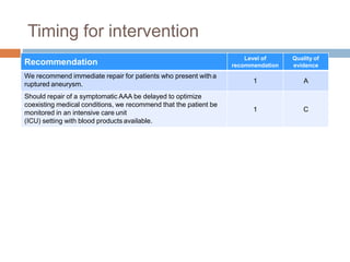
Recommendation Level of
recommendation
Quality of
evidence
In patients with active cardiac conditions, including unstable
angina, decompensated heart failure, severe vulvular disease, and
significant arrythmia, we recommend cardiology consultation before
endovascular aneurysm repair (EVAR) or open surgical repair
(OSR).
1 B
In patients with significant clinical risk factors, such as coronary
artery disease, congestive heart failure, cerebrovascular disease,
diabetes mellitus, chronic renal insufficiency, and unknown or poor
functional capacity (metabolic equivalent [MET] < 4), who are to
undergo OSR or EVAR, we suggest noninvasive stress testing.
2 B
We recommend a preoperative resting 12-lead electrocardiogram
(ECG) in all patients undergoing EVAR or
OSR within 30 days of planned treatment.
1 B
We recommend echocardiography before planned operative repair
in patients with dyspnea of unknown origin or worsening dyspnea
1 A](https://image.slidesharecdn.com/aortoiliacaneurysmsevaluation-220206111519/85/Aortoiliac-aneurysms-evaluation-18-320.jpg)


1) The document discusses aortoiliac aneurysms, including definitions, epidemiology, risk factors, rupture risk, associated aneurysms, pathophysiology, diagnosis, imaging, decision making for treatment, medical management, and indications for intervention. 2) Key risk factors for aneurysm rupture include diameter greater than 5.5 cm, female sex, smoking, and saccular aneurysm morphology. Imaging recommendations include ultrasound screening and CT or MRI for diagnosis. 3) Treatment is generally recommended for aneurysms greater than 5.5 cm in men or 5 cm in women, or those showing rapid growth. Immediate repair is indicated for ruptured aneurysms.


