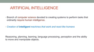
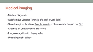
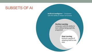
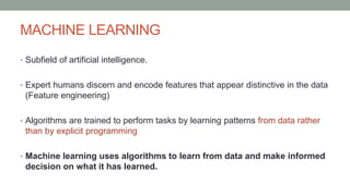
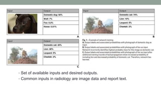
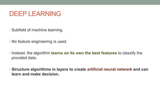
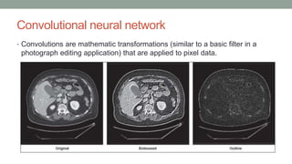
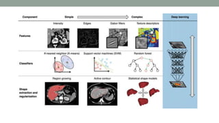
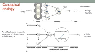
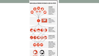
ARTIFICIAL INTELLIGENCE AND
RADIOLOGY
Dr. DEVKANT LAKHERA
ARTIFICIAL INTELLIGENCE
• Branch of computer science devoted to creating systems to perform tasks that
ordinarily require human intelligence.
• Creation of intelligent machines that work and react like humans
Reasoning, planning, learning, language processing, perception and the ability
to move and manipulate objects.
Medical imaging
• Medical diagnosis
• Autonomous vehicles (drones and self-driving cars)
• Search engines (such as Google search), online assistants (such as Siri)
• Creating art ,mathematical theorems
• Image recognition in photographs
• Predicting flight delays
SUBSETS OF AI
MACHINE LEARNING
• Subfield of artificial intelligence.
• Expert humans discern and encode features that appear distinctive in the data
(Feature engineering)
• Algorithms are trained to perform tasks by learning patterns from data rather
than by explicit programming
• Machine learning uses algorithms to learn from data and make informed
decision on what it has learned.
Feature engineering
• Attributes shared by independant units on which analysis is to be done
• Set of available inputs and desired outputs.
• Common inputs in radiology are image data and report text.
• Human versus computer
vision.
Have to apply machine learning
algorithms to distinguish images on
the basis of these features
DEEP LEARNING
• Subfield of machine learning.
• No feature engineering is used.
• Instead, the algorithm learns on its own the best features to classify the
provided data.
• Structure algorithms in layers to create artificial neural network and can
learn and make decision.
Convolutional neural network
• Convolutions are mathematic transformations (similar to a basic filter in a
photograph editing application) that are applied to pixel data.
ARTIFICIAL NEURAL NETWORK
• Artificial neural network is a biologically inspired network of artificial neurons
configured to perform specific task.
• Neural network acquires knowledge through learning.
• Contains large number of artificial neurons arranged in series
• Based on a neural network structure loosely inspired by the human brain –
Convolutional neural network
ARTIFICAL NEURAL NETWORK (ANN)
• ANNs are clusters of interconnected nodes, like brain neurons.
• Feed those blocks into multiple (deep) processing layers, which act as filters,
and then feed data onto further layers with other kinds of filters.
Conceptual
analogy biologic
neurons
artificial
neurons
visual cortex
retina
An artificial neural network is
composed of interconnected
artificial neurons
• Oncology - assisting clinical decision making related to the diagnosis and risk
stratification of different cancers.
• non-small-cell lung cancer (NSCLC) used radiomics to predict distant
metastasis in lung – adenocarcinoma and tumour histological subtypes as well
as disease recurrence, somatic mutations, gene-expression profile and overall
survival.
To train a model, we need data.
• Massive amounts of digital data now available to train algorithms and
modern, powerful computational hardware
• Radiomics: Medical study that aims to extract large amount of
quantitative features from medical images using data-
characterization algorithms
• Radiographic images, coupled with data on clinical outcomes - rapid
expansion of radiomics as a field of medical research
DEEP LEARNING SYSTEM
• DEEP MIND GOOGLE
• Struck a deal with NHS
• 700 scans of head and neck cancer to be given to system so that areas to be
treated and avoided during radiotherapy can be delineated.
• 1 million eye scans with information to be given to teach it to recognize eye
illness.
• Massachusetts General Hospital (MGH) and Brigham and Women’s Hospital
(BWH) spent more than $1 billion on data collection infrastructures.
• Recently, these institutions started a Center for Clinical Data Science to
produce new clinical AI applications, trained with these data,
RECENT USES
• Efforts to enable radiologists to utilize AI as part of their normal PACS workflow.
• Triaging studies that need urgent review by radiologists.
• Facilitating the communication of urgent results.
Detecting
• Intracranial hemorrhage
• Chest Xray diagnosis
• Classifying liver lesions on MRI scans and explaining the findings,
• Characterizing pulmonary nodules on CT
• Helping to avoid unnecessary thyroid nodule biopsies.
AI RADIOLOGY START UPS
• Zebra technologies
• Using million high quality images to
develop DL engine to automatically detect
various medical findings.
• Automatic detection of liver, lung CVS
and bone diseases.
• Deployed at more than 50 hospitals
globally
AI RADIOLOGY START UPS
AIDENCE
• Product provides fully automated
analysis and reporting of pulmonary
nodules.
• Accuracy levels equal and even
surpassing human capabilities.
Artery’s
FDA clearance for product that provides
automated, editable ventricle
segmentations based on cardiac MRI
images as accurate as performed
manually.
AI RADIOLOGY STARTUPS
• Butterfly Network
• Hand held probe connected to
iPhone. Starts 2000 dollars
• DL system will identify
characteristics in image and
make diagnosis.
INDIAN START UPS
QURE.AI
Deep learning based diagnosis
of tuberculosis on chest x ray
and intracerebral hemorrhage
on CT
Processed more than 1.5
million x-rays with accuracy
level near to humans.
Deployed across 4 clinics in
mumbai.
Artificial intelligence in radiology:
Friend or foe?
Artificial intelligence
• Fear has been AI would begin to chip away at jobs.
• While that concern isn’t coming true as yet.
• Radiologists are being urged to accept and incorporate AI into their
interpretations.
Artificial intelligence
• Primary driver - desire for greater efficacy and efficiency in clinical care.
• Studies report that, in some cases, an average radiologist must interpret one image
every 3–4 seconds in an 8-hour workday to meet workload demands
• Involves visual perception as well as independent knowledge, errors are inevitable
• Integrated AI component within the imaging workflow - increase efficiency, reduce
errors and achieve objectives
• providing trained radiologists with pre-screened images and identified features.
• Improves their consistency and quality and potentially lowers operating costs.
• Scientists have shown that few pixels from other image can drastically alter
results.
• Artificial intelligence won’t necessarily replace radiologists, but it will replace
radiologists who don’t use artificial intelligence in future.
THANK YOU

Artificial intelligence in radiology

  • 1.
  • 2.
    ARTIFICIAL INTELLIGENCE • Branchof computer science devoted to creating systems to perform tasks that ordinarily require human intelligence. • Creation of intelligent machines that work and react like humans Reasoning, planning, learning, language processing, perception and the ability to move and manipulate objects.
  • 3.
    Medical imaging • Medicaldiagnosis • Autonomous vehicles (drones and self-driving cars) • Search engines (such as Google search), online assistants (such as Siri) • Creating art ,mathematical theorems • Image recognition in photographs • Predicting flight delays
  • 4.
  • 5.
    MACHINE LEARNING • Subfieldof artificial intelligence. • Expert humans discern and encode features that appear distinctive in the data (Feature engineering) • Algorithms are trained to perform tasks by learning patterns from data rather than by explicit programming • Machine learning uses algorithms to learn from data and make informed decision on what it has learned.
  • 6.
    Feature engineering • Attributesshared by independant units on which analysis is to be done
  • 7.
    • Set ofavailable inputs and desired outputs. • Common inputs in radiology are image data and report text.
  • 8.
    • Human versuscomputer vision. Have to apply machine learning algorithms to distinguish images on the basis of these features
  • 9.
    DEEP LEARNING • Subfieldof machine learning. • No feature engineering is used. • Instead, the algorithm learns on its own the best features to classify the provided data. • Structure algorithms in layers to create artificial neural network and can learn and make decision.
  • 10.
    Convolutional neural network •Convolutions are mathematic transformations (similar to a basic filter in a photograph editing application) that are applied to pixel data.
  • 12.
    ARTIFICIAL NEURAL NETWORK •Artificial neural network is a biologically inspired network of artificial neurons configured to perform specific task. • Neural network acquires knowledge through learning. • Contains large number of artificial neurons arranged in series • Based on a neural network structure loosely inspired by the human brain – Convolutional neural network
  • 13.
    ARTIFICAL NEURAL NETWORK(ANN) • ANNs are clusters of interconnected nodes, like brain neurons. • Feed those blocks into multiple (deep) processing layers, which act as filters, and then feed data onto further layers with other kinds of filters.
  • 14.
    Conceptual analogy biologic neurons artificial neurons visual cortex retina Anartificial neural network is composed of interconnected artificial neurons
  • 16.
    • Oncology -assisting clinical decision making related to the diagnosis and risk stratification of different cancers. • non-small-cell lung cancer (NSCLC) used radiomics to predict distant metastasis in lung – adenocarcinoma and tumour histological subtypes as well as disease recurrence, somatic mutations, gene-expression profile and overall survival.
  • 18.
    To train amodel, we need data. • Massive amounts of digital data now available to train algorithms and modern, powerful computational hardware • Radiomics: Medical study that aims to extract large amount of quantitative features from medical images using data- characterization algorithms
  • 19.
    • Radiographic images,coupled with data on clinical outcomes - rapid expansion of radiomics as a field of medical research
  • 21.
    DEEP LEARNING SYSTEM •DEEP MIND GOOGLE • Struck a deal with NHS • 700 scans of head and neck cancer to be given to system so that areas to be treated and avoided during radiotherapy can be delineated. • 1 million eye scans with information to be given to teach it to recognize eye illness.
  • 22.
    • Massachusetts GeneralHospital (MGH) and Brigham and Women’s Hospital (BWH) spent more than $1 billion on data collection infrastructures. • Recently, these institutions started a Center for Clinical Data Science to produce new clinical AI applications, trained with these data,
  • 23.
    RECENT USES • Effortsto enable radiologists to utilize AI as part of their normal PACS workflow. • Triaging studies that need urgent review by radiologists. • Facilitating the communication of urgent results.
  • 24.
    Detecting • Intracranial hemorrhage •Chest Xray diagnosis • Classifying liver lesions on MRI scans and explaining the findings, • Characterizing pulmonary nodules on CT • Helping to avoid unnecessary thyroid nodule biopsies.
  • 27.
    AI RADIOLOGY STARTUPS • Zebra technologies • Using million high quality images to develop DL engine to automatically detect various medical findings. • Automatic detection of liver, lung CVS and bone diseases. • Deployed at more than 50 hospitals globally
  • 28.
    AI RADIOLOGY STARTUPS AIDENCE • Product provides fully automated analysis and reporting of pulmonary nodules. • Accuracy levels equal and even surpassing human capabilities. Artery’s FDA clearance for product that provides automated, editable ventricle segmentations based on cardiac MRI images as accurate as performed manually.
  • 29.
    AI RADIOLOGY STARTUPS •Butterfly Network • Hand held probe connected to iPhone. Starts 2000 dollars • DL system will identify characteristics in image and make diagnosis.
  • 30.
    INDIAN START UPS QURE.AI Deeplearning based diagnosis of tuberculosis on chest x ray and intracerebral hemorrhage on CT Processed more than 1.5 million x-rays with accuracy level near to humans. Deployed across 4 clinics in mumbai.
  • 31.
    Artificial intelligence inradiology: Friend or foe?
  • 32.
    Artificial intelligence • Fearhas been AI would begin to chip away at jobs. • While that concern isn’t coming true as yet. • Radiologists are being urged to accept and incorporate AI into their interpretations.
  • 33.
    Artificial intelligence • Primarydriver - desire for greater efficacy and efficiency in clinical care. • Studies report that, in some cases, an average radiologist must interpret one image every 3–4 seconds in an 8-hour workday to meet workload demands • Involves visual perception as well as independent knowledge, errors are inevitable • Integrated AI component within the imaging workflow - increase efficiency, reduce errors and achieve objectives • providing trained radiologists with pre-screened images and identified features.
  • 34.
    • Improves theirconsistency and quality and potentially lowers operating costs. • Scientists have shown that few pixels from other image can drastically alter results. • Artificial intelligence won’t necessarily replace radiologists, but it will replace radiologists who don’t use artificial intelligence in future.
  • 35.

Editor's Notes

  • #3 Capabilities as such
  • #5 Two subsets
  • #6 More amount of data is fed to the machine – it will learn and make a more acuurate diagnosis
  • #7  take various attributes and program it to an algorithm based on featues such as edges, gradients, and textures. // Statistical analysis of the presence of these features in a given image can then be used to classify or interpret the image.//
  • #8 And make associations and form output statistical probabilities, also known as nodes.
  • #9  A human expert easily classifies this image as an image of the right kidney. // computer “sees” a matrix of numbers representing pixel brightness. Computer vision typically involves computing the presence of numerical patterns (features) in this matrix, then applying machine learning algorithms to distinguish images on the basis of these features.
  • #10 Complex algorithms , which use multiple lays of processing
  • #12 Gabor filters related to texture -
  • #15 The imaging featues are known as evidence .. Many such evidence s are evaluated in layers which gets more complex.. and are integrated-- outputs a decision signal based on a weighted sum of evidences, and an activation function, which integrates signals from previous neurons. Hundreds of these basic computing units are assembled together to build an artificial neural network computing device. \
  • #18 Fig. 3 |. Artificial intelligence impact areas within oncology imaging. This schematic outlines the various tasks within radiology where artificial intelligence (AI) implementation is likely to have a large impact. a | The workflow comprises the following steps: preprocessing of images after acquisition, image-based clinical tasks (which usually involve the quantification of features either using engineered features with traditional machine learning or deep learning), reporting results through the generation of textual radiology reports and, finally, the integration of patient information from multiple data sources. b | AI is expected to impact image-based clinical tasks, including the detection of abnormalities; the characterization of objects in images using segmentation, diagnosis and staging; and the monitoring of objects for diagnosis and assessment of treatment response. TNM, tumour–node–metastasis.
  • #19 Deep learning is a subset of machine learning that is
  • #20 Radiomics studies have incorporated deep learning techniques to learn feature representations automatically from example images
  • #21 Brain Tumor Image Segmentation (BRATS)
  • #27 Much new radiology conferences have been focusing on artificial intellingenc eas the
  • #28 Deep learning
  • #33 In latest conferences have been urged .. Keep utptodated
  • #35 Though advanced it is liable to make mistakes