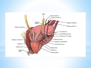
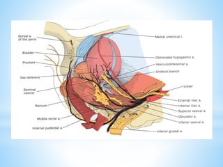
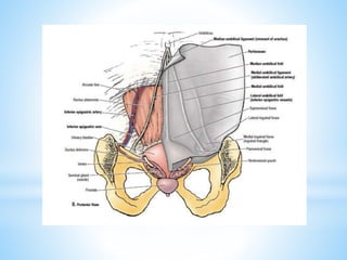
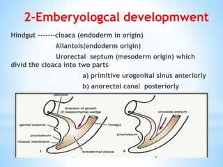
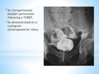
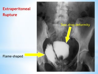
Prepared By : Dr.Lutfi Ahmed Al-Bawri
2017
alaobthani@gmail.com
*
โ€ซุงู„ุฑุญูŠู…โ€ฌ โ€ซุงู„ุฑุญู…ู†โ€ฌ โ€ซู‡ู„ู„ุงโ€ฌ โ€ซุจุณู…โ€ฌ
1-Anatomy of U.B .
2-Emberyologcal development of UB.
3-Etiology of the U.B Trauma.
4-Types of the U.B Trauma.
5- Clinical Signs and Symptoms of UB trauma.
6- Radiographic Imaging of UB trauma.
7- Management of UB trauma.
8- Complications of UB trauma.
The Objectives :-
The site:-
At birth: it lies in the abdominal cavity
(Pelviabdominal organ)
in adult: it occupies the anterior part of the pelvis
(Pelvic organ), when it is full it expand upward into
the abdominal cavity.
4 ANGLES:
Apix :- attached to median umbilical ligament.
lf remains patent, it may cause: Urachal (sinus, fistula,
cyst) or Congenital diverticulum
Neck:- Lying on prostate (male) or pelvic fascia
(femal), it gives the urethra Surrounded by smooth
muscle fibers constituting the sphincter vesicae
Two posterosuperior angles:- receiving the 2 ureters
1-Anatomy of U.B .
Peritoneal covering :-
Male: superior surface and upper part of base .but in Female:
superior surface ONLY.
Relation :-
1. Anterior: pubic bone ,puboprostatic ligment in male and
pubovesical ligment in femal
They fix bladder in pelvis and They are present in Cave Of
Retzius (retropubic space)
2. Inferolateral surface: pubis, retropubic fat, obturator
internus & levator ani muscles.
3-Supperior surface:- ilum and segmoid colon in male .
Uterus and retrovesical pouch in femal.
4. Posterior surface: in mle :two seminal veiscles ,two vasa
defferintia and rectum .In femal:cervix and vagina
Cavity of the urinary bladder
-Trigone:- Triangular part of the base between the 2 openings of the
ureter & the opening of the urethra
Gharacters of trigone:
1.No rugae 2.Pink (rich blood supply) 3.No submucosa
4. lt is richly supplied by nerves so(lt is more sensitive to pain).
-Ureteric orificer: - They lie at the posterolateral angles of the
trigone - The 2 orifices are separated from each other by a transverse
ridge called the interureteric crest
-Uvula of the bladder: - lt is produced by the median lobe of the
prostate.
-lnternal urethral orifice:
- lt is placed at the lowest part of the bladder.
- lt is surrounded by an involuntary non-striated muscle forming the
sphincter vesicae.
-Intramural part of the ureter - The lowermost part of the ureter
passes obliquely through the wall ofthe bladder
Blood supply:-
*Artries: superior and inf.vesical artery which come from
ant.devision of Internal iliac artery .
*Vesical veins:the lateral plexuses, the prostatovesical
plexus (pudendal plexus, Santorini) empty into the internal
iliac veins.
Lymph node:-
Perivesical LN , internal & external iliac LNs , common iliac
LNs, para-aortic LNs.
Nenrve Supply :-
Sympathetic ;T11,12 and L1,2
Parasympathetic; S2,3,4
2-Emberyologcal developmwent
Hindgut -------cloaca (endoderm in origin)
Allantois(endoderm origin)
Urorectal septum (mesoderm origin) which
divid the cloaca into two parts
a) primitive urogenital sinus anteriorly
b) anorectal canal posteriorly
Mesonephric ducts connect to the primitive urogenital sinus
dividing it into upper part (vasicourethral canal) and the lower
part (definitive urogenital sinus)
U.B formed of 1- Vasicourethral canal--------------major body of UB
2- Allantois(proximal part)-----------Median umbilical
ligament and Apex of BU
3- Mesonephric ducts-----------------trigon of UB
Situations in Which the
Bladder May Be Injured
1-(TURBT)
2- Cystoscopic bladder
biopsy
3-(TURP)
4-Cystolitholapaxy
5-Penetrating trauma to
the lower abdomen or
back
6-Caesarean section,
especially as an
emergency
7-Blunt pelvic traumaโ€”in
association with pelvic
fracture or
9-Total hip replacement
(very rare)
10-Rapid deceleration
injuryโ€”seat belt injury with
full bladder in the absence
of a pelvic fracture
11-Spontaneous rupture
after bladder augmentation
3-Etiology of the U.B Trauma.
4-Types of the U.B Trauma.
10% of male and 5% of female pelvic fractures are associated
with a bladder injury.
1-Blunt Trauma
2-perforation (penetrating)
1- Intraperitoneal perforation 30%: the
peritoneum overlying the bladder is breached,
allowing urine to escape into the peritoneal
cavity.
2-Extraperitoneal perforation 60%:
the peritoneum is intact and urine escapes into
the space around the bladder, but not into the
peritoneal cavity.
3-Compained intra and extraperitoneal 10%.
*Grad 1 : contusion, intramural hematoma ,or partial thickness
laceration
*Grad 2 : Extra-peritoneal bladder wall laceration less than 2 cm
*Grad 3: Extra-peritoneal bladder wall laceration more than 2 cm or
intra-peritoneal bladder wall laceration less than 2 cm
*Grad4 : intra-peritoneal bladder wall laceration more than 2 cm
*Grad 5 : intra- or extra-peritoneal bladder wall laceration extending
into bladder neck or ureteral orifices (trigon)
Classification of U.B Trauma
5- Clinical Signs and Symptoms.
- Blood at meatusโ€”in 40โ€“50% of patients (no blood
at meatus in 50โ€“60%).
- Gross haematuria.
- Inability to pass urine.
- Perineal or scrotal bruising.
- โ€˜High ridingโ€™ prostate.
- Inability to pass a urethral catheter.
6- Radiographic Imaging.
Intra peritoneal rupture
Sunburst appearance
*An intraperitoneal
bladder perforation
following a TURBT,
*As demonstrated on a
cystogram
(anteroposterior view).
Extraperitoneal
Rupture
Flame-shaped
Tear drop deformity
7- Management
Extraperitoneal rupture :
bladder drainage with a urethral catheter for 2 weeks
followed by a cystogram to confirm the perforation has healed.
Indications for surgical repair of extraperitoneal bladder perforation:
- If you have opened the bladder to place a suprapubic catheter for a
urethral injury or involvement of bladder neck.
- A bone spike protruding into the bladder on CT.
- Associated rectal or vaginal perforation.
- Where the patient is undergoing open fixation of a pelvic fracture.
Intraperitoneal rupture : usually repaired surgically to prevent
complications from leakage of urine into the peritoneal cavity.
8-COMPLICATIONS OF BLADDER INJURY
*Cystitis & pyelonephritis
*Peritonitis
*Pelvic abscess
*Vesiculovaginal or retrovesical fistula
*Paralytic ileus
*Haemorrhage
*Mortality is 100% without surgical
intervention
*References
*Hinman Atlas of Urosurgical Anatomy - 2nd edition 2012
*Campbell-Walsh Urology, 11th ed 2016
*snell clinical anatomy by regions 9th ed 2012
*Matary-Surgical_Anatomy_2013
*Hinman Atlas of Urologic Surgery 3rd Edition-2012
*Oxford specialist Handbook in Urological Surgery
Urinary bladder trauma 2017

Urinary bladder trauma 2017

  • 1.
    Prepared By :Dr.Lutfi Ahmed Al-Bawri 2017 alaobthani@gmail.com * โ€ซุงู„ุฑุญูŠู…โ€ฌ โ€ซุงู„ุฑุญู…ู†โ€ฌ โ€ซู‡ู„ู„ุงโ€ฌ โ€ซุจุณู…โ€ฌ
  • 2.
    1-Anatomy of U.B. 2-Emberyologcal development of UB. 3-Etiology of the U.B Trauma. 4-Types of the U.B Trauma. 5- Clinical Signs and Symptoms of UB trauma. 6- Radiographic Imaging of UB trauma. 7- Management of UB trauma. 8- Complications of UB trauma. The Objectives :-
  • 3.
    The site:- At birth:it lies in the abdominal cavity (Pelviabdominal organ) in adult: it occupies the anterior part of the pelvis (Pelvic organ), when it is full it expand upward into the abdominal cavity. 4 ANGLES: Apix :- attached to median umbilical ligament. lf remains patent, it may cause: Urachal (sinus, fistula, cyst) or Congenital diverticulum Neck:- Lying on prostate (male) or pelvic fascia (femal), it gives the urethra Surrounded by smooth muscle fibers constituting the sphincter vesicae Two posterosuperior angles:- receiving the 2 ureters 1-Anatomy of U.B .
  • 4.
    Peritoneal covering :- Male:superior surface and upper part of base .but in Female: superior surface ONLY. Relation :- 1. Anterior: pubic bone ,puboprostatic ligment in male and pubovesical ligment in femal They fix bladder in pelvis and They are present in Cave Of Retzius (retropubic space) 2. Inferolateral surface: pubis, retropubic fat, obturator internus & levator ani muscles. 3-Supperior surface:- ilum and segmoid colon in male . Uterus and retrovesical pouch in femal. 4. Posterior surface: in mle :two seminal veiscles ,two vasa defferintia and rectum .In femal:cervix and vagina
  • 8.
    Cavity of theurinary bladder -Trigone:- Triangular part of the base between the 2 openings of the ureter & the opening of the urethra Gharacters of trigone: 1.No rugae 2.Pink (rich blood supply) 3.No submucosa 4. lt is richly supplied by nerves so(lt is more sensitive to pain). -Ureteric orificer: - They lie at the posterolateral angles of the trigone - The 2 orifices are separated from each other by a transverse ridge called the interureteric crest -Uvula of the bladder: - lt is produced by the median lobe of the prostate. -lnternal urethral orifice: - lt is placed at the lowest part of the bladder. - lt is surrounded by an involuntary non-striated muscle forming the sphincter vesicae. -Intramural part of the ureter - The lowermost part of the ureter passes obliquely through the wall ofthe bladder
  • 9.
    Blood supply:- *Artries: superiorand inf.vesical artery which come from ant.devision of Internal iliac artery . *Vesical veins:the lateral plexuses, the prostatovesical plexus (pudendal plexus, Santorini) empty into the internal iliac veins. Lymph node:- Perivesical LN , internal & external iliac LNs , common iliac LNs, para-aortic LNs. Nenrve Supply :- Sympathetic ;T11,12 and L1,2 Parasympathetic; S2,3,4
  • 13.
    2-Emberyologcal developmwent Hindgut -------cloaca(endoderm in origin) Allantois(endoderm origin) Urorectal septum (mesoderm origin) which divid the cloaca into two parts a) primitive urogenital sinus anteriorly b) anorectal canal posteriorly
  • 14.
    Mesonephric ducts connectto the primitive urogenital sinus dividing it into upper part (vasicourethral canal) and the lower part (definitive urogenital sinus) U.B formed of 1- Vasicourethral canal--------------major body of UB 2- Allantois(proximal part)-----------Median umbilical ligament and Apex of BU 3- Mesonephric ducts-----------------trigon of UB
  • 16.
    Situations in Whichthe Bladder May Be Injured 1-(TURBT) 2- Cystoscopic bladder biopsy 3-(TURP) 4-Cystolitholapaxy 5-Penetrating trauma to the lower abdomen or back 6-Caesarean section, especially as an emergency 7-Blunt pelvic traumaโ€”in association with pelvic fracture or 9-Total hip replacement (very rare) 10-Rapid deceleration injuryโ€”seat belt injury with full bladder in the absence of a pelvic fracture 11-Spontaneous rupture after bladder augmentation 3-Etiology of the U.B Trauma.
  • 17.
    4-Types of theU.B Trauma. 10% of male and 5% of female pelvic fractures are associated with a bladder injury. 1-Blunt Trauma 2-perforation (penetrating) 1- Intraperitoneal perforation 30%: the peritoneum overlying the bladder is breached, allowing urine to escape into the peritoneal cavity. 2-Extraperitoneal perforation 60%: the peritoneum is intact and urine escapes into the space around the bladder, but not into the peritoneal cavity. 3-Compained intra and extraperitoneal 10%.
  • 18.
    *Grad 1 :contusion, intramural hematoma ,or partial thickness laceration *Grad 2 : Extra-peritoneal bladder wall laceration less than 2 cm *Grad 3: Extra-peritoneal bladder wall laceration more than 2 cm or intra-peritoneal bladder wall laceration less than 2 cm *Grad4 : intra-peritoneal bladder wall laceration more than 2 cm *Grad 5 : intra- or extra-peritoneal bladder wall laceration extending into bladder neck or ureteral orifices (trigon) Classification of U.B Trauma
  • 20.
    5- Clinical Signsand Symptoms. - Blood at meatusโ€”in 40โ€“50% of patients (no blood at meatus in 50โ€“60%). - Gross haematuria. - Inability to pass urine. - Perineal or scrotal bruising. - โ€˜High ridingโ€™ prostate. - Inability to pass a urethral catheter.
  • 21.
    6- Radiographic Imaging. Intraperitoneal rupture Sunburst appearance
  • 22.
    *An intraperitoneal bladder perforation followinga TURBT, *As demonstrated on a cystogram (anteroposterior view).
  • 23.
  • 24.
    7- Management Extraperitoneal rupture: bladder drainage with a urethral catheter for 2 weeks followed by a cystogram to confirm the perforation has healed. Indications for surgical repair of extraperitoneal bladder perforation: - If you have opened the bladder to place a suprapubic catheter for a urethral injury or involvement of bladder neck. - A bone spike protruding into the bladder on CT. - Associated rectal or vaginal perforation. - Where the patient is undergoing open fixation of a pelvic fracture. Intraperitoneal rupture : usually repaired surgically to prevent complications from leakage of urine into the peritoneal cavity.
  • 25.
    8-COMPLICATIONS OF BLADDERINJURY *Cystitis & pyelonephritis *Peritonitis *Pelvic abscess *Vesiculovaginal or retrovesical fistula *Paralytic ileus *Haemorrhage *Mortality is 100% without surgical intervention
  • 26.
    *References *Hinman Atlas ofUrosurgical Anatomy - 2nd edition 2012 *Campbell-Walsh Urology, 11th ed 2016 *snell clinical anatomy by regions 9th ed 2012 *Matary-Surgical_Anatomy_2013 *Hinman Atlas of Urologic Surgery 3rd Edition-2012 *Oxford specialist Handbook in Urological Surgery