HEART
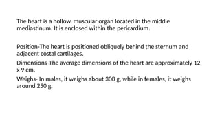
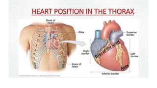
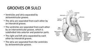
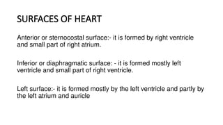
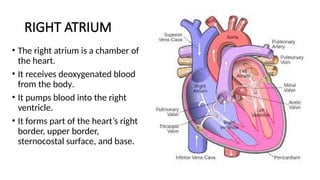
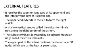
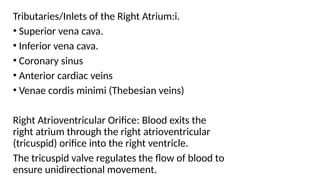
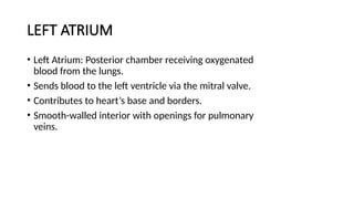
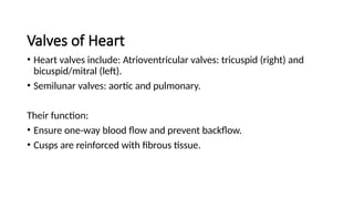
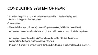
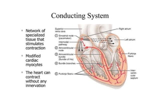
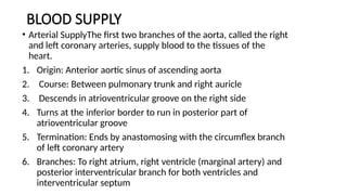
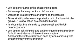
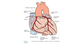
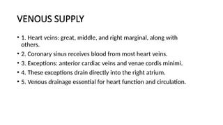
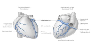
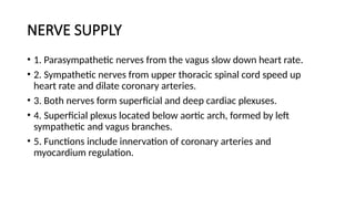
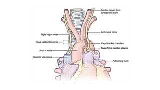
The heart is a hollow, muscular organ located in the middle
mediastinum. It is enclosed within the pericardium.
Position-The heart is positioned obliquely behind the sternum and
adjacent costal cartilages.
Dimensions-The average dimensions of the heart are approximately 12
x 9 cm.
Weighs- In males, it weighs about 300 g, while in females, it weighs
around 250 g.
EXTERNAL FEATURES
• The human heart consists of four
chambers: the right atrium, left
atrium, right ventricle, and left
ventricle.
• An apex directed downwards,
forwards, and to the left, with a
base directed backwards.
Additionally, it has anterior,
inferior, and left surfaces.
GROOVES OR SULCI
• Ventricles and atria separated by
atrioventricular groove.
• The atria are separated from each other by
an interatrial groove.
• The ventricles are separated from each other
by an interventricular groove, which is
subdivided into anterior and posterior parts.
• The right and left atria separated by each
other by interatrial groove.
• The atria are separated from the ventricles
by atrioventricular groove.
APEX OF HEART
• Apex of the heart is a conical area formed entirely by the left
ventricle situated in the left fifth intercostal space
• 9cm lateral to the midsternal line.
BASE OF HEART
• Base of the heart is also called its posterior surface formed
mainly by the left atrium and small part of the right atrium.
BORDERS OF HEART
Upper border: - it is slightly oblique and is
formed by the right and left atrium.
Right border:- it is formed by right atrium. It
extends from the right side of the opening of
SVC to the IVC
Inferior border:- it is formed mainly by the right
ventricle. It extends from the opening of IVC to
the apex of heart.
Left border:- it is formed mainly by left
ventricle and partly by the eft auricle.
SURFACES OF HEART
Anterior or sternocostal surface:- it is formed by right ventricle
and small part of right atrium.
Inferior or diaphragmatic surface: - it is formed mostly left
ventricle and small part of right ventricle.
Left surface:- it is formed mostly by the left ventricle and partly by
the left atrium and auricle
RIGHT ATRIUM
• The right atrium is a chamber of
the heart.
• It receives deoxygenated blood
from the body.
• It pumps blood into the right
ventricle.
• It forms part of the heart’s right
border, upper border,
sternocostal surface, and base.
EXTERNAL FEATURES
• It receives the superior vena cava at its upper end and
the inferior vena cava at its lower end.
• The upper end extends to the left to form the right
auricle.
• A shallow vertical groove called the sulcus terminalis
runs along the right border of the atrium.
• The sulcus terminalis is created by an internal muscular
ridge called the crista terminalis.
• The upper part of the sulcus contains the sinuatrial or SA
node, which acts as the heart’s pacemaker.
Tributaries/Inlets of the Right Atrium:i.
• Superior vena cava.
• Inferior vena cava.
• Coronary sinus
• Anterior cardiac veins
• Venae cordis minimi (Thebesian veins)
Right Atrioventricular Orifice: Blood exits the
right atrium through the right atrioventricular
(tricuspid) orifice into the right ventricle.
The tricuspid valve regulates the flow of blood to
ensure unidirectional movement.
Smooth Posterior Part or Sinus Venarum:
Most tributaries, except the anterior cardiac
veins, open into this region.
The openings include the superior vena cava,
inferior vena cava, coronary sinus (guarded by
the valve of the coronary sinus), and venae cordis
minimi.
Rough Anterior Part or Pectinate Part,
Including the Auricle.
Features transverse muscular ridges called
musculi pectinati.
Interatrial Septum:Contains the fossa ovalis, a
shallow saucer-shaped depression, in the
lower part.
The annulus ovalis or limbus fossa ovalis is the
prominent margin of the fossa ovalis.
RIGHT VENTRICLE
• Triangular chamber receiving blood from the right atrium and
pumping it to the lungs via the pulmonary trunk and arteries.
• Forms the inferior border and a large part of the sternocostal
surface of the heart.
External Features: Two surfaces: anterior (sternocostal) and
inferior (diaphragmatic).
Internal Features
• Divided into two parts:i. Inflowing part: rough with muscular ridges
(trabeculae carneae).ii. Outflowing part (infundibulum): smooth,
giving rise to the pulmonary trunk.
• Separated by the supraventricular crest (infundibuloventricular crest)
between the tricuspid and pulmonary orifices.
Orifices: Right atrioventricular (tricuspid) orifice, guarded by the
tricuspid valve.
Pulmonary orifice, guarded by the pulmonary valve.
Trabeculae Carneae: Three types: ridges, bridges, and pillars (papillary
muscles).
Three papillary muscles: anterior, posterior, and septal, attached to
cusps of the tricuspid valve by chordae tendineae.
Cavity: Crescentic in section due to the forward bulge of the
interventricular septum.
Wall Thickness: Thinner than the left ventricle, in a ratio of 1:3.
LEFT ATRIUM
• Left Atrium: Posterior chamber receiving oxygenated
blood from the lungs.
• Sends blood to the left ventricle via the mitral valve.
• Contributes to heart’s base and borders.
• Smooth-walled interior with openings for pulmonary
veins.
Left Ventricle
Left Ventricle: Receives blood from the left atrium, pumping it into the
aorta.
•Forms heart’s apex.
•Three external surfaces: sternocostal, diaphragmatic, and left.
•Interior has lower rough part and upper smooth part (aortic
vestibule).
•Features two orifices: mitral and aortic, guarded by respective valves.
•Contains papillary muscles and thicker walls compared to the right
ventricle.
Valves of Heart
• Heart valves include: Atrioventricular valves: tricuspid (right) and
bicuspid/mitral (left).
• Semilunar valves: aortic and pulmonary.
Their function:
• Ensure one-way blood flow and prevent backflow.
• Cusps are reinforced with fibrous tissue.
CONDUCTING SYSTEM OF HEART
• Conducting system: Specialized myocardium for initiating and
transmitting cardiac impulses.
Components:
• Sinuatrial node (SA node): Heart’s pacemaker, initiates heartbeat.
• Atrioventricular node (AV node): Located in lower part of atrial septum.
• Atrioventricular bundle (AV bundle or bundle of His): Muscular
connection between atria and ventricles.
• Purkinje fibers: Descend from AV bundle, forming subendocardial plexus.
BLOOD SUPPLY
• Arterial SupplyThe first two branches of the aorta, called the right
and left coronary arteries, supply blood to the tissues of the
heart.
1. Origin: Anterior aortic sinus of ascending aorta
2. Course: Between pulmonary trunk and right auricle
3. Descends in atrioventricular groove on the right side
4. Turns at the inferior border to run in posterior part of
atrioventricular groove
5. Termination: Ends by anastomosing with the circumflex branch
of left coronary artery
6. Branches: To right atrium, right ventricle (marginal artery) and
posterior interventricular branch for both ventricles and
interventricular septum
• Left posterior aortic sinus of ascending aorta
• Between pulmonary trunk and left auricle
• Descends in atrioventricular groove on the left side
• Turns at left border to run in posterior part of atrioventricular
groove. It is now called as circumflex branch
• Its circumflex branch ends by anastomosing with right
coronary artery
• Left atrium, left ventricle and anterior interventricular branch
for both ventricles and interventricular septum
Anterior interventricular branch ends by anastomosing with
posterior interventricular branch
VENOUS SUPPLY
• 1. Heart veins: great, middle, and right marginal, along with
others.
• 2. Coronary sinus receives blood from most heart veins.
• 3. Exceptions: anterior cardiac veins and venae cordis minimi.
• 4. These exceptions drain directly into the right atrium.
• 5. Venous drainage essential for heart function and circulation.
NERVE SUPPLY
• 1. Parasympathetic nerves from the vagus slow down heart rate.
• 2. Sympathetic nerves from upper thoracic spinal cord speed up
heart rate and dilate coronary arteries.
• 3. Both nerves form superficial and deep cardiac plexuses.
• 4. Superficial plexus located below aortic arch, formed by left
sympathetic and vagus branches.
• 5. Functions include innervation of coronary arteries and
myocardium regulation.
Heart presentation by Dr Ela kinra…………..

Heart presentation by Dr Ela kinra…………..

  • 1.
  • 2.
    The heart isa hollow, muscular organ located in the middle mediastinum. It is enclosed within the pericardium. Position-The heart is positioned obliquely behind the sternum and adjacent costal cartilages. Dimensions-The average dimensions of the heart are approximately 12 x 9 cm. Weighs- In males, it weighs about 300 g, while in females, it weighs around 250 g.
  • 4.
    EXTERNAL FEATURES • Thehuman heart consists of four chambers: the right atrium, left atrium, right ventricle, and left ventricle. • An apex directed downwards, forwards, and to the left, with a base directed backwards. Additionally, it has anterior, inferior, and left surfaces.
  • 5.
    GROOVES OR SULCI •Ventricles and atria separated by atrioventricular groove. • The atria are separated from each other by an interatrial groove. • The ventricles are separated from each other by an interventricular groove, which is subdivided into anterior and posterior parts. • The right and left atria separated by each other by interatrial groove. • The atria are separated from the ventricles by atrioventricular groove.
  • 6.
    APEX OF HEART •Apex of the heart is a conical area formed entirely by the left ventricle situated in the left fifth intercostal space • 9cm lateral to the midsternal line. BASE OF HEART • Base of the heart is also called its posterior surface formed mainly by the left atrium and small part of the right atrium.
  • 7.
    BORDERS OF HEART Upperborder: - it is slightly oblique and is formed by the right and left atrium. Right border:- it is formed by right atrium. It extends from the right side of the opening of SVC to the IVC Inferior border:- it is formed mainly by the right ventricle. It extends from the opening of IVC to the apex of heart. Left border:- it is formed mainly by left ventricle and partly by the eft auricle.
  • 8.
    SURFACES OF HEART Anterioror sternocostal surface:- it is formed by right ventricle and small part of right atrium. Inferior or diaphragmatic surface: - it is formed mostly left ventricle and small part of right ventricle. Left surface:- it is formed mostly by the left ventricle and partly by the left atrium and auricle
  • 9.
    RIGHT ATRIUM • Theright atrium is a chamber of the heart. • It receives deoxygenated blood from the body. • It pumps blood into the right ventricle. • It forms part of the heart’s right border, upper border, sternocostal surface, and base.
  • 10.
    EXTERNAL FEATURES • Itreceives the superior vena cava at its upper end and the inferior vena cava at its lower end. • The upper end extends to the left to form the right auricle. • A shallow vertical groove called the sulcus terminalis runs along the right border of the atrium. • The sulcus terminalis is created by an internal muscular ridge called the crista terminalis. • The upper part of the sulcus contains the sinuatrial or SA node, which acts as the heart’s pacemaker.
  • 11.
    Tributaries/Inlets of theRight Atrium:i. • Superior vena cava. • Inferior vena cava. • Coronary sinus • Anterior cardiac veins • Venae cordis minimi (Thebesian veins) Right Atrioventricular Orifice: Blood exits the right atrium through the right atrioventricular (tricuspid) orifice into the right ventricle. The tricuspid valve regulates the flow of blood to ensure unidirectional movement.
  • 13.
    Smooth Posterior Partor Sinus Venarum: Most tributaries, except the anterior cardiac veins, open into this region. The openings include the superior vena cava, inferior vena cava, coronary sinus (guarded by the valve of the coronary sinus), and venae cordis minimi.
  • 14.
    Rough Anterior Partor Pectinate Part, Including the Auricle. Features transverse muscular ridges called musculi pectinati. Interatrial Septum:Contains the fossa ovalis, a shallow saucer-shaped depression, in the lower part. The annulus ovalis or limbus fossa ovalis is the prominent margin of the fossa ovalis.
  • 16.
    RIGHT VENTRICLE • Triangularchamber receiving blood from the right atrium and pumping it to the lungs via the pulmonary trunk and arteries. • Forms the inferior border and a large part of the sternocostal surface of the heart. External Features: Two surfaces: anterior (sternocostal) and inferior (diaphragmatic).
  • 17.
    Internal Features • Dividedinto two parts:i. Inflowing part: rough with muscular ridges (trabeculae carneae).ii. Outflowing part (infundibulum): smooth, giving rise to the pulmonary trunk. • Separated by the supraventricular crest (infundibuloventricular crest) between the tricuspid and pulmonary orifices. Orifices: Right atrioventricular (tricuspid) orifice, guarded by the tricuspid valve. Pulmonary orifice, guarded by the pulmonary valve.
  • 18.
    Trabeculae Carneae: Threetypes: ridges, bridges, and pillars (papillary muscles). Three papillary muscles: anterior, posterior, and septal, attached to cusps of the tricuspid valve by chordae tendineae. Cavity: Crescentic in section due to the forward bulge of the interventricular septum. Wall Thickness: Thinner than the left ventricle, in a ratio of 1:3.
  • 20.
    LEFT ATRIUM • LeftAtrium: Posterior chamber receiving oxygenated blood from the lungs. • Sends blood to the left ventricle via the mitral valve. • Contributes to heart’s base and borders. • Smooth-walled interior with openings for pulmonary veins.
  • 21.
    Left Ventricle Left Ventricle:Receives blood from the left atrium, pumping it into the aorta. •Forms heart’s apex. •Three external surfaces: sternocostal, diaphragmatic, and left. •Interior has lower rough part and upper smooth part (aortic vestibule). •Features two orifices: mitral and aortic, guarded by respective valves. •Contains papillary muscles and thicker walls compared to the right ventricle.
  • 22.
    Valves of Heart •Heart valves include: Atrioventricular valves: tricuspid (right) and bicuspid/mitral (left). • Semilunar valves: aortic and pulmonary. Their function: • Ensure one-way blood flow and prevent backflow. • Cusps are reinforced with fibrous tissue.
  • 23.
    CONDUCTING SYSTEM OFHEART • Conducting system: Specialized myocardium for initiating and transmitting cardiac impulses. Components: • Sinuatrial node (SA node): Heart’s pacemaker, initiates heartbeat. • Atrioventricular node (AV node): Located in lower part of atrial septum. • Atrioventricular bundle (AV bundle or bundle of His): Muscular connection between atria and ventricles. • Purkinje fibers: Descend from AV bundle, forming subendocardial plexus.
  • 25.
    BLOOD SUPPLY • ArterialSupplyThe first two branches of the aorta, called the right and left coronary arteries, supply blood to the tissues of the heart. 1. Origin: Anterior aortic sinus of ascending aorta 2. Course: Between pulmonary trunk and right auricle 3. Descends in atrioventricular groove on the right side 4. Turns at the inferior border to run in posterior part of atrioventricular groove 5. Termination: Ends by anastomosing with the circumflex branch of left coronary artery 6. Branches: To right atrium, right ventricle (marginal artery) and posterior interventricular branch for both ventricles and interventricular septum
  • 26.
    • Left posterioraortic sinus of ascending aorta • Between pulmonary trunk and left auricle • Descends in atrioventricular groove on the left side • Turns at left border to run in posterior part of atrioventricular groove. It is now called as circumflex branch • Its circumflex branch ends by anastomosing with right coronary artery • Left atrium, left ventricle and anterior interventricular branch for both ventricles and interventricular septum Anterior interventricular branch ends by anastomosing with posterior interventricular branch
  • 28.
    VENOUS SUPPLY • 1.Heart veins: great, middle, and right marginal, along with others. • 2. Coronary sinus receives blood from most heart veins. • 3. Exceptions: anterior cardiac veins and venae cordis minimi. • 4. These exceptions drain directly into the right atrium. • 5. Venous drainage essential for heart function and circulation.
  • 30.
    NERVE SUPPLY • 1.Parasympathetic nerves from the vagus slow down heart rate. • 2. Sympathetic nerves from upper thoracic spinal cord speed up heart rate and dilate coronary arteries. • 3. Both nerves form superficial and deep cardiac plexuses. • 4. Superficial plexus located below aortic arch, formed by left sympathetic and vagus branches. • 5. Functions include innervation of coronary arteries and myocardium regulation.