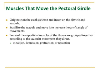
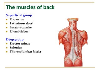
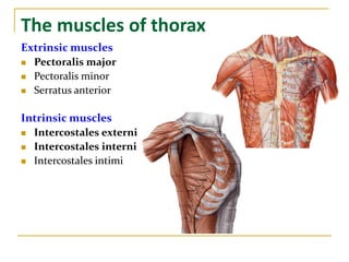
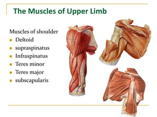
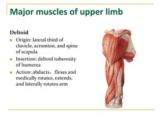
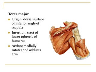
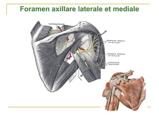
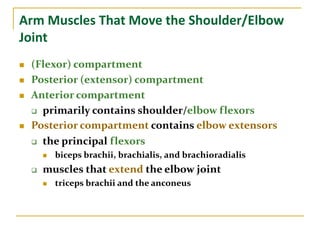
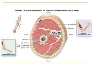
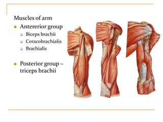
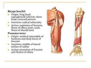
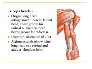
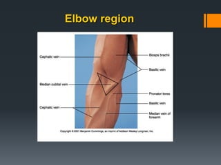
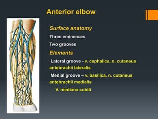
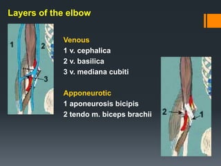
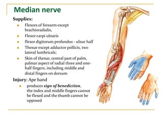
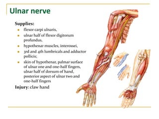
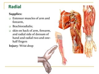
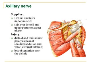
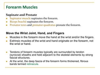
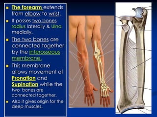
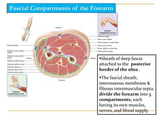
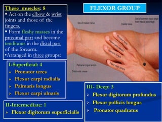
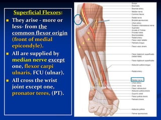
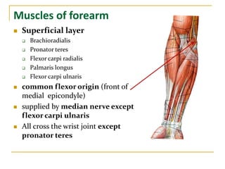
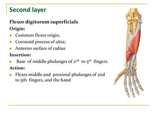
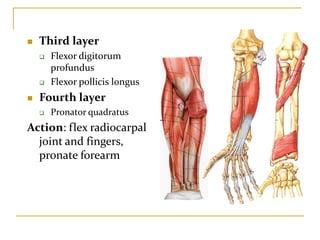
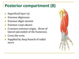
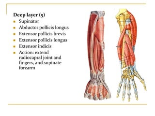
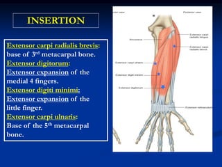
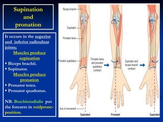
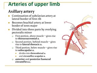
The document describes the muscles of the upper limb. It discusses muscles that move the pectoral girdle, muscles of the back, thorax, shoulder, arm, elbow, and forearm. It provides details on specific muscles like their origin, insertion, and action. It also summarizes the arteries, veins, lymphatic drainage, and nerves of the upper limb including the brachial plexus. The forearm contains muscles that supinate and pronate as well as move the wrist, hand, and fingers. Fascia and membranes in the forearm compartmentalize the muscles.
1. The document describes the muscles of the upper limb including their origins, insertions, and actions. It discusses the muscles of the pectoral girdle, back, thorax, shoulder, arm, forearm, hand, and their roles in moving the respective joints.
2. The arteries and veins of the upper limb are outlined, including the branches of the axillary, brachial, radial, and ulnar arteries. The lymphatic drainage and lymph nodes of the axilla and arm are also described.
3. The formation and branches of the brachial plexus are explained. Key nerves like the median, ulnar, radial, musculocutaneous and axillary nerves
The document describes the anatomy of the upper limb muscles, including:
1) Muscles that move the pectoral girdle and scapula, stabilize the scapula, and direct scapular movement.
2) Muscles of the back, thorax, shoulder, arm, forearm, hand, and their actions.
3) Major nerves of the brachial plexus including the musculocutaneous, median, ulnar, radial, and axillary nerves and their distributions.
The document provides details on the anatomy of the arm, including:
1. The humerus bone has two parts - an upper part for muscle attachments and a lower part for origins. Major muscles attaching to the humerus are described.
2. The brachial artery and accompanying nerves in the arm are outlined. Branches of the axillary artery and surrounding muscles are also noted.
3. Joint movements allowed by different synovial joint types are illustrated, including hinge, pivot, saddle, ball-and-socket, and condyloid joints.
The document summarizes the layers of muscles in the back and neck. It divides the muscles into 4 layers - the first layer includes the trapezius and latissimus dorsi muscles, the second layer includes muscles like the levator scapulae and rhomboideus, the third layer is the erector spinae muscles, and the fourth layer includes small muscles like the multifidus. It also describes the blood supply and innervation of the back muscles and provides details on the origin, insertion, nerve supply and action of some key muscles like the trapezius, latissimus dorsi and erector spinae.
The document describes the anatomy of the shoulder region. It discusses the bones that make up the shoulder girdle, including the clavicle and scapula. It then describes the major muscles of the shoulder girdle, including the deltoid, supraspinatus, infraspinatus, teres minor, teres major, and subscapularis muscles. It provides details on the origins, insertions, nerve supplies and actions of these muscles. It also discusses the rotator cuff and its role in stabilizing the shoulder joint. Finally, it outlines some important neurovascular structures in the shoulder region, including the suprascapular nerve and axillary nerve.
muscels of the upper Limb edited(1).pptxPhebeLois1
This document provides information on the muscles of the upper limb, axilla, and cubital fossa. It describes the origin, insertion, nerve supply, and action of numerous muscles in these regions, including the pectoral region, back, scapula, arm, and forearm (both anterior and posterior sides). The muscles described include the pectoralis major and minor, deltoid, biceps brachii, triceps brachii, brachialis, and many forearm flexors and extensors.
The document summarizes the major arteries and veins of the upper and lower limbs. It describes the branches and flow of the axillary, brachial, radial, ulnar, femoral, popliteal, anterior tibial, posterior tibial arteries and accompanying veins. Key veins include the basilic, cephalic and great saphenous veins.
The neck muscles are divided into 4 layers. The first layer includes the trapezius and latissimus dorsi muscles. The second layer includes the splenius, levator scapulae, rhomboideus major and minor, and serratus posterior superior and inferior muscles. The third layer is the erector spinae muscle. The fourth layer includes the multifidus, rotatores, interspinales, intertransversii and sub occipital muscles. The back muscles are divided into 3 groups: superficial muscles around the shoulder, intermediate muscles for respiration, and deep muscles of the vertebral column.
The document describes the anatomy of the axilla, subclavian artery, axillary artery, brachial artery, radial artery, ulnar artery, and intermuscular spaces in the arm. Key points include:
- The axilla is a pyramidal space between the upper arm and chest wall containing lymph nodes, blood vessels and nerves.
- The subclavian artery becomes the axillary artery in the axilla and then the brachial artery in the arm, with named branches along its course.
- The radial and ulnar arteries are terminal branches of the brachial artery, running in the forearm and hand.
- There are three intermuscular spaces in the arm that contain named
This document provides a table summarizing various muscles of the upper extremity, including their origin, insertion, innervation, main actions, blood supply, and muscle group. It lists over 30 muscles and describes each in a consistent format. The document focuses on describing muscle anatomy and function for medical education purposes.
The arm contains 3 compartments divided by intermuscular septa. The anterior compartment contains flexors like biceps and brachialis innervated by the musculocutaneous nerve. The posterior compartment's extensors include triceps innervated by the radial nerve. The brachial artery travels through the arm giving off branches before dividing into radial and ulnar arteries at the cubital fossa. The cubital fossa also contains the median nerve and radial nerve as they pass through the elbow region.
The document summarizes the arterial supply of the upper limb. It begins with the subclavian artery becoming the axillary artery after crossing the first rib, and the axillary artery becoming the brachial artery after crossing the posterior axillary fold. The brachial artery then divides into the radial and ulnar arteries distal to the elbow. It provides details on the branches and course of these main arteries supplying the upper limb.
15 Vascular anatomy of the upper limb2010 (2).pptAmanuelIbrahim
The document summarizes the arterial supply and venous drainage of the upper limb. It describes the origin and branches of the axillary, brachial, radial, and ulnar arteries. It also outlines the formation of the superficial and deep palmar arches. Regarding veins, it discusses the superficial veins that drain into the basilic and cephalic veins and the deep veins that accompany the arteries.
The thigh region lies between the hip and knee joints. It contains a single bone, the femur. The thigh is divided into anterior, medial, and posterior compartments by intermuscular septa. The anterior compartment contains muscles that flex the hip and extend the knee, while the posterior compartment contains muscles that extend the hip and flex the knee. The femoral triangle located in the upper anterior thigh contains the femoral nerve, artery and vein. The adductor canal on the medial thigh contains the femoral vessels and continues below as they pass into the leg. Major muscles of the thigh include the quadriceps femoris, sartorius, gracilis, and adductors.
The arm is divided into anterior, medial, and posterior compartments by intermuscular septae. The anterior compartment contains the biceps, brachialis, and coracobrachialis muscles. The posterior compartment contains the triceps muscle. Major nerves include the median, ulnar, radial, and musculocutaneous nerves. The brachial artery and its branches supply the arm, and lymph drainage involves the deltopectoral and supratrochlear lymph nodes.
This document provides an overview of the anatomy of the upper limb, including muscles, arteries, veins and innervation. It describes the major flexor and extensor muscles of the arm, forearm and hand. It details the brachial artery and its branches, including the radial and ulnar arteries which form palmar arches in the hand. It outlines the superficial and deep veins that drain the upper limb, including the basilic and cephalic veins. Finally, it maps the brachial plexus and its divisions which provide motor and sensory innervation to the arm.
This document provides an overview of the anatomy of the upper limb, including muscles, arteries, veins and innervation. It describes the major flexor and extensor muscles of the arm, forearm and hand. It discusses the brachial plexus and its formation from cervical and thoracic spinal nerves. The major nerves of the upper limb and their distributions are outlined. The axial arteries including the axillary, brachial, radial and ulnar arteries are summarized. The superficial and deep veins that drain the upper limb back to the heart are also described.
The axilla is the space between the upper arm and chest wall. It is bounded by the clavicle, first rib, and scapula. The axilla contains the axillary artery and vein, brachial plexus nerves, lymph nodes, and loose connective tissue. The brachial plexus is formed by the union of cervical and upper thoracic spinal nerves and provides motor and sensory innervation to the upper limb. Injuries to different parts of the brachial plexus can result in specific neuropathies like Erb's palsy or Klumpke's palsy, characterized by weakness or paralysis of certain muscles.
The thigh is divided into three compartments - anterior, medial, and posterior - by fascial septa. The anterior compartment muscles like the quadriceps femoris extend the knee and flex the hip. The medial compartment adductor muscles like adductor longus and brevis adduct the thigh. The posterior compartment hamstring muscles like biceps femoris and semitendinosus extend the hip and flex the knee. Each compartment is innervated by a specific nerve like the femoral nerve for the anterior compartment. The thigh also contains structures like the femoral triangle and adductor canal.
The brachial plexus is formed by the spinal nerves C5-T1 and divides into supraclavicular and infraclavicular parts. It gives rise to branches that innervate muscles and skin in the upper limb. In the forearm, the median nerve innervates flexor muscles and thenar muscles, and the radial and ulnar nerves innervate extensor muscles. Compression of the median nerve can cause carpal tunnel syndrome.
This document describes the anatomy of several muscles around the shoulder joint, including their origins, insertions, nerve supplies, and actions. It also discusses the rotator cuff muscles, quadrangular space, triangular spaces, axillary nerve, suprascapular nerve, and arterial blood supply around the shoulder joint. Key structures mentioned include the deltoid, trapezius, latissimus dorsi, levator scapulae, rhomboid muscles, and rotator cuff muscles.
Anatomy of brachial plexus explained in detail along with nerve supply of all the muscles of upper limb and various paralysis caused by brachial plexus injury
The document provides an overview of forearm anatomy including bones (ulna, radius), joints (elbow, radioulnar), muscles, nerves, blood vessels, and approaches. Key points include:
- The ulna articulates with the trochlea of the humerus at the elbow joint. The radial head articulates with the capitellum.
- Flexor and extensor muscle groups originate on the humerus, ulna, and radius to flex/extend the elbow, pronate/supinate the forearm, and flex/extend the wrist and fingers.
- The median, radial, and ulnar nerves innervate muscles in the forearm and hand.
-
The document summarizes the muscles of the forelimb in cattle. It describes the extrinsic muscles that connect the forelimb to the trunk, and the intrinsic muscles of the shoulder, arm, carpal/digital joints, and flexor/extensor groups. Key muscles mentioned include the trapezius, rhomboids, latissimus dorsi, pectorales, deltoid, biceps brachii, triceps brachii, and superficial/deep digital flexors. The origin, insertion, action, blood supply and nerve innervation are provided for many of the major muscles.
shoulder Anatomy by ayalew.orthopedic residentpptx,AyalewKomande1
The document provides an overview of shoulder anatomy and physical examination. It describes the bones that make up the shoulder joint including the clavicle, scapula, and proximal humerus. It outlines the fascia, muscles including rotator cuff and axioappendicular muscles, as well as neurovascular structures like the brachial plexus in the axilla. The physical examination section covers inspection, palpation, and range of motion assessment including special tests for shoulder conditions.
The document summarizes the gross anatomy of the forearm, including:
- The bones of the forearm are the radius and ulna. The radius articulates with the humerus proximally and wrist bones distally. The ulna articulates with the humerus proximally.
- The superficial and deep muscles of the anterior compartment are described, including flexor muscles like pronator teres, flexor digitorum superficialis, and flexor digitorum profundus.
- The arteries and nerves of the anterior compartment are outlined, including the ulnar artery, radial artery, median nerve, and ulnar nerve.
- The superficial muscles of the posterior compartment
The document describes the anatomy of the elbow joint. It discusses the muscles that flex and extend the elbow - the biceps brachii, brachialis, and triceps brachii. It describes the bones that make up the elbow joint, the humerus, ulna and radius, as well as the ligaments and synovial membrane that support the joint. The document outlines the movements of flexion and extension at the elbow and the muscles responsible for these movements. It defines the cubital fossa and lists the structures found within this space.
This document summarizes a lecture on inborn errors of metabolism. It discusses various metabolic disorders affecting carbohydrate metabolism, including galactosemia caused by a defect in galactose-1-phosphate uridyltransferase, resulting in hypoglycemia, jaundice and mental retardation. It also discusses disorders of amino acid metabolism, such as phenylketonuria caused by phenylalanine hydroxylase deficiency, and homocystinuria caused by cystathionine synthase deficiency. Finally, it summarizes various fatty acid oxidation disorders caused by defects in fatty acid beta-oxidation pathways.
The root of the neck is the junction between the thorax and neck. It contains several important structures including arteries, veins and nerves.
The document describes the anatomy of the thyroid gland, including its location in the neck, lobes and isthmus, relations to surrounding structures, blood supply from the superior and inferior thyroid arteries, innervation and role in hormone production and calcium metabolism.
The parathyroid glands are usually four in number, located on the posterior surface of the thyroid gland. They are supplied by branches from the inferior thyroid artery and their primary function is to regulate calcium levels through secretion of parathyroid hormone.
The pectoral region contains the pectoral muscles which connect the upper limb to the chest wall. The major pectoral muscle originates from the clavicle, sternum and ribs and inserts on the humerus, adducting and medially rotating the arm. The minor pectoral muscle covers the major and connects the ribs to the coracoid process. The breast overlies the pectoral muscles and contains lobules and ducts draining into the nipple. Lymph from the breast drains primarily to axillary nodes, with the medial breast draining to internal mammary nodes.
The document summarizes the major muscles of the leg and foot. It describes the origin, insertion, innervation, action, and arterial supply of key muscles like the gastrocnemius, soleus, tibialis anterior, flexor hallucis longus, and the intrinsic foot muscles. The muscles are grouped into those of the posterior and anterior leg compartments and the plantar and dorsal aspects of the foot.
This document discusses respiratory physiology topics including high altitude physiology, aviation physiology, and deep sea diving physiology. For high altitude physiology, it describes the stages of hypoxia, acclimatization responses like increased ventilation and hematocrit, and mountain sickness. Aviation physiology addresses positive and negative G-forces and their effects. Deep sea diving physiology discusses issues like nitrogen narcosis, oxygen toxicity, and decompression sickness prevention through techniques like slow ascent and decompression tanks.
The kidney performs several essential functions: it filters the blood to remove waste and regulate fluid balance. The kidney achieves this through its basic functional unit, the nephron. Each nephron contains a glomerulus that filters the blood and a renal tubule that processes the filtrate, removing waste and regulating electrolyte and water balance in the body through reabsorption and secretion. Precise regulation of blood flow and filtration allow the kidney to maintain homeostasis.
These lecture slides, by Dr Sidra Arshad, offer a quick overview of the physiological basis of a normal electrocardiogram.
Learning objectives:
1. Define an electrocardiogram (ECG) and electrocardiography
2. Describe how dipoles generated by the heart produce the waveforms of the ECG
3. Describe the components of a normal electrocardiogram of a typical bipolar lead (limb II)
4. Differentiate between intervals and segments
5. Enlist some common indications for obtaining an ECG
6. Describe the flow of current around the heart during the cardiac cycle
7. Discuss the placement and polarity of the leads of electrocardiograph
8. Describe the normal electrocardiograms recorded from the limb leads and explain the physiological basis of the different records that are obtained
9. Define mean electrical vector (axis) of the heart and give the normal range
10. Define the mean QRS vector
11. Describe the axes of leads (hexagonal reference system)
12. Comprehend the vectorial analysis of the normal ECG
13. Determine the mean electrical axis of the ventricular QRS and appreciate the mean axis deviation
14. Explain the concepts of current of injury, J point, and their significance
Study Resources:
1. Chapter 11, Guyton and Hall Textbook of Medical Physiology, 14th edition
2. Chapter 9, Human Physiology - From Cells to Systems, Lauralee Sherwood, 9th edition
3. Chapter 29, Ganong’s Review of Medical Physiology, 26th edition
4. Electrocardiogram, StatPearls - https://www.ncbi.nlm.nih.gov/books/NBK549803/
5. ECG in Medical Practice by ABM Abdullah, 4th edition
6. Chapter 3, Cardiology Explained, https://www.ncbi.nlm.nih.gov/books/NBK2214/
7. ECG Basics, http://www.nataliescasebook.com/tag/e-c-g-basics
TEST BANK For An Introduction to Brain and Behavior, 7th Edition by Bryan Kol...rightmanforbloodline
TEST BANK For An Introduction to Brain and Behavior, 7th Edition by Bryan Kolb, Ian Q. Whishaw, Verified Chapters 1 - 16, Complete Newest Versio
TEST BANK For An Introduction to Brain and Behavior, 7th Edition by Bryan Kolb, Ian Q. Whishaw, Verified Chapters 1 - 16, Complete Newest Version
Osteoporosis - Definition , Evaluation and Management .pdfJim Jacob Roy
Osteoporosis is an increasing cause of morbidity among the elderly.
In this document , a brief outline of osteoporosis is given , including the risk factors of osteoporosis fractures , the indications for testing bone mineral density and the management of osteoporosis
share - Lions, tigers, AI and health misinformation, oh my!.pptxTina Purnat
• Pitfalls and pivots needed to use AI effectively in public health
• Evidence-based strategies to address health misinformation effectively
• Building trust with communities online and offline
• Equipping health professionals to address questions, concerns and health misinformation
• Assessing risk and mitigating harm from adverse health narratives in communities, health workforce and health system
Basavarajeeyam is a Sreshta Sangraha grantha (Compiled book ), written by Neelkanta kotturu Basavaraja Virachita. It contains 25 Prakaranas, First 24 Chapters related to Rogas& 25th to Rasadravyas.
8 Surprising Reasons To Meditate 40 Minutes A Day That Can Change Your Life.pptxHolistified Wellness
We’re talking about Vedic Meditation, a form of meditation that has been around for at least 5,000 years. Back then, the people who lived in the Indus Valley, now known as India and Pakistan, practised meditation as a fundamental part of daily life. This knowledge that has given us yoga and Ayurveda, was known as Veda, hence the name Vedic. And though there are some written records, the practice has been passed down verbally from generation to generation.
Muktapishti is a traditional Ayurvedic preparation made from Shoditha Mukta (Purified Pearl), is believed to help regulate thyroid function and reduce symptoms of hyperthyroidism due to its cooling and balancing properties. Clinical evidence on its efficacy remains limited, necessitating further research to validate its therapeutic benefits.
TEST BANK For Community Health Nursing A Canadian Perspective, 5th Edition by...Donc Test
TEST BANK For Community Health Nursing A Canadian Perspective, 5th Edition by Stamler, Verified Chapters 1 - 33, Complete Newest Version Community Health Nursing A Canadian Perspective, 5th Edition by Stamler, Verified Chapters 1 - 33, Complete Newest Version Community Health Nursing A Canadian Perspective, 5th Edition by Stamler Community Health Nursing A Canadian Perspective, 5th Edition TEST BANK by Stamler Test Bank For Community Health Nursing A Canadian Perspective, 5th Edition Pdf Chapters Download Test Bank For Community Health Nursing A Canadian Perspective, 5th Edition Pdf Download Stuvia Test Bank For Community Health Nursing A Canadian Perspective, 5th Edition Study Guide Test Bank For Community Health Nursing A Canadian Perspective, 5th Edition Ebook Download Stuvia Test Bank For Community Health Nursing A Canadian Perspective, 5th Edition Questions and Answers Quizlet Test Bank For Community Health Nursing A Canadian Perspective, 5th Edition Studocu Test Bank For Community Health Nursing A Canadian Perspective, 5th Edition Quizlet Test Bank For Community Health Nursing A Canadian Perspective, 5th Edition Stuvia Community Health Nursing A Canadian Perspective, 5th Edition Pdf Chapters Download Community Health Nursing A Canadian Perspective, 5th Edition Pdf Download Course Hero Community Health Nursing A Canadian Perspective, 5th Edition Answers Quizlet Community Health Nursing A Canadian Perspective, 5th Edition Ebook Download Course hero Community Health Nursing A Canadian Perspective, 5th Edition Questions and Answers Community Health Nursing A Canadian Perspective, 5th Edition Studocu Community Health Nursing A Canadian Perspective, 5th Edition Quizlet Community Health Nursing A Canadian Perspective, 5th Edition Stuvia Community Health Nursing A Canadian Perspective, 5th Edition Test Bank Pdf Chapters Download Community Health Nursing A Canadian Perspective, 5th Edition Test Bank Pdf Download Stuvia Community Health Nursing A Canadian Perspective, 5th Edition Test Bank Study Guide Questions and Answers Community Health Nursing A Canadian Perspective, 5th Edition Test Bank Ebook Download Stuvia Community Health Nursing A Canadian Perspective, 5th Edition Test Bank Questions Quizlet Community Health Nursing A Canadian Perspective, 5th Edition Test Bank Studocu Community Health Nursing A Canadian Perspective, 5th Edition Test Bank Quizlet Community Health Nursing A Canadian Perspective, 5th Edition Test Bank Stuvia
Histololgy of Female Reproductive System.pptxAyeshaZaid1
Dive into an in-depth exploration of the histological structure of female reproductive system with this comprehensive lecture. Presented by Dr. Ayesha Irfan, Assistant Professor of Anatomy, this presentation covers the Gross anatomy and functional histology of the female reproductive organs. Ideal for students, educators, and anyone interested in medical science, this lecture provides clear explanations, detailed diagrams, and valuable insights into female reproductive system. Enhance your knowledge and understanding of this essential aspect of human biology.
2. Muscles That Move the Pectoral Girdle
Originate on the axial skeleton and insert on the clavicle and
scapula.
Stabilize the scapula and move it to increase the arm’s angle of
movements.
Some of the superficial muscles of the thorax are grouped together
according to the scapular movement they direct.
elevation, depression, protraction, or retraction
3. The muscles of back
Superficial group
Trapezius
Latissimus dorsi
Levator scapulae
Rhomboideus
Deep group
Erector spinae
Splenius
Thoracolumbar fascia
4. The muscles of thorax
Extrinsic muscles
Pectoralis major
Pectoralis minor
Serratus anterior
Intrinsic muscles
Intercostales externi
Intercostales interni
Intercostales intimi
5. The Muscles of Upper Limb
Muscles of shoulder
Deltoid
supraspinatus
Infraspinatus
Teres minor
Teres major
subscapularis
6. Major muscles of upper limb
Deltoid
Origin: lateral third of
clavicle, acromion, and spine
of scapula
Insertion: deltoid tuberosity
of humerus
Action: abducts,flexes and
medically rotates, extends,
and laterally rotates arm
7. Teres major
Origin: dorsal surface
of inferior angle of
scapula
Insertion: crest of
lesser tubercle of
humerus
Action: medially
rotates and adducts
arm
9. Arm Muscles That Move the Shoulder/Elbow
Joint
(Flexor) compartment
Posterior (extensor) compartment
Anterior compartment
primarily contains shoulder/elbow flexors
Posterior compartment contains elbow extensors
the principal flexors
biceps brachii, brachialis, and brachioradialis
muscles that extend the elbow joint
triceps brachii and the anconeus
10.
11. Muscles of arm
Antererior group
Biceps brachii
Coracobrachialis
Brachialis
Posterior group –
triceps brachii
12. Biceps brachii
Origin: long head,
supraglenoid tubercle; short
head, coracoid process
Insertion: radical tuberosity
Action: supinator of forearm,
flexor of elbow joint, weak
flexor of should joint
Pronator teres
Origin: medical epicondyle of
humerus and deep fascia of
forearm
Insertion: middle of lateral
surface of radius
Action: pronation of forearm
and flexion of elbow
13. Triceps brachii
Origin: long head,
infraglenoid tubercle; lateral
head, above groove for
radical n., medical head,
below groove for radical n.
Insertion: olecranon of ulna
Action: extends elbow joint),
long head can extend and
adduct shoulder joint
14. Surface anatomy
M. triceps brachii
caput longum, medialis, lateralis
Surface elements
Skin thick and movable
Subcutaneous tissue well expressed
n. cutaneus brachii posterior
n. cutaneus brachii lateralis inferior
n. cutaneus antebrachii posterior
Posterior of the arm
15. Deep structures
А. Fascia brachialis
- Thick, thins distally
B. Neurovascular bundles
1. Upper - in canalis humero-muscularis, between:
- sulcus n. radialis
- medial and lateral head of m. triceps brachii
1.1. n. radialis – lies on the humerus
- n. cutaneus brachii lateralis inerior
- n. cutaneus antebrachii posterior
- rr. musculres
1.2. a. profunda brachii
- a. collateralis media
- collateralis radialis
19. Anterior elbow
Surface anatomy
Three eminences
Two grooves
Elements
Lateral groove - v. cephalica, n. cutaneus
antebrachii lateralis
Medial groove – v. basilica, n. cutaneus
antebrachii medialis
V. mediana cubiti
20. Deep structures
А. Muscles
Flexors of forearm – medial eminence
Extensors of forearm – lateral eminence
Brachial muscles, mm. biceps, brachialis –
middle eminence
21. Deep structures
B. Vessels and nerves
Medial bundle - between m. biceps brachii and pronator
teres
- a.v. brachialis, n. medianus
Lateral bundle - between m. brachialis and
brachioradialis
- n. radialis, a. collateralis radialis, a. recurrens radialis
C. Cubital fossa
Boundaries
- m. brachioradialis (lateral), m. pronator teres (medial)
Content
- tendo m. brachialis, m. biceps brachii, bursa bicipitoradialis
22. Layers of the elbow
Venous
1 v. cephalica
2 v. basilica
3 v. mediana cubiti
Apponeurotic
1 aponeurosis bicipis
2 tendo m. biceps brachii
23. Neurovascular
1 a. brachialis
2 n. medianus
Muscle
1 m. supinator
2 m. brachialis
3 tendo m. biceps brachii
Bone
1 humerus
2 radius
3 ulna
27. Arteries of upper limb
Axillary artery
Continuation of subclavian artery at
lateral border of first rib
Becomes brachial artery at lower
border of teres major
Divided into three parts by overlying
pectoralis minor
First portion, above muscle-gives rise
to thoracoacromial a.
Second portion, behind muscle-gives
rise to lateral thoracic a.
Third portion, below muscle-gives rise
to subscapular a.
divides into throcodorsal a.
and circumflex scapular a.
anterior and posterior humeral
circumflex a.;
28.
29. Brachial artery
Continuation of axillary artery
Divides into radial and ulnar
arteries at level of neck of radius
Branches
Deep brachial a. accompanies
radial nerve
Superior ulnar collaeral a.
accompanies ulnar nerve
Inferior ulnar collateral a.
30. Radial artery and branches
Radial recurrent a.
Superfical palmar branch
Principal artery of thumb
Ulnar artery and branches
Ulnar recurrent a.
Common interosseous artery
Anterior interossous a.
Posterior interosseous a.
Deep palmar branch
31. Basilic vein
Arises from the medial side of the
dorsal venous rete of hand
Ascends on the ulnar side of forearm
to the elbow and then in the medial
bicepital brachii furrow to middle of
the arm where it pierces the deep
fascia and joins the brachial vein or
axillary vein
Median cubital vein
links cephalic vein and basilic vein in
the cubital fossa. It is a frequent site
for venipuncture to remove a sample
of blood or add fluid to the blood
32. The lymphatic drainage of upper limb
Lymphatic vessels
Superficial-follow the
superficial veins, drain into
supratrochlear and axillary
lymph nodes
Deep-accompany main vessels,
end in axillary lymph nodes
Lymph nodes
Cubital lymph node: lies above
medial epicondyle of humerus
Axillary lymph node-arranged
in five groups
33. Axillary lymph nodes
Arranged in five groups
Lateral lymph nodes lie around
the distal end of axillary vein ,
receiving drainage from the arm,
forearm, and hand
Pectoral lymph nodes lie along
lateral thoracic vessels, receive
afferents from anterior thoracic wall
including central and lateral portion of
mamma
Subscapular lymph node along
subscapular vessels, receive lymph
from nape and scapular region
Efferents above three groups pass to
central lymph node
34. Brachial plexus
Formation:
Five roots: anterior rami of C5-C8
and T1 spinal nerves, roots C5-
C7give rise to long thoracic n.
Three trunks
Upper trunk formed by the joining of
roots C4,C5,C6.
Middle trunk continuation of root C7.
Lower trunk formed by the joining of
roots C8 and T1.
Six divisions: above clavicle,
trunks form anterior and posterior
divisions
Three cords: below clavicle,
divisions form three cords that
surround the second portion of
axillary a.
35. Position:
passes through the scalene fissure to
posterosuperior of subclavian artery, then
enters the axilla to form lateral, medial and
posterior cords
Main branches
Lateral cord
Musculocutaneous n.
Lateral root to median n.
Medial cord
Medial root to median n.
Ulnar n.
Medial brachial cutaneous n.
Medial antebrachial cutaneous n.
38. Median nerve
Supplies:
Flexors of forearm except
brachioradialis,
Flexor carpi ulnaris
flexor digitorum profundus - ulnar half
Thenar except adductor pollicis, two
lateral lumbricals;
Skin of thenar, central part of palm,
palmar aspect of radial three and one-
half fingers, including middle and
distal fingers on dorsum
Injury: Ape hand
produces sign of benediction,
the index and middle fingers cannot
be flexed and the thumb cannot be
opposed
39. Ulnar nerve
Supplies:
flexor carpi ulnaris,
ulnar half of flexor digitorum
profundus,
hypothenar muscles, interossei,
3rd and 4th lumbricals and adductor
pollicis;
skin of hypothenar, palmar surface
of ulnar one and one-half fingers,
ulnar half of dorsum of hand,
posterior aspect of ulnar two and
one-half fingers
Injury: claw hand
40. Radial
Supplies:
Extensor muscles of arm and
forearm,
Brachioradialis;
skin on back of arm, forearm,
and radial side of dorsum of
hand and radial two and one-
half fingers
Injury: Wrist drop
41. Axillary nerve
Supplies:
Deltoid and teres
minor muscle;
skin over deltoid and
upper posterior aspect
of arm
Injury:
deltoid and teres minor
paralysis (loss of
shoulder abdution and
wheel external rotation)
loss of sensation over
the deltoid
42. Forearm Muscles
Supinate and Pronate
Supinator muscle supinates the forearm.
Biceps brachii supinates the forearm.
Pronator teres and pronator quadratus pronate the forearm.
Move the Wrist Joint, Hand, and Fingers
Muscles in the forearm move the hand at the wrist and/or the fingers.
Extrinsic muscles of the wrist and hand originate on the forearm, not
the wrist or hand.
Tendons of forearm muscles typically are surrounded by tendon
(synovial) sheaths and held adjacent to the skeletal elements by strong
fascial structures.
At the wrist, the deep fascia of the forearm forms thickened, fibrous
bands termed retinacula.
43. The forearm extends
from elbow to wrist.
It posses two bones
radius laterally & Ulna
medially.
The two bones are
connected together
by the interosseous
membrane.
This membrane
allows movement of
Pronation and
Supination while the
two bones are
connected together.
Also it gives origin for the
deep muscles.
44. Sheath of deep fascia
attached to the posterior
border of the ulna .
The fascial sheath,
interosseous membrane &
fibrous intermuscular septa,
divide the forearm into 3
compartments, each
having its own muscles,
nerves, and blood supply.
Fascial Compartments of the Forearm
45. I-Superficial: 4
Pronator teres
Flexor carpi radialis
Palmaris longus
Flexor carpi ulnaris
II-Intermediate: 1
Flexor digitorum superficialis
These muscles: 8
Act on the elbow & wrist
joints and those of the
fingers.
Form fleshy masses in the
proximal part and become
tendinous in the distal part
of the forearm.
•Arranged in three groups:
III- Deep: 3
Flexor digitorum profundus
Flexor pollicis longus
Pronator quadratus
FLEXOR GROUP
46. Superficial Flexors:
They arise - more or
less- from the
common flexor origin
(front of medial
epicondyle).
All are supplied by
median nerve except
one, flexor carpi
ulnaris, FCU (ulnar).
All cross the wrist
joint except one,
pronator teres, (PT).
47. Muscles of forearm
Superficial layer
Brachioradialis
Pronator teres
Flexor carpi radialis
Palmaris longus
Flexor carpi ulnaris
common flexor origin (front of
medial epicondyle)
supplied by median nerve except
flexor carpi ulnaris
All cross the wrist joint except
pronator teres
48. Second layer
Flexor digitorum superficials
Origin:
Common flexor origin,
Coronoid process of ulna;
Anterior surface of radius
Insertion:
Base of middle phalanges of 2nd to 5th fingers.
Action:
Flexes middle and proximal phalanges of 2nd
to 5th fingers, and the hand
50. Lateral compartment (3)
Brachioradialis
Extensor carpi radialis longus
Extensor carpi radialis brevis
Common extensor origin,
(front of lateral epicondyle of
the humerus), EXCEPT, 2 (BR
& ECRL).
Cross the wrist EXCEPT
brachioradialis.
Supplied by deep branch of
radial nerve
51. Posterior compartment (8)
Superficial layer (3)
Extensor digitorum
Extensor digiti minimi
Extensor carpi ulnaris
Common extensor origin, (front of
lateral epicondyle of the humerus),
Cross the wrist.
Supplied by deep branch of radial
nerve
53. Extensor carpi radialis brevis:
base of 3rd metacarpal bone.
Extensor digitorum:
Extensor expansion of the
medial 4 fingers.
Extensor digiti minimi:
Extensor expansion of the
little finger.
Extensor carpi ulnaris:
Base of the 5th metacarpal
bone.
INSERTION
54. Supination
and
pronation
It occurs in the superior
and inferior radioulnar
joints;
Muscles produce
supination
Biceps brachii.
Supinator.
Muscles produce
pronation
Pronator teres.
Pronator quadratus.
NB. Brachioradialis put
the forearm in midprone-
position.
56. Muscles of hand (palm)
Lateral group thenar (4)
Abductor pollicis brevis
Flexor pollicis brevis
Opponens pollicis
Adductor pollicis
Action: flex, abduct, adduct and
oppose thumb
Medial group hypothenar (3)
Abductor digiti minimi
Flexor digiti minimi brevis
Opponens digiti minimi
Action: flex, abduct , and oppose
little finger
57. Intermedial group
Lumbricals (4) flex
fingers at MP joints;
extend fingers at IP
joints
Palmar interossei (3)
adduct fingers towards
middle finger at MP
joints
Dorsal interossei (3)
abduct fingers away
from middle finger at
MP joints
58. Arteries of upper limb
Axillary artery
Continuation of subclavian artery at
lateral border of first rib
Becomes brachial artery at lower
border of teres major
Divided into three parts by overlying
pectoralis minor
First portion, above muscle-gives rise
to thoracoacromial a.
Second portion, behind muscle-gives
rise to lateral thoracic a.
Third portion, below muscle-gives rise
to subscapular a.
divides into throcodorsal a.
and circumflex scapular a.
anterior and posterior humeral
circumflex a.;
59.
60. Brachial artery
Continuation of axillary artery
Divides into radial and ulnar
arteries at level of neck of radius
Branches
Deep brachial a. accompanies
radial nerve
Superior ulnar collaeral a.
accompanies ulnar nerve
Inferior ulnar collateral a.
61. Radial artery and branches
Radial recurrent a.
Superfical palmar branch
Principal artery of thumb
Ulnar artery and branches
Ulnar recurrent a.
Common interosseous artery
Anterior interossous a.
Posterior interosseous a.
Deep palmar branch
62. Superficial palmar arch
Formed by ulnar artery and
superficial palmar branch
of radial artery
Curve of arch lies across
the palm, level with the
distal border of fully
extended thumb
Gives rise to three common
palmar digital arteries each
then divides into two
proper palmar digital
arteries
63. Deep palmar arch
Formed by radial artery
and deep palmar branch of
ulnar artery
Curve of arch lies across
upper part of palmar at
level with proximal border
of extended thumb
Gives rise to three palmar
metacarpal arteries
64. Veins of the upper limb
Deep veins: accompany the arteries of
the same region and bear similar
names
Superficial veins
Cephalic vein
Arises from the lateral side of the
dorsal venous rete of hand
Ascends on radial side of the forearm
to the elbow and then in the lateral
side of biceps brachii furrow,
continues up the arm in the
deltopectoral groove and then to the
infraclavicular fossa, where it pierces
clavipectoral fascia to drain into
axillary vein