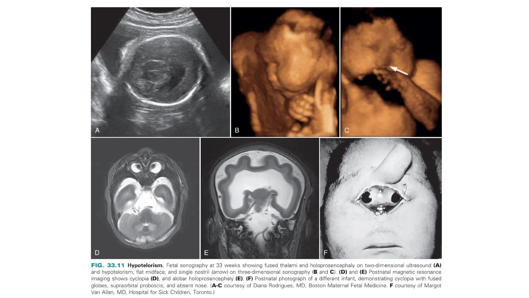
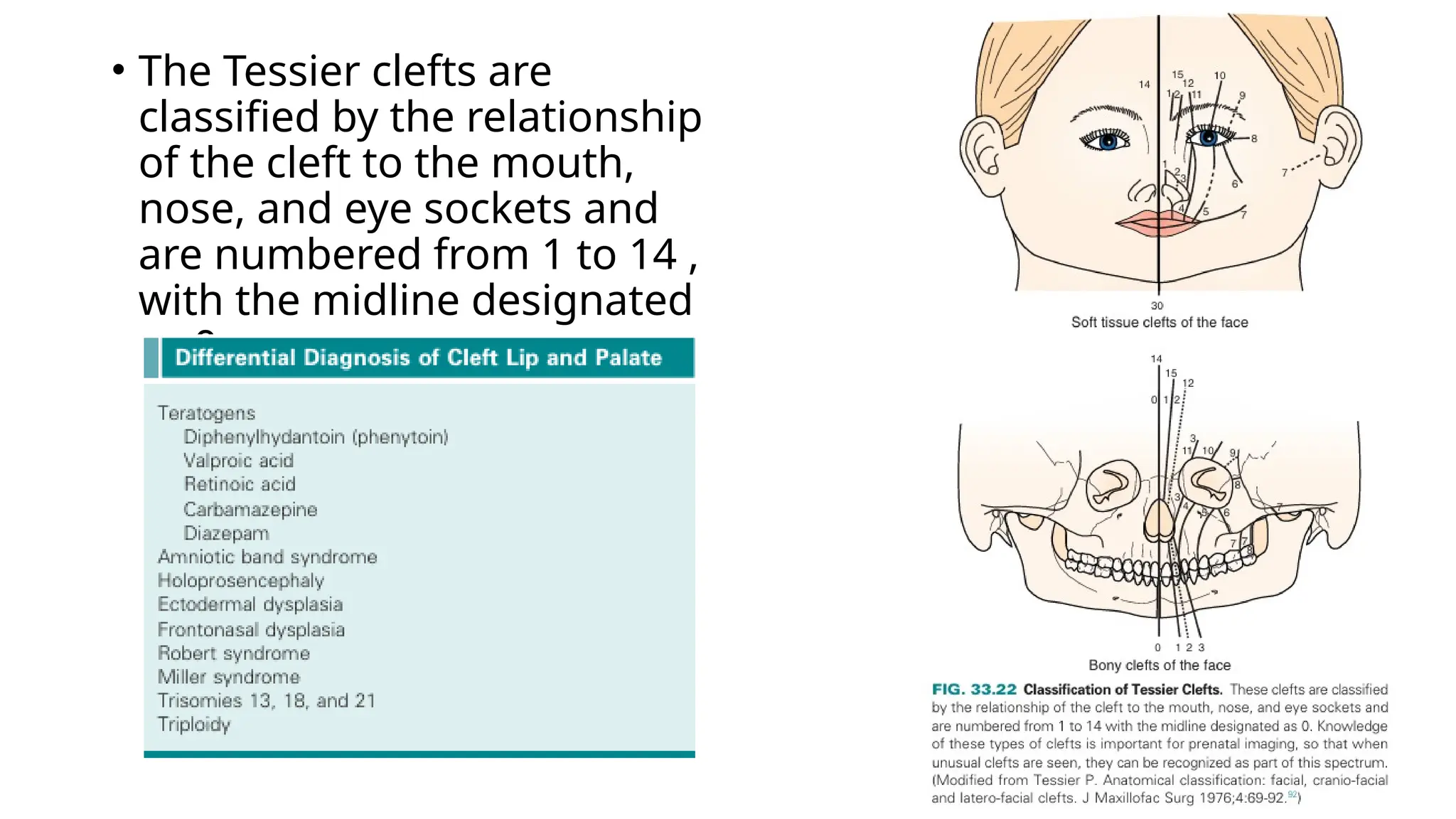
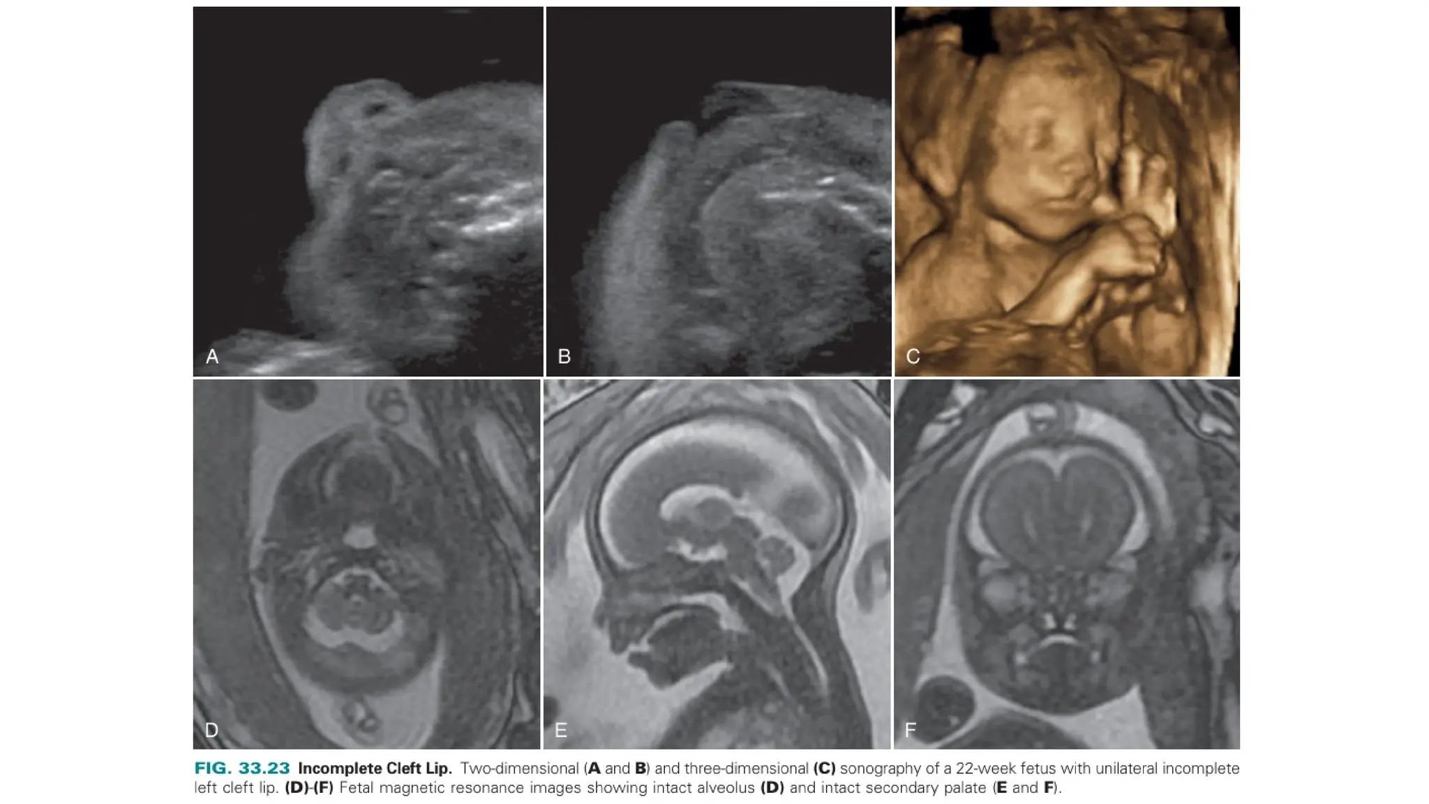
FOETAL HEAD AND NECK
- A. Abdul Ansari Ghani (Junior Resident)
Moderator – Dr. Mohan S (Assistant
professor)
FOETAL HEAD
• Introduction:
• The fetal face starts to develop at 4th
week of gestation, with
completion of major events by 8th
week.
• Ectoderm, mesoderm, endoderm and neural crest cells are
involved in this complex process.
• Ectoderm – Surrounds primitive mouth.
• Brachial arches (Composed of central mesenchyme, outer
ecto and inner endo)– Fuses in the mid-line.
• Neural crest cells – Give rise to connective tissues of the face.
PROMINENCES:
• Five main tissue buds form the fetal face.
• They are as follows:
Frontonasal prominence - Forehead and dorsum apex of
the face
Medial prominence - Nasal septum.
Lateral prominence – Nasal ala.
Maxillary prominence – Upper cheeks and upper lip.
Mandible prominence – Lower cheeks, lower lip and chin
USG FEATURES OF NORMAL FOETAL
FACE:
• The sagittal facial profile view is obtained possible and
should demonstrate the presence of normal configuration –
nasal bone, lip, chin and forehead.
• Axial views of orbit are obtained to visualize the presence
of globe, its size and the normal distance between the
orbits.
• In case of clefts of primary palate , axial views of maxilla
and alveolar ridge is obtained.
• Secondary palate is not much visualized in 2-D view, but
better appreciated in 3-D views.
ORBIT ABNORMALITIES:
• Axial and coronal views are best preferred, which
helps visualize both orbits, size, shape and the
distance between them.
• Sagittal view may help in case of abnormal anterior
displacement of the globes like in case of proptosis
and exorbitism.
• The orbits should be symmetrical in size and the
outer and inner interorbital distances within a
normal range.
• Hypotelorism: It is defined as the small distance
between the orbits and is often associated with
other anomalies.
• Holoprosencephaly associated cyclopia.
• Ethmocephaly (Hypotelorism with falied
development of nose and proboscis).
• Cebocephaly (Hypotelorism and poorly developed
nose with single nostril).
• Hypertelorism: It is defined as widely spaced eyes.
• Microphtalmia (abnormally small orbits) and
Anopthalmia (Absence of orbits).
COLOBOMA:
• It results from incomplete closure of optic fissure and most
often affects the iris inferiorly.
• It can affect any structure from eyelid to optic nerve or
retina.
• Depending on the abnormalities and the extent of the
structures, vision may or may not be affected.
• Diagnosis depends on visualization of focal bulge in the
posterior aspect of the globe.
• In case of artifacts – 3D USG and MRI might be helpful.
• It is a rarely anomaly and may be associated with other
anomalies and syndromes such as CHARGE syndrome.
DACRYOCYSTOCELE:
• It results from obstruction of nasolacrimal ducts.
• These appear as cystic masses that may contain low-
level internal echoes, located inferomedial to the
orbit in the expected location of the nasolacrimal duct.
• No mass effect or vascularity on color doppler.
• Diagnosis made after 30 weeks of gestation.
• Differentials include – Teratomas (often solid or mi ed
cystic and solid, and may contain calcifications) and
Haemangiomas (solid, echogenic sonographic
appearance, with increased vascular flow).
CONGENITAL CATARACTS:
• It may be diagnosed on prenatal
sonography, Which will show a rounded
echogenic mass in the anterior
portion of the globe.
• This is a rare disorder, with a reported
incidence of approximately 3 per 10,000
births.
• Causes of congenital cataracts include
genetic disorders, infection, syndromes,
and microphthalmia.
• Some cases are inherited by either
autosomal dominant or autosomal
recessive transmission.
EAR ABNORMALITIES:
• Abnormalities of the ears can be very difficult to
diagnose on fetal sonography, but low-set ears are
associated with multiple syndromes, including Noonan
syndrome and certain trisomies.
• Low-set ears are described as the helix joining the
cranium at a level below a horizontal plane through the
inner canthi of the eyes.
• It is easily detectable in 3D sonography.
• Microtia (Small ears): It is a rare anomaly with an
incidence of approximately 1 per 10,000 live births and
is often associated with syndromes and aneuploidy.
• Otocephly: It is a condition with union of the ears on the
front of the neck and is caused by failure of ascent of the
auricles during embryologic development.
• The most severe form of otocephaly may be associated with
absence of the eyes, forebrain, and mouth.
• It is a fatal anomaly often associated with agnathia or
micrognathia, as ell as holoprosencephaly.
MIDFACE ABNORMALITIES:
• The midface is the area between the upper lip and
forehead.
• Hypoplasia:
• Midface hypoplasia arises from a variety of causes,
including syndromes such as Apert, Crouzon, Treacher
Collins, Wolf-Hirschhorn, Pfeiffer, Turner, and trisomy
21.
• Midface hypoplasia may also result from facial clefts,
cranio-synostosis, and skeletal dysplasia's.
• It can be demonstrated at sagittal mid-line views.
ABSENT NASAL BONE:
• It can be assessed in the 1st
trimester, Hypoplastic or absent
nasal bone is seen with increased incidence in foetuses with
trisomy 21.
• It must be evaluated at mid-sagittal section.
• Some state that combining the findings of absence of nasal
bone and NT measurement, trisomy 21 detection
improves at 1st
trimester.
• The absence of a nasal bone at second trimester
sonography, or abnormally short nasal bone
measurements in combination with other markers of
aneuploidy, increases detection of aneuploidy.
OTHER NASAL ABNORMALITIES:
• Fetal nasal masses are rare, but can be identified in sonography.
• Differentials include glioma, dermoid, haemangioma and
encephalocele.
• Nasal glioma – located on the bridge or sides of the nose, may be
extra nasal(60%) , intranasal (30%) and both (10%). No vascularity
on colour doppler.
• Dermoid – Solid, cystic and heterogenous.
• Haemangiomas – Frequently echogenic and demonstrate vascular
flow on colour doppler.
• Encephalocele – may appears cystic (meninges or CSF) or
heterogenous (brain tissue).
• Fetal MRI is helpful to look for intracranial extension in
encphalocele and may be in case of nasal gliomas also.
CLEFT LIP AND PALATE
• It results as a failure of fusion of medial prominence and maxillary
prominence
• M>F
• It is seen more common in trisomies 13 and 18, some are familial
depending on the affected parents or siblings.
• Best identified after 20 weeks of gestation.
• Fetal facial clefts should be described using standard craniofacial
terminology and including accurate description and classification
of the cleft in relation to the lip, nostril, alveolus (maxillary tooth
bearing arc), and secondary palate.
• The secondary palate has an anterior, bony segment and a
posterior, soft tissue segment. Both cleft lip and cleft palate may be
unilateral or bilateral.
• A complete cleft lip is defined as a cleft that fully divides the lip
and extends completely through the base of the ala of the nose
and is associated with a cleft of the underlying tooth-bearing
alveolus.
• An incomplete cleft lip involves a portion of the lip, but at least a
band of sot tissue spans the cleft. Incomplete cleft lip does not
involve the ipsilateral underlying bony tooth-bearing alveolus.
• Sonographic signs of cleft palate include the following :
Abnormally high position of the fetal tongue,
Hypertelorism,
Deviation of the vomer (a triangular bone in the nasal septum
forming the posterior and inferior portion of the septum),
Micrognathia.
• In order to describe the type of cleft , two embryogenic
structures are considered as follows:
• The primary palate, formed by the prolabium, premaxilla,
and columella, which includes the lip, nares, and alveolus.
• The secondary palate, which begins at the incisive foramen
and is formed by a horizontal portion of the maxilla, the
horizontal portion of the palatine bones, and the soft
• Cleft palate may interfere with fetal swallowing and result in
polyhydramnios.
• Infants with cleft palate have difficulty with feeding, are at
increased risk of otitis media, and may have difficulty with
hearing and speech.
• Unilateral cleft lip or palate:
There is an offset between the two sides of the cleft, Lesser
segment at the side of the cleft and greater segment on the
side opposite to the cleft.
It occurs most commonly on the left side.
• Bilateral cleft lip or palate:
It occurs in 10% of the cases.
It may be symmetrical or asymmetrical.
In midsagittal view, abnormal premaxillary protrusion of soft
tissue anterior and above the normal position of upper lip.
• Median cleft lip or palate:
It is a classical finding in alobar holoprosencephaly.
Head size is small compared to menstrual dates and have
hypertelorism.
Some cases represent without holoprosencephaly.
• Unusual facial (Tessier) cleft:
• Asymmetrical clefts in seen in unusual locations, which may be
the result of amniotic band syndrome or may fall into the Tessier
cleft category.
• It is rare.
• 3D sonography is reliable.
• Tessier clefts can involve either the sot tissues (e.g., hairline,
eyebrows, eyelids, nostrils, lips, ears) or the skeleton.
• The Tessier clefts are
classified by the relationship
of the cleft to the mouth,
nose, and eye sockets and
are numbered from 1 to 14 ,
with the midline designated
as 0 .
• Isolated cleft of secondary palate:
• It is rarely identifiable in prenatal scans because of shadowing.
• Sonographic diagnosis is based on secondary signs, such as
abnormal oropharyngeal fluid flow on color Doppler
imaging and high position of the tongue.
• Syndrome associated are Goldenhar syndrome, Pierre-Robin
sequence, Treacher Collins syndrome, Tickler syndrome, and
velocardiofacial syndrome.
• Three-dimensional sonography is often helpful in assessing
the secondary palate, given a favorable fetal position and
gestational age and adequate amniotic fluid.
• Sagittal fetal MRI helps in delineating the normal soft tissues of
the palate and in accurately characterizing palatal clefting, even
when isolated to the posterior secondary (soft) palate.
LOWER FACE ABNORMALITIES:
• Macroglossia and oral
masses:
It is visualized in sagittal or
axial views, as tongue
(abnormally enlarged)
protrudes outside the oral
cavity.
It has variety of aetiology and
is associated with variety of
conditions.
Micrognathia and Retrognathia
• Micrognathia – small chin. It is visualized in mid-sagittal
view.
• Retrognathia – Posteriorly displaced chin.
• These are distinct abnormalities and frequently occurs
together.
• Pierre-Robin sequence is the clinical triad of micrognathia,
glossoptosis (downward displacement of the tongue), and
subsequent airway obstruction, often associated with cleft
soft palate .
• If the abnormality is severe enough to interfere with fetal
swallowing in utero, polyhydramnios may result.
• Micrognathia can lead to substantial feeding difficulties and
problems with airway management after birth. So prenatal
• This finding is associated with syndromes (e.g., tickler,
velocardiofacial, Miller Diecker, Beckwith Wiedemann,
Treacher Collins, Pfeiffer, femoral-facial) chromosomal
anomalies (typically trisomy 18 or 13) and skeletal dysplasias
(e.g., diastrophic, spondyloepiphyseal, congenital,
camptomelic).
• Agnathia is total or partial absence of the lower jaw and is
often associated with holoprosencephaly.
• Microstomia is a small mouth, often associated with
agnathia and otocephaly.
SOFT TISSUE TUMOURS
• Soft tissue or bony tumors can cause alterations in head size or shape.
• Hemangioma is the most common and increases in size during fetal
size and can be infiltrative (does not infiltrate bony structures).
• It appears echogenic and predominantly solid masses with vascularity
on colour doppler.
• It can occur at any location and involves fetal face.
• If lesion is large is size, it causes high cardiac output failure due to
vascular shunting.
• There may also be platelet trapping in the hemangioma, leading to
thrombocytopenia which referred to as “ Kasabach- Merritt
syndrome”.
FOETAL NECK:
• The development of the fetal neck is similarly complete ,
with extensive embryologic events contributing to
development of vascular, neurologic, musculoskeletal,
lymphatic, and endocrine systems.
• The laryngotracheal groove forms during the fourth w eek
of gestation along the floor of the primitive mouth.
• After evagination of this groove, the laryngotracheal
diverticulum forms.
• The distal end forms the lung bud.
• The endoderm of this diverticulum forms the epithelium of
the larynx and trachea.
• The endothelium of the larynx proliferates and temporarily
occludes its lumen.
• Recanalization occurs by 10th
week of gestation with formation
of the laryngeal ventricle, vocal folds, and vestibular folds.
• The fourth and sixth pharyngeal arches form the surrounding
cartilage and muscles.
• At 4 to6 weeks of gestation, right and left jugular lymph sacs
develop as diverticula of the subclavian veins.
• At to eeks’ gestation, right and left jugular lymph sacs develop
as diverticula of the subclavian veins.
• Abnormal connections between the lymphatic sacs and venous
system are thought to contribute to lymphatic malformation
and thickened nuchal translucency (NT) in the first trimester, as
well as a thickened nuchal fold in the second trimester.
ABNORMALITIES OF NECK:
• Nuchal Translucency and Thickening:
• The nuchal translucency (NT) is the fluid collection that
forms posterior to the fetal neck during early development.
• It is visualized at mid-sagittal section.
• The measurements are combined with patient age,
gestational age and serum testing to rule the risk of
aneuploidy.
• The nuchal fold thickness is measured at sub-occiptal
bregmatic plane, if the measurement is >6mm, at 15 to 22
weeks – it denotes an increased risk of trisomy 21.
LYMPHATIC MALFORMATION:
• It is called as Cystic hygroma.
• It is a septated fluid collection behind the fetal neck,
thought to result from early maldevelopment of the
lymphatic system.
• Highly associated with Turner’s syndrome.
• If it occurs with hydrops – foetal mortality is high.
• They are presumed to be a result from obstructed lymphatic
sacs that do not communicate with main lymphatic
channels.
• It can occur in head and neck and can be microcystic or
macrocystic.
• Although benign, morbidity is associated with mass effect
on the fetal airway, fetal MRI is often helpful in such cases.
• Fluid-filled lymphatic malformations are less likely to cause
airway obstruction.
• When these malformations involve the tongue, cystic
hygromas may interfere with swallowing and feeding after
birth.
CERVICAL TERATOMA:
• Teratoma is most common and occurs at sacrum and
coccyx.
• In 5% of cases, it occurs in the neck region with equal
gender predominance.
• Sonogram shows complex masses composed of solid (show
vascular flow) and cystic areas, calcification (in some cases).
• In the neck, they are usually anterolateral in location and
can increases large, often involving the thyroid gland and
may impinge the air way, might inerfere fetal swallowing,
and result in polydramnios.
• Hyperextension of the neck is best viewed in sagittal plane.
• Teratomas of the oropharynx epinathus often protrude from
the mouth.
THYROMEGALY AND GOITER:
• Fetal goiter is rare and most often related to maternal thyroid
disease, such as graves disease or Hashimoto thyroiditis, with
the abtiobiotics that cross and cause fetal thyroid dysfunction.
• Primary fetal thyroid dysfunction may also cause goiter.
• It appears as a midline homogeneous solid mass in the anterior
neck surrounding the trachea and shows increased vascularity .
• Neck hyperextension can lead to fetal malpresentation and can
cause difficulties at delivery.
• Cordocentesis may be necessary to determine if there is fetal
hypothyroidism or hyperthyroidism.
THANK
YOU

fetal head and neck imaging and spotters

  • 1.
    FOETAL HEAD ANDNECK - A. Abdul Ansari Ghani (Junior Resident) Moderator – Dr. Mohan S (Assistant professor)
  • 2.
    FOETAL HEAD • Introduction: •The fetal face starts to develop at 4th week of gestation, with completion of major events by 8th week. • Ectoderm, mesoderm, endoderm and neural crest cells are involved in this complex process. • Ectoderm – Surrounds primitive mouth. • Brachial arches (Composed of central mesenchyme, outer ecto and inner endo)– Fuses in the mid-line. • Neural crest cells – Give rise to connective tissues of the face.
  • 3.
    PROMINENCES: • Five maintissue buds form the fetal face. • They are as follows: Frontonasal prominence - Forehead and dorsum apex of the face Medial prominence - Nasal septum. Lateral prominence – Nasal ala. Maxillary prominence – Upper cheeks and upper lip. Mandible prominence – Lower cheeks, lower lip and chin
  • 5.
    USG FEATURES OFNORMAL FOETAL FACE: • The sagittal facial profile view is obtained possible and should demonstrate the presence of normal configuration – nasal bone, lip, chin and forehead. • Axial views of orbit are obtained to visualize the presence of globe, its size and the normal distance between the orbits. • In case of clefts of primary palate , axial views of maxilla and alveolar ridge is obtained. • Secondary palate is not much visualized in 2-D view, but better appreciated in 3-D views.
  • 7.
    ORBIT ABNORMALITIES: • Axialand coronal views are best preferred, which helps visualize both orbits, size, shape and the distance between them. • Sagittal view may help in case of abnormal anterior displacement of the globes like in case of proptosis and exorbitism. • The orbits should be symmetrical in size and the outer and inner interorbital distances within a normal range.
  • 9.
    • Hypotelorism: Itis defined as the small distance between the orbits and is often associated with other anomalies. • Holoprosencephaly associated cyclopia. • Ethmocephaly (Hypotelorism with falied development of nose and proboscis). • Cebocephaly (Hypotelorism and poorly developed nose with single nostril). • Hypertelorism: It is defined as widely spaced eyes. • Microphtalmia (abnormally small orbits) and Anopthalmia (Absence of orbits).
  • 15.
    COLOBOMA: • It resultsfrom incomplete closure of optic fissure and most often affects the iris inferiorly. • It can affect any structure from eyelid to optic nerve or retina. • Depending on the abnormalities and the extent of the structures, vision may or may not be affected. • Diagnosis depends on visualization of focal bulge in the posterior aspect of the globe. • In case of artifacts – 3D USG and MRI might be helpful. • It is a rarely anomaly and may be associated with other anomalies and syndromes such as CHARGE syndrome.
  • 17.
    DACRYOCYSTOCELE: • It resultsfrom obstruction of nasolacrimal ducts. • These appear as cystic masses that may contain low- level internal echoes, located inferomedial to the orbit in the expected location of the nasolacrimal duct. • No mass effect or vascularity on color doppler. • Diagnosis made after 30 weeks of gestation. • Differentials include – Teratomas (often solid or mi ed cystic and solid, and may contain calcifications) and Haemangiomas (solid, echogenic sonographic appearance, with increased vascular flow).
  • 19.
    CONGENITAL CATARACTS: • Itmay be diagnosed on prenatal sonography, Which will show a rounded echogenic mass in the anterior portion of the globe. • This is a rare disorder, with a reported incidence of approximately 3 per 10,000 births. • Causes of congenital cataracts include genetic disorders, infection, syndromes, and microphthalmia. • Some cases are inherited by either autosomal dominant or autosomal recessive transmission.
  • 20.
    EAR ABNORMALITIES: • Abnormalitiesof the ears can be very difficult to diagnose on fetal sonography, but low-set ears are associated with multiple syndromes, including Noonan syndrome and certain trisomies. • Low-set ears are described as the helix joining the cranium at a level below a horizontal plane through the inner canthi of the eyes. • It is easily detectable in 3D sonography. • Microtia (Small ears): It is a rare anomaly with an incidence of approximately 1 per 10,000 live births and is often associated with syndromes and aneuploidy.
  • 21.
    • Otocephly: Itis a condition with union of the ears on the front of the neck and is caused by failure of ascent of the auricles during embryologic development. • The most severe form of otocephaly may be associated with absence of the eyes, forebrain, and mouth. • It is a fatal anomaly often associated with agnathia or micrognathia, as ell as holoprosencephaly.
  • 23.
    MIDFACE ABNORMALITIES: • Themidface is the area between the upper lip and forehead. • Hypoplasia: • Midface hypoplasia arises from a variety of causes, including syndromes such as Apert, Crouzon, Treacher Collins, Wolf-Hirschhorn, Pfeiffer, Turner, and trisomy 21. • Midface hypoplasia may also result from facial clefts, cranio-synostosis, and skeletal dysplasia's. • It can be demonstrated at sagittal mid-line views.
  • 26.
    ABSENT NASAL BONE: •It can be assessed in the 1st trimester, Hypoplastic or absent nasal bone is seen with increased incidence in foetuses with trisomy 21. • It must be evaluated at mid-sagittal section. • Some state that combining the findings of absence of nasal bone and NT measurement, trisomy 21 detection improves at 1st trimester. • The absence of a nasal bone at second trimester sonography, or abnormally short nasal bone measurements in combination with other markers of aneuploidy, increases detection of aneuploidy.
  • 28.
    OTHER NASAL ABNORMALITIES: •Fetal nasal masses are rare, but can be identified in sonography. • Differentials include glioma, dermoid, haemangioma and encephalocele. • Nasal glioma – located on the bridge or sides of the nose, may be extra nasal(60%) , intranasal (30%) and both (10%). No vascularity on colour doppler. • Dermoid – Solid, cystic and heterogenous. • Haemangiomas – Frequently echogenic and demonstrate vascular flow on colour doppler. • Encephalocele – may appears cystic (meninges or CSF) or heterogenous (brain tissue). • Fetal MRI is helpful to look for intracranial extension in encphalocele and may be in case of nasal gliomas also.
  • 29.
    CLEFT LIP ANDPALATE • It results as a failure of fusion of medial prominence and maxillary prominence • M>F • It is seen more common in trisomies 13 and 18, some are familial depending on the affected parents or siblings. • Best identified after 20 weeks of gestation. • Fetal facial clefts should be described using standard craniofacial terminology and including accurate description and classification of the cleft in relation to the lip, nostril, alveolus (maxillary tooth bearing arc), and secondary palate. • The secondary palate has an anterior, bony segment and a posterior, soft tissue segment. Both cleft lip and cleft palate may be unilateral or bilateral.
  • 31.
    • A completecleft lip is defined as a cleft that fully divides the lip and extends completely through the base of the ala of the nose and is associated with a cleft of the underlying tooth-bearing alveolus. • An incomplete cleft lip involves a portion of the lip, but at least a band of sot tissue spans the cleft. Incomplete cleft lip does not involve the ipsilateral underlying bony tooth-bearing alveolus. • Sonographic signs of cleft palate include the following : Abnormally high position of the fetal tongue, Hypertelorism, Deviation of the vomer (a triangular bone in the nasal septum forming the posterior and inferior portion of the septum), Micrognathia.
  • 32.
    • In orderto describe the type of cleft , two embryogenic structures are considered as follows: • The primary palate, formed by the prolabium, premaxilla, and columella, which includes the lip, nares, and alveolus. • The secondary palate, which begins at the incisive foramen and is formed by a horizontal portion of the maxilla, the horizontal portion of the palatine bones, and the soft • Cleft palate may interfere with fetal swallowing and result in polyhydramnios. • Infants with cleft palate have difficulty with feeding, are at increased risk of otitis media, and may have difficulty with hearing and speech.
  • 34.
    • Unilateral cleftlip or palate: There is an offset between the two sides of the cleft, Lesser segment at the side of the cleft and greater segment on the side opposite to the cleft. It occurs most commonly on the left side. • Bilateral cleft lip or palate: It occurs in 10% of the cases. It may be symmetrical or asymmetrical. In midsagittal view, abnormal premaxillary protrusion of soft tissue anterior and above the normal position of upper lip.
  • 35.
    • Median cleftlip or palate: It is a classical finding in alobar holoprosencephaly. Head size is small compared to menstrual dates and have hypertelorism. Some cases represent without holoprosencephaly. • Unusual facial (Tessier) cleft: • Asymmetrical clefts in seen in unusual locations, which may be the result of amniotic band syndrome or may fall into the Tessier cleft category. • It is rare. • 3D sonography is reliable. • Tessier clefts can involve either the sot tissues (e.g., hairline, eyebrows, eyelids, nostrils, lips, ears) or the skeleton.
  • 36.
    • The Tessierclefts are classified by the relationship of the cleft to the mouth, nose, and eye sockets and are numbered from 1 to 14 , with the midline designated as 0 .
  • 37.
    • Isolated cleftof secondary palate: • It is rarely identifiable in prenatal scans because of shadowing. • Sonographic diagnosis is based on secondary signs, such as abnormal oropharyngeal fluid flow on color Doppler imaging and high position of the tongue. • Syndrome associated are Goldenhar syndrome, Pierre-Robin sequence, Treacher Collins syndrome, Tickler syndrome, and velocardiofacial syndrome. • Three-dimensional sonography is often helpful in assessing the secondary palate, given a favorable fetal position and gestational age and adequate amniotic fluid. • Sagittal fetal MRI helps in delineating the normal soft tissues of the palate and in accurately characterizing palatal clefting, even when isolated to the posterior secondary (soft) palate.
  • 42.
    LOWER FACE ABNORMALITIES: •Macroglossia and oral masses: It is visualized in sagittal or axial views, as tongue (abnormally enlarged) protrudes outside the oral cavity. It has variety of aetiology and is associated with variety of conditions.
  • 44.
    Micrognathia and Retrognathia •Micrognathia – small chin. It is visualized in mid-sagittal view. • Retrognathia – Posteriorly displaced chin. • These are distinct abnormalities and frequently occurs together. • Pierre-Robin sequence is the clinical triad of micrognathia, glossoptosis (downward displacement of the tongue), and subsequent airway obstruction, often associated with cleft soft palate . • If the abnormality is severe enough to interfere with fetal swallowing in utero, polyhydramnios may result. • Micrognathia can lead to substantial feeding difficulties and problems with airway management after birth. So prenatal
  • 45.
    • This findingis associated with syndromes (e.g., tickler, velocardiofacial, Miller Diecker, Beckwith Wiedemann, Treacher Collins, Pfeiffer, femoral-facial) chromosomal anomalies (typically trisomy 18 or 13) and skeletal dysplasias (e.g., diastrophic, spondyloepiphyseal, congenital, camptomelic). • Agnathia is total or partial absence of the lower jaw and is often associated with holoprosencephaly. • Microstomia is a small mouth, often associated with agnathia and otocephaly.
  • 48.
    SOFT TISSUE TUMOURS •Soft tissue or bony tumors can cause alterations in head size or shape. • Hemangioma is the most common and increases in size during fetal size and can be infiltrative (does not infiltrate bony structures). • It appears echogenic and predominantly solid masses with vascularity on colour doppler. • It can occur at any location and involves fetal face. • If lesion is large is size, it causes high cardiac output failure due to vascular shunting. • There may also be platelet trapping in the hemangioma, leading to thrombocytopenia which referred to as “ Kasabach- Merritt syndrome”.
  • 50.
    FOETAL NECK: • Thedevelopment of the fetal neck is similarly complete , with extensive embryologic events contributing to development of vascular, neurologic, musculoskeletal, lymphatic, and endocrine systems. • The laryngotracheal groove forms during the fourth w eek of gestation along the floor of the primitive mouth. • After evagination of this groove, the laryngotracheal diverticulum forms. • The distal end forms the lung bud. • The endoderm of this diverticulum forms the epithelium of the larynx and trachea.
  • 51.
    • The endotheliumof the larynx proliferates and temporarily occludes its lumen. • Recanalization occurs by 10th week of gestation with formation of the laryngeal ventricle, vocal folds, and vestibular folds. • The fourth and sixth pharyngeal arches form the surrounding cartilage and muscles. • At 4 to6 weeks of gestation, right and left jugular lymph sacs develop as diverticula of the subclavian veins. • At to eeks’ gestation, right and left jugular lymph sacs develop as diverticula of the subclavian veins. • Abnormal connections between the lymphatic sacs and venous system are thought to contribute to lymphatic malformation and thickened nuchal translucency (NT) in the first trimester, as well as a thickened nuchal fold in the second trimester.
  • 52.
    ABNORMALITIES OF NECK: •Nuchal Translucency and Thickening: • The nuchal translucency (NT) is the fluid collection that forms posterior to the fetal neck during early development. • It is visualized at mid-sagittal section. • The measurements are combined with patient age, gestational age and serum testing to rule the risk of aneuploidy. • The nuchal fold thickness is measured at sub-occiptal bregmatic plane, if the measurement is >6mm, at 15 to 22 weeks – it denotes an increased risk of trisomy 21.
  • 53.
    LYMPHATIC MALFORMATION: • Itis called as Cystic hygroma. • It is a septated fluid collection behind the fetal neck, thought to result from early maldevelopment of the lymphatic system. • Highly associated with Turner’s syndrome. • If it occurs with hydrops – foetal mortality is high. • They are presumed to be a result from obstructed lymphatic sacs that do not communicate with main lymphatic channels. • It can occur in head and neck and can be microcystic or macrocystic.
  • 54.
    • Although benign,morbidity is associated with mass effect on the fetal airway, fetal MRI is often helpful in such cases. • Fluid-filled lymphatic malformations are less likely to cause airway obstruction. • When these malformations involve the tongue, cystic hygromas may interfere with swallowing and feeding after birth.
  • 56.
    CERVICAL TERATOMA: • Teratomais most common and occurs at sacrum and coccyx. • In 5% of cases, it occurs in the neck region with equal gender predominance. • Sonogram shows complex masses composed of solid (show vascular flow) and cystic areas, calcification (in some cases). • In the neck, they are usually anterolateral in location and can increases large, often involving the thyroid gland and may impinge the air way, might inerfere fetal swallowing, and result in polydramnios.
  • 57.
    • Hyperextension ofthe neck is best viewed in sagittal plane. • Teratomas of the oropharynx epinathus often protrude from the mouth.
  • 58.
    THYROMEGALY AND GOITER: •Fetal goiter is rare and most often related to maternal thyroid disease, such as graves disease or Hashimoto thyroiditis, with the abtiobiotics that cross and cause fetal thyroid dysfunction. • Primary fetal thyroid dysfunction may also cause goiter. • It appears as a midline homogeneous solid mass in the anterior neck surrounding the trachea and shows increased vascularity . • Neck hyperextension can lead to fetal malpresentation and can cause difficulties at delivery. • Cordocentesis may be necessary to determine if there is fetal hypothyroidism or hyperthyroidism.
  • 60.

Editor's Notes

  • #2 Primitive mouth - stumodeum
  • #9 Holoprosencephaly – Failure of cerebrum to separate. Cyclopia – single mid-line eye with failed development of nose with or without proboscis.
  • #15 CHARGE – Coloboma, Heart defects, choanal Atresia, Restricted growth and development, genital and Ear anomalies.
  • #23 midface hypoplasia is best demonstrated on sagittal midline views of the face, where one can see abnormal concavity of the midface, between the lower margin of the orbits and the upper jaw
  • #24 Mid face Hypoplasiaa (A)midface hypoplasia in association with cleft lip and palate. sagittal view of the face shows midface retrusion in a 34 week fetus with unilateral complete left cleft lip and palate. Note the absent palate above the tongue (T) (B)-(D) midface hypoplasia in association with microphthalmia seen with sagittal ultrasound three-dimensional ultrasound and MRI respectively. (E) and (F) Midface hypoplasia in a fetus with hypertelorism seen in same fetus. (E) sagittal sonogram and (F) MRI show an abnormally shaped skull and midface hypoplasia.
  • #39 Unilateral Complete Cleft Lip and Palate (A) Coronal three-dimensional sonogram of the fetal lip and nose shows a normal right nostril and lip cleft left lip (arrow) and a downwards sloping left nostril at 29 weeks’ gestation. (B) Coronal MRI shows complete left cleft lip with deformation of left nostril (arrow) (C) Axial MRI shows complete cleft left alveolus and lip arrow heads indicate lateral margins of bony tooth bearing alveolus with greater segment on the griht and lesser segment on the left. (D) Axial MRI shows deviation of the tip of the bony nasal septum vomer away from the side of the cleft (arrow) (E) Coronal MRI shows an intact right horizontal palatal shelf and absence of the left palatal shelf above the tongue (T) note how a small amount of amniotic fluid in the oral cavity is helpful in making this determination. (F) Sagittal MRI shows an abnormally high position of the tongue in keeping with a complete left cleft lip and palate.
  • #40 Bilateral Complete Cleft lip and palate: Axial sonogram of 18 week fetus with bilateral complete cleft lip and palate shows bilateral clefts in the tooth bearing alveolus of the maxilla with two tooth buds (T) displaced anteriorly in the intermaxillary segment. (B) Sagittal sonogram shows protrusion of the intermaxillary segment of the maxilla (arrow) (C) and (D) Oblique axial and sagittal MRI images show bilateral cleft lip and the anteriorly displaced intermaxillary segment of the maxilla (arrow). (E) In a different fetus at 32 weeks with Pfeiffer syndrome axial MRI shows bilateral cleft palate with displaced intermaxillary segment of the maxilla (arrow) . (F) Postnatal three-dimensional computed tomography reconstruction shows coronal craniosynostosis and the marked anterior position of the intermaxillary segment (arrow)
  • #49 Hemangioma a 30 Week ’ of gestation: Axial sonogram shows a large, lateral and posterior soft tissue scalp mass (arrows) (B) High-frequency coned-down view illustrates the heterogeneous echoenicity of the mass. (C) Power doppler view shows the vascularity of the mass. (D) Oblique sonogram of the fetal chest and neck show a greatly enlarged superior vena cava (arrow heads) The heart was also enlarged and the fetus was in heart failure from the volume of blood circulation through the scalp mass. (E) Coronal prenatal and (F) postnatal magnetic resonance images show the left scalp mass (arrows)