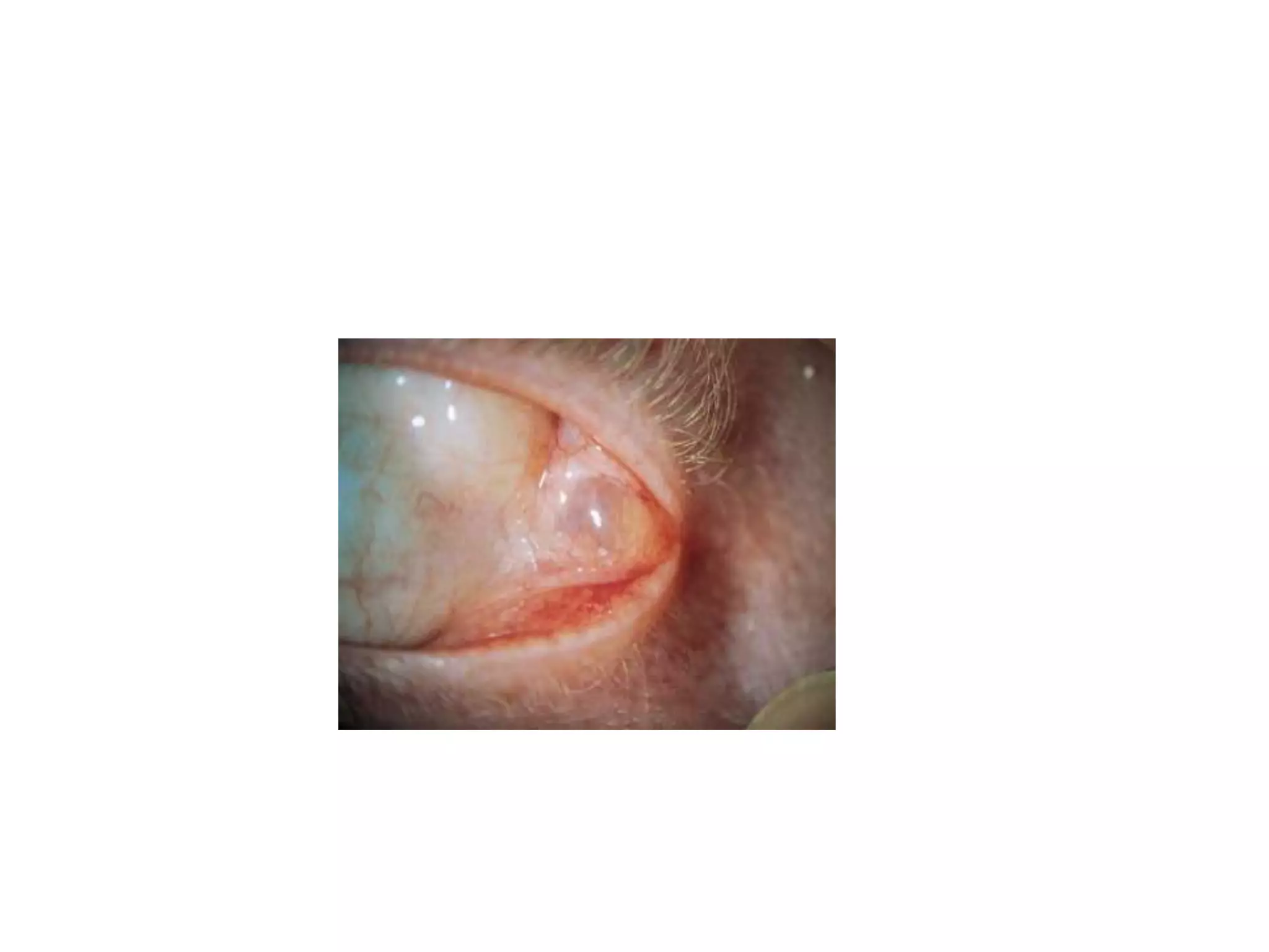
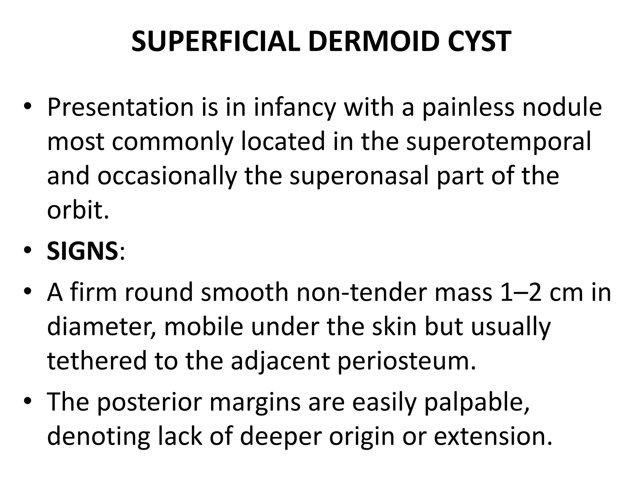
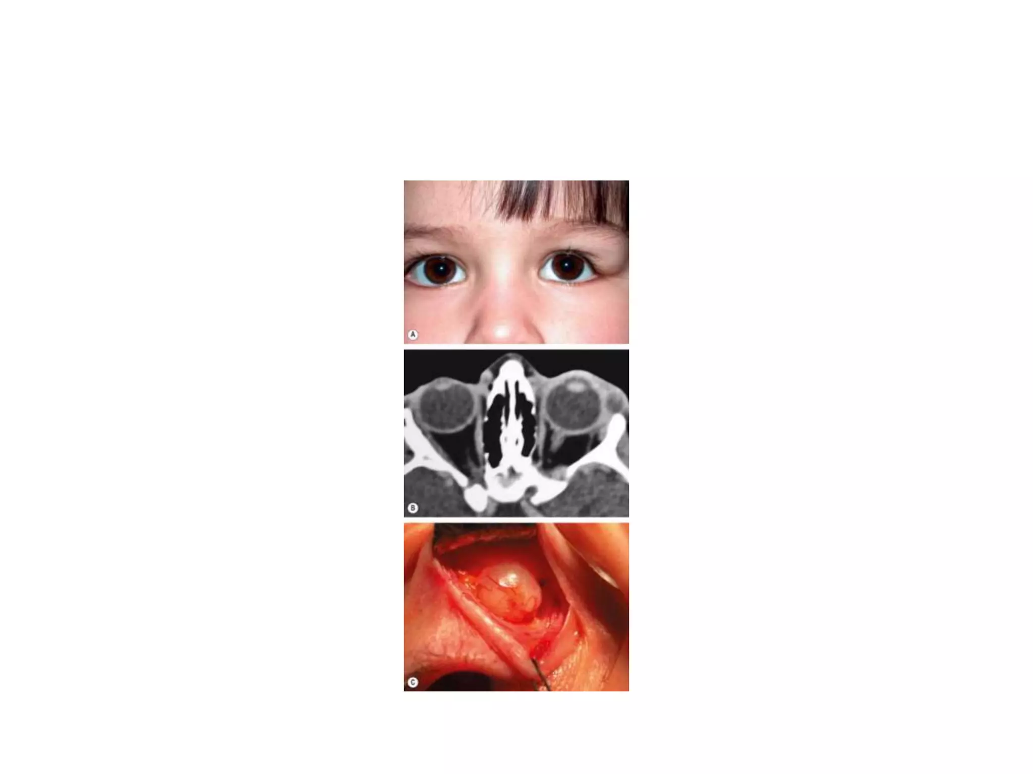
CYSTIC LEISIONS OF ORBIT 
BY 
DR. M. UZAIR HAFEEZ
DACRYOPS 
• A dacryops is a ductal cyst of the lacrimal gland. 
• Relatively uncommon orbital cystic lesion. 
• Frequently bilateral. 
• SIGNS: 
• A round, cystic lesion, originating from the palpebral 
portion of the lacrimal gland, which protrudes into 
the superior fornix. 
• TREATMENT: 
• Excision or marsupialization.
DERMOID CYST 
• An orbital dermoid cyst is a choristoma derived 
from displacement of ectoderm to a 
subcutaneous location along embryonic lines of 
closure. 
• Lined by keratinized stratified squamous 
epithelium (like skin), have a fibrous wall and 
contain dermal appendages such as sweat glands, 
sebaceous glands and hair follicles. 
• Dermoids may be (a) superficial or (b) deep, 
located anterior or posterior to the orbital 
septum respectively.
SUPERFICIAL DERMOID CYST 
• Presentation is in infancy with a painless nodule 
most commonly located in the superotemporal 
and occasionally the superonasal part of the 
orbit. 
• SIGNS: 
• A firm round smooth non-tender mass 1–2 cm in 
diameter, mobile under the skin but usually 
tethered to the adjacent periosteum. 
• The posterior margins are easily palpable, 
denoting lack of deeper origin or extension.
• CT shows a heterogeneous well-circumscribed 
lesion. 
• Treatment is by excision, taking care not to 
rupture the lesion, since leaking of keratin into 
the surrounding tissue results in severe 
granulomatous inflammation.
DEEP DERMOID CYST 
• Presentation is in adolescence or adult life. 
• SIGNS: 
• Proptosis, dystopia or a mass lesion with indistinct 
posterior margins. 
• Rupture may incite an inflammatory reaction. 
• Some deep dermoids, associated with bony 
defects, may extend into the inferotemporal fossa 
or intracranially.
• CT shows a well-circumscribed cystic lesion. 
• Treatment by excision in toto is advisable 
because deep dermoids enlarge and may leak 
their contents into adjacent tissues inducing a 
painful granulomatous inflammation, often 
followed by fibrosis. If incompletely excised, 
dermoids may recur and cause persistent low-grade 
inflammation.
SINUS MUCOCELE 
• A mucocele develops when the drainage of normal 
para-nasal sinus secretions is obstructed due to 
infection, allergy, trauma, tumour or congenital 
narrowing. 
• A slowly expanding cystic accumulation of mucoid 
secretions and epithelial debris develops and 
gradually erodes the bony walls of the sinuses, 
causing symptoms by encroaching upon surrounding 
tissues. 
• Orbital invasion occurs usually from frontal or 
ethmoidal mucoceles, and rarely from those arising in 
the maxillary sinus.
• Presentation is in adult life with proptosis or 
dystopia, diplopia or epiphora. Pain is uncommon 
unless secondary infection develops (mucopyocele). 
• CT shows a soft tissue mass with thinning or erosion 
of the bony walls of the sinus (Fig. 3.28B). 
• Treatment involves complete removal of the 
mucocele.
ENCEPHALOCELE 
• An encephalocele is formed by herniation of 
intracranial contents through a congenital defect 
of the base of the skull. 
• A meningocele contains only dura whilst a 
meningoencephalocele also contains brain tissue. 
• Orbital encephalocele may be (a) anterior (fronto-ethmoidal) 
or (b) posterior, which is associated 
with dysplasia of the sphenoid bone.
• Presentation is usually during infancy. 
• SIGNS: 
• Anterior encephaloceles involve the superomedial 
part of the orbit and displace the globe forwards 
and laterally. 
• Posterior encephaloceles displace the globe 
forwards and downwards . 
• The displacement increases on straining or crying 
and may be reduced by manual pressure. 
• Pulsating proptosis may occur due to 
communication with the subarachnoid space but, 
because the communication is not vascular, there is 
neither a thrill nor a bruit.
• CT shows the bony defect responsible for the 
herniation. 
• Differential diagnosis 
• a Of anterior encephaloceles includes other 
causes of medial canthal swellings such as dermoid 
cysts and amniontoceles of the lacrimal sac. 
• b Of posterior encephaloceles includes other 
orbital lesions that present during early life such as 
capillary haemangioma, juvenile xanthogranuloma, 
teratoma and microphthalmos with cyst.
• Associations 
• a Other bony abnormalities such as 
hypertelorism , broad nasal bridge and cleft 
palate . 
• b Ocular associations include 
microphthalmos, orbital varices, colobomas and 
the morning glory syndrome . 
• c Neurofibromatosis type 1 (NF1) is 
• frequently associated with posterior 
encephalocele.
• THANK YOU

Cystic leisions of orbit

  • 1.
    CYSTIC LEISIONS OFORBIT BY DR. M. UZAIR HAFEEZ
  • 2.
    DACRYOPS • Adacryops is a ductal cyst of the lacrimal gland. • Relatively uncommon orbital cystic lesion. • Frequently bilateral. • SIGNS: • A round, cystic lesion, originating from the palpebral portion of the lacrimal gland, which protrudes into the superior fornix. • TREATMENT: • Excision or marsupialization.
  • 4.
    DERMOID CYST •An orbital dermoid cyst is a choristoma derived from displacement of ectoderm to a subcutaneous location along embryonic lines of closure. • Lined by keratinized stratified squamous epithelium (like skin), have a fibrous wall and contain dermal appendages such as sweat glands, sebaceous glands and hair follicles. • Dermoids may be (a) superficial or (b) deep, located anterior or posterior to the orbital septum respectively.
  • 5.
    SUPERFICIAL DERMOID CYST • Presentation is in infancy with a painless nodule most commonly located in the superotemporal and occasionally the superonasal part of the orbit. • SIGNS: • A firm round smooth non-tender mass 1–2 cm in diameter, mobile under the skin but usually tethered to the adjacent periosteum. • The posterior margins are easily palpable, denoting lack of deeper origin or extension.
  • 6.
    • CT showsa heterogeneous well-circumscribed lesion. • Treatment is by excision, taking care not to rupture the lesion, since leaking of keratin into the surrounding tissue results in severe granulomatous inflammation.
  • 8.
    DEEP DERMOID CYST • Presentation is in adolescence or adult life. • SIGNS: • Proptosis, dystopia or a mass lesion with indistinct posterior margins. • Rupture may incite an inflammatory reaction. • Some deep dermoids, associated with bony defects, may extend into the inferotemporal fossa or intracranially.
  • 9.
    • CT showsa well-circumscribed cystic lesion. • Treatment by excision in toto is advisable because deep dermoids enlarge and may leak their contents into adjacent tissues inducing a painful granulomatous inflammation, often followed by fibrosis. If incompletely excised, dermoids may recur and cause persistent low-grade inflammation.
  • 11.
    SINUS MUCOCELE •A mucocele develops when the drainage of normal para-nasal sinus secretions is obstructed due to infection, allergy, trauma, tumour or congenital narrowing. • A slowly expanding cystic accumulation of mucoid secretions and epithelial debris develops and gradually erodes the bony walls of the sinuses, causing symptoms by encroaching upon surrounding tissues. • Orbital invasion occurs usually from frontal or ethmoidal mucoceles, and rarely from those arising in the maxillary sinus.
  • 12.
    • Presentation isin adult life with proptosis or dystopia, diplopia or epiphora. Pain is uncommon unless secondary infection develops (mucopyocele). • CT shows a soft tissue mass with thinning or erosion of the bony walls of the sinus (Fig. 3.28B). • Treatment involves complete removal of the mucocele.
  • 14.
    ENCEPHALOCELE • Anencephalocele is formed by herniation of intracranial contents through a congenital defect of the base of the skull. • A meningocele contains only dura whilst a meningoencephalocele also contains brain tissue. • Orbital encephalocele may be (a) anterior (fronto-ethmoidal) or (b) posterior, which is associated with dysplasia of the sphenoid bone.
  • 15.
    • Presentation isusually during infancy. • SIGNS: • Anterior encephaloceles involve the superomedial part of the orbit and displace the globe forwards and laterally. • Posterior encephaloceles displace the globe forwards and downwards . • The displacement increases on straining or crying and may be reduced by manual pressure. • Pulsating proptosis may occur due to communication with the subarachnoid space but, because the communication is not vascular, there is neither a thrill nor a bruit.
  • 16.
    • CT showsthe bony defect responsible for the herniation. • Differential diagnosis • a Of anterior encephaloceles includes other causes of medial canthal swellings such as dermoid cysts and amniontoceles of the lacrimal sac. • b Of posterior encephaloceles includes other orbital lesions that present during early life such as capillary haemangioma, juvenile xanthogranuloma, teratoma and microphthalmos with cyst.
  • 17.
    • Associations •a Other bony abnormalities such as hypertelorism , broad nasal bridge and cleft palate . • b Ocular associations include microphthalmos, orbital varices, colobomas and the morning glory syndrome . • c Neurofibromatosis type 1 (NF1) is • frequently associated with posterior encephalocele.
  • 19.