This study assessed the diagnostic accuracy of lung comet-tail images detected by ultrasound compared to chest radiography, wedge pressure, and extravascular lung water (EVLW) measured by the PiCCO system. 20 patients undergoing cardiac surgery were examined before, immediately after, and 24 hours following surgery using ultrasound to detect comet-tail images, chest radiography, pulmonary artery catheterization, and the PiCCO system. Significant positive correlations were found between the number of comet-tail images and EVLW, wedge pressure, and radiographic lung water score, indicating ultrasound detection of comet-tail images provides reliable information about interstitial pulmonary edema.
Ultrasonography of the lungs can detect "comet-tail images" originating from water-thickened interlobular septa, which may indicate pulmonary edema. This study assessed the diagnostic accuracy of lung comet-tail images compared to chest radiography, pulmonary wedge pressure, and extravascular lung water (EVLW) measured by transpulmonary thermodilution. In 60 patients studied before and after cardiac surgery:
1) The number of comet-tail images correlated positively with EVLW, wedge pressure, radiographic lung water score.
2) A negative comet test result accurately detected EVLW < 500 mL, while a positive test accurately detected EVLW > 500 mL.
3) Ul
Ultrasound is useful for both diagnosing and guiding procedures involving the pleura. It can detect pleural effusions and pneumothoraces more sensitively than chest x-rays. Ultrasound improves the accuracy of pleural procedures by identifying the best puncture site and reducing complications. Evidence shows ultrasound-guided thoracenteses reduce the risk of pneumothorax compared to procedures without ultrasound. The document discusses using ultrasound to characterize pleural effusions, detect pneumothoraces, identify pleural thickening, and guide biopsies and drainage of pleural tumors or collections.
This document provides an overview of pulmonary anatomy, physiology, and lung scintigraphy principles. It describes the positioning of the heart and lungs in the thorax, including the lobes and segments of the lungs. It discusses the pulmonary circulation and flow of oxygenated and deoxygenated blood. Key concepts covered include ventilation, regulation of breathing, and matching of ventilation and perfusion. Clinical indications for lung scintigraphy include evaluating for pulmonary embolism, COPD, and lung tumors. The document reviews radiopharmaceuticals, administration techniques, and normal scan findings for lung perfusion and ventilation imaging.
ERS London 2016: Poster Discussion:Ultrasound Guided Pleural Brushing: a New ...Gamal Agmy
Ultrasound guided pleural brushing is a new technique for obtaining pleural specimens in patients with malignant pleural effusions. This study evaluated the diagnostic yield of ultrasound guided pleural brushing in 22 patients with suspected malignant pleural effusions. Pleural brushing alone yielded a positive diagnosis in 41% of patients and was the sole diagnostic technique in 14% of patients. When combined with pleural fluid cytology and biopsy forceps, ultrasound guided pleural brushing provided a positive diagnosis in 86% of patients. The study demonstrates that ultrasound guided pleural brushing is a simple, relatively safe procedure that provides additional diagnostic value in evaluating malignant pleural effusions.
1. Several imaging modalities can provide detailed assessment of lung structure and function in asthmatic patients, including CT, MRI, PET, OCT, and EBUS.
2. Measurements from CT such as airway wall thickness, air trapping, and ventilation defects have been shown to correlate with disease severity and control.
3. Imaging measurements can serve as biomarkers to evaluate responses to new therapies like inhaled corticosteroids and anti-IL5 monoclonal antibodies, and determine if treatments are modifying the disease course.
This document provides an overview of ultrasound applications in the intensive care unit and emergency department. It discusses using ultrasound at the bedside for lung imaging as an alternative to radiography. It describes the signs of pneumothorax, pulmonary edema, pulmonary embolism, and lung consolidation seen on ultrasound. The document also reviews using ultrasound to evaluate the inferior vena cava, heart, and pericardial space. Protocols for lung ultrasound and applications in critically ill patients are presented.
Evaluation of lung ultrasound for the diagnosis of pneumonia in the EDMario Robusti
Lung ultrasound was performed on 49 patients suspected of having pneumonia who presented to the emergency department. Pneumonia was confirmed in 32 cases (65.3%). Lung ultrasound detected pneumonia (consolidation with air bronchograms) in 31 of these 32 cases (96.9%), while chest x-ray detected pneumonia in 24 cases (75%). In 8 cases (25%), lung ultrasound detected pneumonia when chest x-ray did not. Computed tomography scans confirmed the lung ultrasound results in these 8 cases. Lung ultrasound took less than 5 minutes to perform and could visualize all areas of the chest, while chest x-rays were only able to image both views in 66% of cases. This study suggests lung ultrasound may be
Diagnostic Accuracy of Transthoracic Sonography in Patients with Pneumonia an...Gamal Agmy
Trans-thoracic ultrasonography (TUS) has gained interest in diagnosing pneumonia and pulmonary embolism. This study assessed the diagnostic accuracy of TUS for these conditions compared to CT and chest x-ray. TUS had a sensitivity of 88.2% and specificity of 87.5% for pneumonia, and 71.4% sensitivity and 80.9% specificity for pulmonary embolism. TUS also detected pleural effusion with higher sensitivity than chest x-ray. The study concludes that TUS is a useful rapid diagnostic tool for pneumonia and pulmonary embolism that may be superior to chest x-ray.
Ultrasound has many advantages for critically ill patients in the ICU. It enables rapid, repeated, and inexpensive bedside evaluation. There are two main probe types: B-mode produces 2D images while M-mode shows motion over time, analogous to video. Ultrasound can assess volume status by measuring the diameter and collapse of the inferior vena cava. It can diagnose pneumothorax by lung sliding signs or stratosphere and seashore artifacts. Ultrasound is also used for vascular access, intubation, diaphragm assessment, and identifying pleural effusions and hemothorax. Critical care physicians should receive training to utilize ultrasound's benefits for critically ill patients.
This document discusses lung ultrasound patterns and artifacts that can be identified using ultrasound in intensive care and critically ill patients. It outlines ultrasound frequencies used for chest/lung ultrasound. Key normal and abnormal ultrasound findings are described, including pleural sliding, A-lines, B-lines, lung consolidation, pleural effusions, and pneumothorax patterns. A clinical case example is provided of a current smoker presenting with fever, cough and chest pain. Lung ultrasound findings are correlated with CT scans.
An Educational material showing Chest Imaging and describing NORMAL IMAGING-VOLUME LOSS-LOSS OF PARENCHYMA-ALVEOLAR PROCESSES-BRONCHIECTASIS
PLEURAL ABNORMALITIES
NODULES AND MASSES
Thoracic Imaging in critically ill patientsGamal Agmy
Chest radiography remains the primary imaging modality for critically ill patients, however images are often limited quality due to patient movement and positioning challenges. Mistakes can occur in assessing conditions like pleural effusions or infiltrates. Routine daily chest x-rays are not recommended for ICU patients unless clinically indicated. Ultrasound is a useful bedside tool for evaluating the lungs, IVC, heart, and detecting pneumothorax. Computed tomography can also be used but requires transporting unstable patients.
ICN Victoria presents Dr Andrew Hilton, Intensivist at the Austin Hospital in Melbourne, talking on the use of ultrasound in ICU to evaluate and treat lung pathology. Recorded at our November 2014 ICN Victoria meeting.
Ultrasound can replace chest x-rays in the emergency care of obese patients. Ultrasound is more sensitive than chest x-rays at detecting certain common conditions like pneumothorax and traumatic hemothorax. While fat tissue attenuates ultrasound, thoracic ultrasound uses low frequencies that are less affected. Conditions like pleural effusions, pulmonary embolism, and pneumonia can be diagnosed with ultrasound with sensitivity and specificity comparable or superior to chest x-rays. Ultrasound also enables rapid diagnosis and guided procedures without radiation exposure. With training, ultrasound can become the front-line imaging method for obese emergency patients, providing advantages over chest x-rays.
Radiology chs 3101 cardiovascular and respiratory system disordersWEEKLYMEDIC
This document outlines a presentation on cardiovascular and respiratory disorders demonstrated through radiological imaging. It discusses the various imaging modalities used, including their indications. Common terminology used in radiography, CT, and ultrasound is defined. Examples of the radiological appearance of several respiratory disorders are illustrated, such as pneumonia, tuberculosis, lung metastases, cavities, pleural effusion, and pneumothorax.
This document provides an overview of transthoracic sonography and its applications in evaluating the lungs and chest. It includes descriptions of normal lung anatomy as seen on ultrasound and discusses various pathologies such as pneumonia, pulmonary embolism, COPD and lung cancer. Imaging techniques like assessing lung sliding and the presence of comet tail artifacts are described for evaluating diseases like pneumonia. Other applications covered include using ultrasound to identify pleural effusions, pneumothorax, chest wall abnormalities and for guiding procedures. In summary, the document outlines the role of ultrasound in evaluating pulmonary and chest wall diseases and conditions.
1) Lung ultrasound is a useful technique for evaluating pulmonary conditions at the bedside with several advantages over other imaging modalities.
2) Normal lung ultrasound findings include lung sliding, the seashore sign, A-lines, and the lung pulse. Absence of lung sliding can indicate a pneumothorax.
3) B-lines appear as laser-like artifacts that arise from the pleural line and indicate excess fluid or interstitial syndrome. A higher number of B-lines correlates with decompensated heart failure.
Endobronchial Ultrasound Guidance of TBNA. Current Approach To Lung Cancer St...Bassel Ericsoussi, MD
EBUS-TBNA, EUS-FNA or their combination have finally gained acceptance as the tests of first choice in mediastinal staging. In suspected non-small cell lung cancer, endobronchial ultrasound may be preferred in the histologic sampling of paratracheal and subcarinal mediastinal adenopathy because the diagnostic yield can surpass mediastinoscopy
The document summarizes advances in pulmonary embolism imaging. It reviews the importance of clinical prediction scores and various imaging modalities for diagnosing pulmonary embolism such as CT pulmonary angiography, ventilation-perfusion scanning, ultrasound, and MRI. It also discusses findings on CT imaging including signs of right ventricular strain and thrombus burden. New techniques such as low-dose CT and dual-energy CT are introduced.
Emergency sonography in Pediatrics has evolved to become one of the most versatile
modalities for diagnosing and guiding
treatment of critically ill patients.
Ultrasound has many useful applications in critical care. It can reinvigorate the physical exam by providing valuable information about patients with limited mobility. Basic ultrasound skills should be part of critical care training, as brief training allows intensivists to perform limited transthoracic echocardiography and change patient management in many cases. Ultrasound is portable, avoids radiation, and can be repeated as needed at the bedside. It is useful for diagnosing problems like venous thrombosis, pulmonary diseases, and acute respiratory failure. The BLUE protocol allows rapid ultrasound evaluation of the lungs. Bedside echocardiography also has applications in critical care for assessing hemodynamics, infections, and postoperative complications.
Imaging of Pulmonary Vascular Lesions ``Gamal Agmy
This document contains a series of questions and answers about the management of pulmonary embolism from Gamal Agmy, a professor of chest diseases. It discusses issues such as when to use thrombolysis for intermediate risk PE, the risks of thrombolysis for patients with recent surgery or stroke, and alternatives to thrombolysis if it is deemed too unsafe. It also addresses questions about managing PE in pregnant patients, PE with right atrial thrombus, and the appropriate use of IVC filters.
This document provides an overview of lung ultrasound and discusses various lung pathologies that can be identified using ultrasound. It begins with background on lung anatomy and ultrasound principles. Various normal and abnormal findings are then described, including pneumothorax, pulmonary edema, consolidation, pleural effusions, and lung tumors. Case studies are presented to demonstrate ultrasound identification of conditions like emphysema, pneumonia, pulmonary edema, pneumothorax, and lung cancer. The document emphasizes that lung ultrasound allows accurate diagnosis of many lung conditions at the point of care based on visualization of artifacts, B-lines, lung sliding, and consolidations.
Diaphragm Ultrasound as a Novel Guide of Weaning from Invasive VentilationGamal Agmy
This study evaluated diaphragm thickness (DT) measured by ultrasound as a predictor of successful weaning from mechanical ventilation, compared to the rapid shallow breathing index (RSBI). 78 COPD patients undergoing ventilation were assessed with ultrasound during a spontaneous breathing trial to measure DT changes. A DT over 40% was highly accurate in predicting successful weaning, with sensitivity of 88% and specificity of 92%. RSBI under 105 was also found to be highly accurate in predicting success. The study concluded that DT assessed by ultrasound is an excellent predictor of weaning outcome in COPD patients.
Endobronchial Ultrasound - dr deepak talwar best pulmonologist in IndiaMetro Hospital
Dr. Deepak Talwar
Director & Chair, Pulmonary,
Sleep & Critical Care Medicine,
Metro Group of Hospitals, Noida http://www.metrohospitals.com/doctors/deepak-talwar
This document provides an overview of acute decompensated heart failure (ADHF), including its pathophysiology, classification, and pharmacotherapy. ADHF accounts for most of the $39 billion spent annually on chronic heart failure in the US. It results from exacerbation of chronic cardiac, pulmonary, or renal dysfunction. Pharmacotherapy for ADHF is guided by understanding the patient's hemodynamic status and Forrester classification. The goals of treatment include relieving symptoms of fluid overload or hypoperfusion. Intravenous loop diuretics are first-line to treat fluid overload, while vasodilators may be used to improve cardiac output and relieve symptoms of hypoperfusion. Proper inpatient treatment combined with coordinated discharge and
The Seattle Heart Failure Model was developed by researchers at the University of Washington to accurately predict 1, 2, and 3-year survival rates for patients with heart failure based on simple clinical variables. The model also estimates how a patient's survival would be impacted by various heart failure medications and devices. Validation testing found the model provided excellent accuracy. It is available online and allows clinicians to estimate how adding different treatment options could extend a patient's predicted lifespan. The researchers hope this model will encourage both patients and doctors to more frequently use proven life-saving heart failure medications and devices.
Physiologic volume redistribution and acute heart failure management (printer...drucsamal
This document provides information about a continuing medical education (CME) activity on physiologic volume redistribution and acute heart failure management. The activity aims to increase healthcare providers' knowledge of caring for patients with acutely decompensated heart failure. It outlines learning objectives, credits available, accreditation statements, faculty disclosures, and instructions for participating and receiving credit. The faculty includes experts from the US, Switzerland, Sweden, and Poland who will discuss pathophysiological mechanisms, treatment selection, and emerging therapies for common acute heart failure phenotypes.
This clinical trial studied the effects of cinaciguat, a soluble guanylate cyclase activator, in patients with acute decompensated heart failure. The trial found that cinaciguat significantly reduced pulmonary capillary wedge pressure and right atrial pressure compared with placebo after 8 hours. However, cinaciguat also caused a significant decrease in blood pressure. The trial was terminated prematurely due to an increased risk of hypotension at cinaciguat doses of 200 mg/h or higher. While cinaciguat showed potential for unloading the heart, high doses were associated with hypotension, demonstrating a narrow therapeutic window.
This document summarizes research on risk assessment of patients presenting to the emergency department (ED) with acute heart failure (AHF). Nearly 700,000 ED visits each year are due to AHF, with over 80% resulting in hospital admission. Existing risk prediction tools for AHF have not impacted admission rates. The authors hypothesize that evaluating both physiological risk factors and barriers to self-care, along with strategies to overcome barriers and shared decision making between providers and patients, could allow more patients to be safely discharged from the ED or observation units rather than admitted. This approach may help reduce hospital admissions, readmissions, and costs while improving long-term management of heart failure.
Vascular effects of urocortins 2 and 3 in healthy volunteersdrucsamal
Urocortins 2 and 3 are endogenous peptides that have roles in cardiovascular physiology. This study examined the direct vascular effects of urocortins 2 and 3 in the forearms of 18 healthy male volunteers using plethysmography. The study found that both urocortins caused potent and prolonged arterial vasodilation in a dose-dependent manner mediated partly through endothelial nitric oxide and cytochrome P450 metabolites of arachidonic acid. The roles of urocortins 2 and 3 in heart failure remain to be explored, but they have potential therapeutic benefits.
1) Sixty-two patients with heart failure and reduced ejection fraction were given ascending doses of human stresscopin (JNJ-39588146) or placebo through intravenous infusion to examine safety, pharmacokinetics, and effects on hemodynamics.
2) Statistically significant increases in cardiac index and reductions in systemic vascular resistance were seen with the 15 and 30 ng/kg/min doses of JNJ-39588146 without significant changes in heart rate or blood pressure.
3) No statistically significant reductions in pulmonary capillary wedge pressure were observed with any dose, though a trend toward reduction was seen. The drug was well tolerated with no safety concerns observed.
1) This study uses data from 39,372 heart failure patients from 30 studies to develop a risk score for predicting mortality. 13 factors were identified as independent predictors of mortality including age, ejection fraction, NYHA class, creatinine, diabetes, medications, blood pressure, body mass, time since diagnosis, smoking status, and comorbidities.
2) A risk score was developed that assigns points to each risk factor level and quantifies a patient's 3-year mortality risk. Mortality risk ranged from 10% for the lowest risk quintile to 70% for the highest risk decile.
3) The risk score can be used clinically and has potential for widespread implementation via a website to help target
This document discusses the management of acute heart failure. It notes that current therapies are based on improving hemodynamics and symptoms but lack evidence. There is heterogeneity in treatment approaches and outcomes. Biomarkers can help diagnosis but accuracy is still limited. The paradigm is that patients receive diuretics and vasodilators in the emergency department to relieve symptoms, but often still have residual congestion on discharge. This leads to high readmission rates. A shift in approach may be needed to better address the underlying disease progression.
Novel treatment options for acute hf a multidisciplinary approach (printer f...drucsamal
This document discusses a CME activity on novel treatment options for acute heart failure using a multidisciplinary approach. It provides learning objectives, faculty disclosures, and instructions for participating physicians to earn CME credits. The activity aims to evaluate current and emerging therapies for acute heart failure and summarize strategies for multidisciplinary management from the emergency department through discharge and at-home care. The discussion will focus on improving outcomes for the over 1 million patients hospitalized annually for acute heart failure in the United States.
Current Modalities for Invasive and Non Invasive Monitoring of Volume status ...drucsamal
Heart failure represents a major health burden worldwide. New monitoring strategies aim to detect worsening heart failure early by assessing fluid status. Home monitoring of weight and symptoms has shown mixed results, failing to consistently reduce hospitalizations. Thoracic impedance monitoring via implantable devices shows promise, as impedance correlates inversely with fluid levels and may predict worsening heart failure before symptoms occur. Invasive hemodynamic monitoring also demonstrates benefits but requires device implantation. Overall, fluid monitoring strategies hold potential to improve heart failure outcomes but require further study.
Urocortin-2 Infusion in Acute Decompensated Heart Failure
The study investigated the effects of urocortin-2 infusion compared to placebo in patients with acute decompensated heart failure. Fifty-three patients were randomly assigned to receive either 5 ng/kg/min of urocortin-2 or placebo infusion for 4 hours in addition to standard therapy. Urocortin-2 produced greater increases in cardiac output and decreases in blood pressure and total peripheral resistance compared to placebo. Renal indices fell transiently during urocortin-2 infusion but returned to above baseline levels after infusion. Further studies are needed to understand the full potential of urocortin-2 for treating acute
Prognosis and treatment of cardiogenic shock complicating acute myocardial in...drucsamal
This document discusses the prognosis and treatment of cardiogenic shock complicating acute myocardial infarction. It notes that while the mortality rate for cardiogenic shock used to be 80-90%, studies now report in-hospital mortality rates between 42-74%. Several factors predict higher mortality, such as increasing age, prior heart attack, and low blood pressure/cardiac output. The document recommends general measures like aspirin, heparin, and vasopressors to treat cardiogenic shock. It advises against beta blockers and favors early revascularization when possible to improve outcomes for patients experiencing this complication of a heart attack.
Risk prediction models for mortality in ambulatorydrucsamal
This document summarizes a systematic review of risk prediction models for mortality in ambulatory heart failure patients. It found 20 models described in 34 studies but only 5 models were validated in independent cohorts, including the Heart Failure Survival Score and Seattle Heart Failure Model. Both demonstrated modest discrimination and questionable calibration. The review concludes currently validated models have inconsistent performance and a new model derived from contemporary patients may be needed for improved prognosis.
This study analyzed data from over 30,000 patients with heart failure (HF) to identify predictors of adverse outcomes based on left ventricular ejection fraction (LVEF). Patients were categorized as having preserved LVEF (≥50%), borderline LVEF (41-49%), or reduced LVEF (≤40%). Over a median follow up of 1.8 years, 26.8% of patients died, 26.9% were hospitalized for HF, and 67.4% were hospitalized for any reason. Multivariable models found that nearly all tested predictors, such as demographics, medical history, and comorbidities, performed similarly for predicting death and hospitalization across the different LVEF
Evaluation of acute decompensated heart failure2drucsamal
- The patient presented with acute decompensated heart failure (ADHF), characterized by acute dyspnea and accumulation of fluid in the lungs.
- An evaluation was performed which included physical exam, chest x-ray, ECG, lab tests, and echocardiogram to diagnose ADHF and identify potential precipitating factors such as nonadherence, infection, arrhythmias, or myocardial ischemia.
- Invasive hemodynamic monitoring with a Swan-Ganz catheter is not routinely recommended but may help in select cases to guide management of worsening renal function or need for advanced therapies.
This study examined urinary levels of C-type natriuretic peptide (CNP) in patients with acute decompensated heart failure (ADHF) and healthy controls. The study found that ADHF patients had higher levels of three CNP molecular forms (CNP22, CNP53, and NT-CNP53) in their urine than controls. Higher urinary levels of NT-CNP53 in ADHF patients predicted higher mortality and rehospitalization rates. NT-CNP53 was a better predictor of outcomes than other urinary biomarkers of kidney injury and provided additional predictive value when combined with plasma levels of NT-proBNP. The findings suggest urinary CNP levels have clinical utility as biomarkers
This document provides evidence-based recommendations for point-of-care lung ultrasound from an international panel of 28 experts. The panel reviewed literature from 1966 to 2011 using the GRADE method to determine evidence quality and develop recommendations. They discussed 73 proposed statements over three conferences using a modified Delphi technique and anonymous voting. Strong recommendations were made for 65 statements, and weak recommendations for 2 statements. The recommendations aim to standardize the application of lung ultrasound in clinical settings and provide a framework to guide its future use and research.
Ultrasonography in Critically Ill PatientsGamal Agmy
This document discusses the use of chest sonography in critically ill patients. It notes that bedside chest radiography has limitations in critically ill patients. Chest sonography can help diagnose various lung conditions at the bedside including pulmonary consolidation, atelectasis, edema, effusions, and pneumothorax. It reviews the sonographic signs and patterns associated with these conditions. The document also discusses using lung ultrasound and IVC views to assess shock states and guide treatment. Overall, it promotes the use of bedside lung ultrasound as a valuable tool to complement radiography in critically ill patients.
Sudanese chest sonography workshop (Sonography in critical ill patients)Gamal Agmy
This document summarizes the use of chest sonography in critically ill patients. It notes that while bedside chest radiography is commonly used, images are often of limited quality due to patient movement and technical factors. Chest sonography allows clinicians to diagnose various lung conditions at the bedside including pulmonary consolidation, atelectasis, alveolar-interstitial syndrome, pneumonia, and pleural effusions. It describes the normal ultrasound anatomy of the lungs and identifies sonographic signs that are diagnostic of various lung pathologies without transporting critically ill patients for other imaging.
This document provides an overview of chest ultrasonography. Some key points:
- Chest ultrasound can evaluate a wide range of chest abnormalities and is well-suited for bedside use. It has benefits of being easily accessible, low cost, allowing real-time evaluation, and lacking radiation exposure.
- A curvilinear probe is used to visualize deeper structures and a linear probe provides detailed images of superficial abnormalities. Lung sliding and the bat sign indicate normal findings.
- Chest ultrasound can detect and characterize pleural effusions and pleural thickening. It is more sensitive than chest x-rays for small pleural effusions. Effusions appear anechoic, complex, or echogenic depending on their nature
Brief discussion on ultrasonography of the chest: Benefits, Techniques and Instrumentation, Normal Anatomy, Diagnostic US of the chest, Limitations of Thoracic US, US based differential diagnosis, Take home points.
Thoracic Ultrasound For The Respiratory System In Critically Ill PatientsBassel Ericsoussi, MD
Thoracic ultrasound can be used to diagnose pneumothorax in critically ill patients. It is more sensitive than chest x-ray and can detect even very small pneumothoraces. Normal lung ultrasound shows the sliding of the visceral and parietal pleura and A-lines, while a pneumothorax is identified by the absence of sliding, A-lines only, and the lung point sign. Ultrasound can also assess endotracheal tube position and risk of post-extubation stridor.
1) Interventional bronchoscopy is an evolving field that focuses on providing diagnostic and procedural services for malignant and non-malignant airway disorders using advanced bronchoscopic techniques.
2) Key techniques discussed include autofluorescence bronchoscopy, endobronchial ultrasound, electromagnetic navigation bronchoscopy, and optical coherence tomography, which provide high resolution imaging and improve detection of early lung cancers and peripheral lesions.
3) Endobronchial ultrasound guided biopsy of mediastinal lymph nodes and peripheral lung lesions has high diagnostic accuracy for staging and diagnosis of lung cancers.
This study compared carotid-femoral pulse wave velocity (cfPWV) measurements obtained using a tonometer-based device and a cuff-based device with and without an adjustment algorithm. 88 participants across 4 centers underwent triplicate cfPWV measurement with each device. The unadjusted cuff-based method yielded lower cfPWV values than the tonometer-based method. Application of an algorithm to adjust for additional distance and transit time in the cuff-based method resulted in cfPWV values similar to the tonometer-based method. Analysis showed the adjusted cuff-based method provided comparable results to the tonometer-based method, validating the novel cuff-based assessment of cfPWV.
Demonstrate how to do and use chest ultrasound for diagnosis and management of different pulmonary and pleural diseases also for taken.lung biopsy and insertion of central venous line differential diagnosis of interstetial lung disease .pleural biopsy and diaphragmatic movement .vascular abnormality cardic disease and oericardial effusion
The document summarizes the anatomy and radiographic investigation of the thorax. It describes the structures that make up the thoracic wall and cavities. It then discusses various imaging modalities used to examine the thorax, including plain radiography, CT, MRI, PET, and others. It provides details on technical factors and positioning for chest x-rays and interpreting chest x-ray findings based on relative tissue densities.
diagnostic workup of the the thoracic surgery patientAkin Balci
This document discusses the diagnostic workup and various imaging modalities used for thoracic surgery patients, including chest radiographs, computed tomography (CT), positron emission tomography (PET), magnetic resonance imaging (MRI), and ultrasound. CT is often the primary imaging method and can evaluate lungs, mediastinum, chest wall and vasculature. PET/CT provides functional and anatomical data for staging cancers. MRI is also useful for soft tissue contrast while ultrasound effectively images pleural effusions. Accurate staging of thoracic neoplasms with these imaging tools is important for determining appropriate therapy.
Role of Sonography in Respiratory EmergenciesGamal Agmy
1) Chest sonography can be used in respiratory emergencies to assess both superficial and deep structures using high and low frequency probes respectively.
2) Common signs seen on sonography include B-lines indicating pulmonary edema, the bat sign of normal lung, and the seashore sign indicating a pneumothorax.
3) Sonography can also assess volume status by measuring the inferior vena cava diameter and calculating the caval index, evaluate lung consolidations and air bronchograms, and detect pulmonary embolism.
Ultrasound can be useful in the evaluation and diagnosis of patients presenting in shock. Integrating bedside ultrasound allows for a more accurate initial diagnosis and earlier treatment. The RUSH protocol assesses the heart, IVC, pericardial space and lungs to help classify the type of shock. Ultrasound findings of a dilated and collapsing IVC along with evidence of free fluid suggest the patient has hypovolemic shock likely due to internal bleeding.
Walif Chbeir: Medical Imaging of PneumoThorax (PNO)–3Walif Chbeir
Dr. Walif Chbeir outlines in detail the medical imaging practice and diagnostic approach of pneumothorax (also known as PNO). This is the third in a four-part piece on PNO by Chbeir.
Ultrasonography evaluation during the weaning processFadel Omar
Ultrasonography can be useful for assessing cardiac function, diaphragm mobility, pleural effusions, and lung aeration during the mechanical ventilation weaning process. Assessment of left ventricular diastolic function, diaphragm excursion and thickening fraction, size of pleural effusions, and lung ultrasound score can provide information on risks of weaning failure and identify issues like cardiogenic pulmonary edema. Removal of moderate or large pleural effusions may improve chances of successful weaning in patients with respiratory dysfunction. Lung ultrasound before and after spontaneous breathing trials can detect loss of lung aeration associated with post-extubation respiratory issues.
This document provides an overview of intensive care monitoring systems. It defines monitoring as repeated observations of a patient's physiological functions and life support equipment to guide treatment decisions. It discusses the history of vital sign measurements and various types of monitoring systems, including respiratory monitoring methods like pulse oximetry and transcutaneous monitoring, and cardiovascular monitoring like ECG, blood pressure, and cardiac output monitoring. Computers are now widely used in intensive care to acquire, store, analyze, and report physiological data from multiple sources to guide patient care.
Dr. Escobar’s CMC X-Ray Mastery Project: December CasesSean M. Fox
Drs. Daniel Escobar, Angela Pikus, and Alex Blackwell are Emergency Medicine Residents and interested in medical education. With the guidance of Dr. Michael Gibbs, a notable Professor of Emergency Medicine, they aim to help augment our understanding of emergent imaging. Follow along with the EMGuideWire.com team as they post these educational, self-guided radiology slides. This set will cover:
- Blunt hemothorax
- Pulmonary contusion
- Lung Cancer with Bone Metastases
- Pneumomediastinum
- Pneumopericardium
This case report describes a patient who developed a non-stump bronchopleural fistula (BPF) after a right lower lobectomy for lung cancer. On re-exploration, the bronchial stump was intact but the membranous portion of the bronchus intermedius was necrotic, causing a large fistula. The patient underwent a completion sleeve bilobectomy, removing the remaining middle lobe and performing a sleeve resection of the involved portion of the main bronchus. The upper lobe bronchus was re-anastomosed, and the patient recovered uneventfully with an intact bronchial anastomosis. This novel procedure successfully treated a rare case of non-stump BPF.
Should functional mr be fixed in heart failuredrucsamal
This document discusses functional mitral regurgitation (FMR) in heart failure patients. It presents evidence that even mild FMR results in poor survival outcomes, and that FMR is not just a late marker but also a cause of worse prognosis. Surgical mitral repair using a small, complete, rigid ring to reduce the mitral annulus has been shown to improve survival, ventricular remodeling, and functional status compared to no repair or incomplete repairs that do not fully correct FMR. Ongoing studies are exploring newer percutaneous approaches to treating FMR, but surgical repair remains the standard treatment when anatomically feasible to fully correct FMR.
Aortic Valve Stenosis with low EF : TAVR versus Replacementdrucsamal
1) Patients with low ejection fraction (EF < 50%) and severe aortic stenosis who undergo transcatheter aortic valve replacement (TAVI) have similar mortality at 1 year compared to those with higher EF, despite being higher risk.
2) TAVI is associated with significant improvements in EF, symptoms, and quality of life over 1 year in patients with very low EF (≤30%). However, mortality remains higher compared to those with EF >30%.
3) Both TAVI and surgical aortic valve replacement (SAVR) are associated with improvements in EF at 3 months in propensity matched populations with low EF. Short term outcomes are similar, but TAVI is associated with more pacemakers
When is less more minimally invasive surgery in low efdrucsamal
The document discusses treatment options for patients with reduced ejection fraction and secondary mitral regurgitation, including the use of minimally invasive mitral valve surgery which can be considered for elderly patients or those with comorbidities. It presents a case study of a 69-year-old male with severe secondary mitral regurgitation who underwent a minimally invasive mitral valve repair which eliminated his mitral regurgitation and improved his symptoms and ejection fraction. Long-term data on isolated mitral valve surgery in patients with reduced ejection fraction shows improvement in mitral regurgitation and functional status with no difference in survival between repair and replacement
The document discusses when to consider tricuspid valve repair. Tricuspid regurgitation is associated with poor prognosis, especially when secondary to left-sided heart lesions, cardiomyopathy, pulmonary hypertension, or in the setting of LVAD placement or heart transplantation. Tricuspid valve repair is indicated for severe, symptomatic primary tricuspid regurgitation, and may also be considered for significant functional regurgitation concurrent with mitral valve surgery, after isolated mitral valve surgery if regurgitation is severe, or when placing a continuous-flow LVAD. Prophylactic tricuspid annuloplasty during heart transplantation reduces the severity of post-operative regurgitation and is associated with improved long-term survival
Cad and low ef does viability assessment matterdrucsamal
This document discusses the value of viability studies in patients with coronary artery disease (CAD) and low ejection fraction (EF). It summarizes several studies on the topic and discusses their limitations. A key trial was the STICH trial, which found no significant difference in outcomes between revascularization and medical therapy alone, challenging the belief that revascularization benefits those with viable myocardium. The document concludes that while viability concepts are biologically plausible, recent trials create confusion and there is no consensus on how to apply viability testing in practice.
This document discusses the conundrum of managing mitral regurgitation (MR) in patients with heart failure. It highlights the importance of using multimodality imaging to:
1) Assess the severity of MR at rest and with exercise to determine risk and need for intervention.
2) Evaluate left ventricular function, dyssynchrony, viability and ischemia to determine indications for cardiac resynchronization therapy or revascularization.
3) Assess left ventricular remodeling and mitral valve deformation to predict risk of recurrent MR after repair and determine the best repair/replacement option.
Imaging provides essential information to optimize treatment strategies for MR in heart failure.
The complex patient vad transplant exchange or hospicedrucsamal
This document discusses the case of a 76-year-old man presenting with heart failure symptoms and recurrent cough syncope. Testing revealed cardiac amyloidosis due to a TTR gene mutation. For patients with TTR amyloidosis, treatment options include organ transplantation, TTR stabilizers in clinical trials, or enrollment in hospice. Given his age and comorbidities, the man's options included a left ventricular assist device, extended criteria transplant, or a clinical trial for TTR amyloidosis treatment. He was ultimately listed and transplanted, and has since recovered well.
The complex patient vad transplant exchange or hospicedrucsamal
This document discusses the case of a 76-year-old man presenting with heart failure symptoms and recurrent cough syncope. Testing revealed cardiac amyloidosis due to a TTR gene mutation. For patients with TTR amyloidosis, treatment options include organ transplantation, TTR stabilizers in clinical trials, or enrollment in hospice. Given his age and comorbidities, the man's options included a left ventricular assist device, extended criteria transplant, or a clinical trial for TTR amyloidosis treatment. He was ultimately listed and transplanted within a month, and has since recovered well.
Surgical director heart transplant and mechanical assist device programdrucsamal
The document discusses a 55-year-old female patient with a history of rheumatic valve disease and multiple prior heart surgeries who was admitted with recurrent heart failure and an ejection fraction below 10%, outlining her medical history and current status, treatment options, and clinical course including optimization, a redo aortic valve replacement and implantation of a HeartMate II left ventricular assist device.
The complex patient vad ransplant vad exchange or hospicedrucsamal
L.B. is a 62-year-old man with a long history of coronary artery disease and heart failure who has undergone multiple coronary bypass surgeries. He now has an ejection fraction of 25% and intractable ventricular tachycardia despite medical management. Due to the risks of ablation, the team is considering options like hospice care, cardiac transplantation, VAD implantation, or VAD exchange to treat his advanced heart failure. A cardiac catheterization revealed multiple occluded arteries and stenoses. Given his medical history and surgical history, the team must determine the best treatment approach for his condition.
This document discusses the management of mitral regurgitation (MR) in heart failure patients. It explores the differences between primary and functional (secondary) MR, and notes that correcting primary MR may improve outcomes but the benefits are less clear for functional MR which is primarily a ventricular problem. The document reviews potential management options for MR in heart failure including optimal medical therapy, cardiac resynchronization therapy, surgery, and percutaneous techniques such as the MitraClip system. It presents evidence from studies on the acute effects of CRT and the impact of CRT on functional MR severity. It also discusses guidelines on indications for mitral valve surgery in chronic secondary MR and barriers to surgery.
Whom to refer for mitral valve repair and whom notdrucsamal
This document discusses the treatment of mitral regurgitation in patients with heart failure. It describes the mechanisms of functional and ischemic mitral regurgitation. While medical therapy can improve symptoms and survival, cardiac resynchronization therapy may also help reduce mitral regurgitation severity and improve outcomes. Surgery to repair the mitral valve is an option but the risk of recurrence of mitral regurgitation is high, especially with more advanced left ventricular remodeling. Randomized trials are still needed to determine whether surgical correction provides clear benefits over medical therapy alone in high-risk patients. Percutaneous mitral valve repair may be a lower risk option for inoperable patients to reduce symptoms.
Devices and intervention in heart failure.drucsamal
- The document discusses the speaker's receipt of honoraria and research support from numerous pharmaceutical and device companies.
- It summarizes several journal articles and studies related to left ventricular remodeling post-myocardial infarction, baroreflex activation therapy for heart failure, and the effects of bi-ventricular pacing on left ventricular ejection fraction and end-systolic volume.
- Key findings from the PACE trial are highlighted showing improvements in left ventricular ejection fraction and end-systolic volume up to 2 years with bi-ventricular pacing compared to right ventricular pacing alone.
European Journal of Heart Failure's year in Cardiologydrucsamal
This document contains information about Prof. Fausto J. Pinto who is the Head of Cardiology at University Hospital Sta Maria-HPV and University of Lisbon in Portugal. It discloses that he has received consultancy fees and lecture fees from various pharmaceutical companies. It also contains several figures and images from various medical studies and publications related to cardiology.
This document lists the collaborations and conflicts of interest for speakers Thomas F. Lüscher and Marco Metra. It notes that they have received research grants, educational grants, and honoraria from numerous pharmaceutical companies. The rest of the document discusses the European Heart Journal, including new associate editors, submission rates and acceptance rates, impact factors, and plans to launch new open access and supplement journals.
This document summarizes a presentation on cardiology topics including acute and advanced heart failure. It discusses trends in heart failure hospitalizations and mortality. It describes different hemodynamic profiles in acute heart failure patients and their corresponding treatments. It also discusses topics like iron deficiency in heart failure, sleep disordered breathing, and a study showing sleep disordered breathing is common in acute heart failure and predicts mortality.
This document discusses the importance of prevention in treating cardiovascular disease. It outlines stages of heart failure progression from asymptomatic left ventricular dysfunction to refractory heart failure. Clinical trials show benefits of treating hypertension and post-MI left ventricular dysfunction to prevent heart failure. Treatment with ACE inhibitors reduces mortality and morbidity from heart failure. Prevention of risk factors is emphasized as the best strategy to avoid full-blown heart disease.
Can we afford heart failure management in the futuredrucsamal
Heart failure is a major global health problem, affecting 26 million people worldwide. It accounts for 1-3% of hospital admissions in Europe and North America. Hospitalization is the main driver of the high economic costs of heart failure management, which is estimated to rise significantly in the coming decade. To better manage heart failure costs in the future, new models of coordinated and integrated care will need to be implemented, with a focus on preventing hospitalizations and readmissions through improved education, care transitions, and treatment of comorbidities.
This document discusses statistics related to heart failure. It summarizes data on outcomes for hospitalized heart failure patients compared to chronic heart failure patients. Hospitalized patients generally have worse outcomes, with high 1-year mortality rates around 25-27%. Chronic heart failure patients have lower but still significant 1-year mortality rates of around 5-6%. The document also reviews real-world data showing high readmission rates after hospitalization for heart failure. It concludes that while treatments for chronic heart failure with reduced ejection fraction have improved outcomes over decades, more efforts are still needed to improve care and outcomes for hospitalized patients and those with preserved ejection fraction.
The heart failure association global awareness programme.drucsamal
The Global Heart Failure Awareness Programme aims to make the prevention and management of heart failure a global health priority. It is led by the Heart Failure Association of the European Society of Cardiology and supported by educational grants. The programme's objectives are to build a common global approach to raising awareness of heart failure among targeted audiences and to call for heart failure to be a health priority in every country. It plans to achieve this through developing educational content, building advocacy coalitions, and implementing initiatives at the national and local levels between 2015 and 2017.
Exploring the Benefits of Binaural Hearing: Why Two Hearing Aids Are Better T...Ear Solutions (ESPL)
Binaural hearing using two hearing aids instead of one offers numerous advantages, including improved sound localization, enhanced sound quality, better speech understanding in noise, reduced listening effort, and greater overall satisfaction. By leveraging the brain’s natural ability to process sound from both ears, binaural hearing aids provide a more balanced, clear, and comfortable hearing experience. If you or a loved one is considering hearing aids, consult with a hearing care professional at Ear Solutions hearing aid clinic in Mumbai to explore the benefits of binaural hearing and determine the best solution for your hearing needs. Embracing binaural hearing can lead to a richer, more engaging auditory experience and significantly improve your quality of life.
Unlocking the Secrets to Safe Patient Handling.pdfLift Ability
Furthermore, the time constraints and workload in healthcare settings can make it challenging for caregivers to prioritise safe patient handling Australia practices, leading to shortcuts and increased risks.
MYASTHENIA GRAVIS POWER POINT PRESENTATIONblessyjannu21
Myasthenia gravis is a neurological disease. It affects the grave muscles in our body. Myasthenia gravis affects how the nerves communicate with the muscles. Drooping eyelids and/or double vision are often the first noticeable sign. It is involving the muscles controlling the eyes movement, facial expression, chewing and swallowing. It also effects the muscles neck and lip movement and respiration.
It is a neuromuscular disease characterized by abnormal weakness of voluntary muscles that improved with rest and the administration of anti-cholinesterase drugs.
The person may find difficult to stand, lift objects and speak or swallow. Medications and surgery can help the patient to relieve the symptoms of this lifelong illness.
At Malayali Kerala Spa Ajman, Full Service includes individualized care for every client. We specifically design each massage session for the individual needs of the client. Our therapists are always willing to adjust the treatments based on the client's instruction and feedback. This guarantees that every client receives the treatment they expect.
By offering a variety of massage services, our Ajman Spa Massage Center can tackle physical, mental, and emotional illnesses. In addition, efficient identification of specific health conditions and designing treatment plans accordingly can significantly enhance the quality of massaging.
At Malayali Kerala Spa Ajman, we firmly believe that everyone should have the option to experience top-quality massage services regularly. To achieve that goal we offer cheap massage services in Ajman.
If you are interested in experiencing transformative massage treatment at Malayali Kerala Spa Ajman, you can use our Ajman Massage Center WhatsApp Number to schedule your next massage session.
Contact @ +971 529818279
Visit @ https://malayalikeralaspaajman.com/
At Apollo Hospital, Lucknow, U.P., we provide specialized care for children experiencing dehydration and other symptoms. We also offer NICU & PICU Ambulance Facility Services. Consult our expert today for the best pediatric emergency care.
For More Details:
Map: https://cutt.ly/BwCeflYo
Name: Apollo Hospital
Address: Singar Nagar, LDA Colony, Lucknow, Uttar Pradesh 226012
Phone: 08429021957
Opening Hours: 24X7
About this webinar: This talk will introduce what cancer rehabilitation is, where it fits into the cancer trajectory, and who can benefit from it. In addition, the current landscape of cancer rehabilitation in Canada will be discussed and the need for advocacy to increase access to this essential component of cancer care.
TEST BANK FOR Health Assessment in Nursing 7th Edition by Weber Chapters 1 - ...rightmanforbloodline
TEST BANK FOR Health Assessment in Nursing 7th Edition by Weber Chapters 1 - 34.
R3 Stem Cell Therapy: A New Hope for Women with Ovarian FailureR3 Stem Cell
Discover the groundbreaking advancements in stem cell therapy by R3 Stem Cell, offering new hope for women with ovarian failure. This innovative treatment aims to restore ovarian function, improve fertility, and enhance overall well-being, revolutionizing reproductive health for women worldwide.
Get Covid Testing at Fit to Fly PCR TestNX Healthcare
A Fit-to-Fly PCR Test is a crucial service for travelers needing to meet the entry requirements of various countries or airlines. This test involves a polymerase chain reaction (PCR) test for COVID-19, which is considered the gold standard for detecting active infections. At our travel clinic in Leeds, we offer fast and reliable Fit to Fly PCR testing, providing you with an official certificate verifying your negative COVID-19 status. Our process is designed for convenience and accuracy, with quick turnaround times to ensure you receive your results and certificate in time for your departure. Trust our professional and experienced medical team to help you travel safely and compliantly, giving you peace of mind for your journey.
International Cancer Survivors Day is celebrated during June, placing the spotlight not only on cancer survivors, but also their caregivers.
CANSA has compiled a list of tips and guidelines of support:
https://cansa.org.za/who-cares-for-cancer-patients-caregivers/
CANSA support - Caring for Cancer Patients' Caregivers
Ultrasound comet tail image
1. “Ultrasound Comet-Tail Images”:
A Marker Of Pulmonary Edema*
A Comparative Study With Wedge Pressure And
Extravascular Lung Water
Eustachio Agricola, MD; Tiziana Bove, MD; Michele Oppizzi, MD;
Giovanni Marino, MD; Alberto Zangrillo, MD; Alberto Margonato, MD; and
Eugenio Picano, MD
Background: Echographic examination of the lung surface may reveal multiple “comet-tail
images” originating from water-thickened interlobular septa. These images could be useful for
noninvasive assessment of interstitial pulmonary edema.
Study objective: The purpose of this study was to assess the diagnostic accuracy of lung comet-tail
images compared with chest radiography, wedge pressure, and extravascular lung water (EVLW)
quantified by the indicator dilution method (PiCCO System, version 4.1; Pulsion Medical Systems;
Munich, Germany).
Methods and patients: We enrolled 20 patients (mean age, 62.6 ؎ 11.5 years [؎ SD]). Patients
were studied before, immediately after, and 24 h following cardiac surgery with chest ultrasound,
chest radiography, pulmonary artery catheterization, and the PiCCO system. Performing echo
scanning (right and left hemithorax, from second to fourth intercostal space, from parasternal to
midaxillary line), an individual patient comet score was obtained by summing the number of
comets in each scanned space.
Results: A total of 60 comparisons were obtained. Significant positive linear correlations were
found between comet score and EVLW determined by the PiCCO System (r ؍ 0.42, p ؍ 0.001),
between comet score and wedge pressure (r ؍ 0.48, p ؍ 0.01), and between comet score and
radiologic lung water score (r ؍ 0.60, p ؍ 0.0001).
Conclusions: The presence and the number of comet-tail images provide reliable information on
interstitial pulmonary edema. Therefore, ultrasonography represent an attractive, easy-to-use,
bedside diagnostic tool for assessing cardiac function and pulmonary congestion.
(CHEST 2005; 127:1690–1695)
Key words: alveolar interstitial syndrome; chest ultrasound; comet-tail artifact; extravascular lung water
Abbreviations: CO ϭ cardiac output; EVLW ϭ extravascular lung water
The objective diagnosis of interstitial pulmonary
edema is traditionally based on chest radio-
graphic findings, which when performed at the
bedside may be difficult to interpret, and can have
weak correlations with extravascular lung water
(EVLW).1,2 The lung is considered poorly accessible
using ultrasound because air prevents the progres-
sion of the ultrasound beam with production of
reverberation artifacts under the lung surface.3 The
“comet-tail image” is an echographic image detect-
able with a cardiac ultrasound probe positioned over
the chest.4 This image consists of multiple comet
tails fanning out from the lung surface originating
from water-thickened interlobular septa, and could
provide useful information on EVLW.
The correlation between comet-tail images and
interstitial edema has been documented by CT
scanning,5,6 but not validated by quantitative mea-
surements of EVLW.7,8 The purpose of this study
was to assess the correlation between lung comet-tail
*From the Divisions of Non-Invasive Cardiology (Drs. Agricola,
Oppizzi, and Margonato) and Anesthesiology and Intensive Care
(Drs. Bove, Marino, and Zangrillo), Cardiothoracic Department,
San Raffaele Hospital, Istituto di Richerche e Cura a carattere
Scientifico, Milano; and Institute of Clinical Physiology (Dr.
Picano), Consiglio Nazionale delle Richerche, Pisa, Italy.
Manuscript received May 11, 2004; revision accepted September
15, 2004.
Reproduction of this article is prohibited without written permission
from the American College of Chest Physicians (www.chestjournal.
org/misc/reprints.shtml).
Correspondence to: Eustachio Agricola, MD, Division of Non-
Invasive Cardiology, Cardiothoracic Department, San Raffaele
Hospital, IRCCS, Via Olgettina 60, 20132 Milano, Italy; e-mail:
agricola.eustachio@hsr.it
1690 Clinical Investigations
Downloaded From: http://journal.publications.chestnet.org/ on 11/28/2013
2. images with chest radiographic findings, wedge pres-
sure, and EVLW measured by the indicator dilution
method (PiCCO System, version 4.1; Pulsion Medi-
cal Systems; Munich, Germany).
Materials and Methods
Patients
We enrolled 20 patients (16 men and 4 women; mean age,
62.6 Ϯ 11.5 years) who underwent cardiac surgery with cardio-
pulmonary bypass (Table 1). Patients with lung diseases were
excluded. The patients were assessed with chest ultrasonography,
chest radiography, pulmonary artery catheterization, and the
PiCCO System at baseline, immediately after surgical operation,
and after 24 h. All examinations were performed within a few
minutes and were read by independent operators unaware of the
results of the other tests. All patients gave their informed
consent.
Chest Ultrasound
The echographic examinations were performed with patients in
the supine position. The ultrasound scanning of the anterior and
lateral chest was obtained on both the right and left hemithorax,
the second to fourth (on the right side to the fifth) intercostal
space, and the parasternal to midaxillary line. In each intercostal
space, the number of comet-tail images was registered at the
parasternal, midclavear, anterior, and middle axillary lines as
previously described.9 The sum of the comet-tail images was
provided as an echo comet score of the extravascular fluid of the
lung. Zero was defined as a complete absence of comet-tail
images on the investigated area. The intraobserver and interob-
server variabilities of the echo comet score were assessed by two
independent observers (E.A. and T.B.) in a set of 10 consecutive
cases, and were 3.1% and 4.4%, respectively. The comet-tail
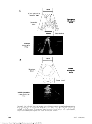
image was defined as a hyperechogenic, coherent bundle with
narrow basis spreading from the transducer to the further border
of the screen.5,6 The comet-tail image described here extends to
the edge of the screen (whereas short comet-tail artifacts may
exist in other regions), and arises only from the pleural line.6
Comet-tail images arising from the pleural line can be localized
or disseminated to the whole lung surface, or again isolated or
multiple (when at least three artifacts are visible in a frozen image
in one longitudinal scan), with a distance Յ 7 mm between two
artifacts) [Fig 1, top, A].6 A positive (or pathologic) test result was
defined as bilateral multiple comet-tail images, either dissemi-
nated (defined as all over the anterolateral lung surface) or lateral
(defined as limited to the lateral lung surface). A negative test
result was defined as an absence of comet-tail images, replaced
by the horizontal artifact (Fig 1, bottom, B), or when rare, isolated
comet-tail images were visible or when multiple comet-tail
images were confined laterally to the last intercostal space above
the diaphragm.6 The examinations were performed using an
ultrasound system (Sonos 5500; Phillips Medical Systems; An-
dover, MA) equipped with 1.8- to 3.6-MHz probe.
Chest Radiography
The patients underwent chest radiography in the supine
position with specific assessment of EVLW using a commercially
available radiograph machine and a standard technique. A pre-
viously validated radiologic score of EVLW was used incorporat-
ing assessment of hilar vessels (dimension, density, blurring),
Kerley lines (A, B, and C), micronoduli, widening of interlobar
fissures, peribronchial and perivascular cuffs, subpleural effusion,
and diffuse increase in density (Table 2).10–12 The intraobserver
and interobserver reproducibility of radiologic scoring of EVLW
among experienced observers was very high, as previously de-
scribed.10–12
PiCCO System
The PiCCO System is a device for cardiac output (CO)
measurement combined with cardiac preload volume and lung
water monitoring. It computes the CO utilizing an arterial pulse
contour analysis algorithm after calibration by means of a
transpulmonary thermodilution method.
In all patients, a 5F thermistor-tipped catheter (Pulsiocath
PV8115; Pulsion Medical Systems) was placed into the right
femoral artery, and connected to the PiCCO System for moni-
toring. To calibrate this system, individual arterial input imped-
ance to arterial pressure is calculated by simultaneously deter-
mining the area under the systolic portion of the arterial pulse
wave. A 10-mL bolus of cold 5% dextrose solution is injected
through central venous catheter, and the thermodilution curve is
evaluated with arterial catheter inserted in the femoral artery.
The mean of three consecutive boluses was used. If an injection
had to be rejected, more injections were made to obtain three
measurements after rejecting the lowest and the highest value.
From the CO we can obtain the intrathoracic thermal volume
and the intrathoracic blood volume; from the difference of these
two parameters, we can obtain the value of EVLW. Normally,
EVLW is Ͻ 500 mL13–15; the alveolar flooding appears usually
when the EVLW is Ͼ 75% above normal limit.15,16
Pulmonary Artery Pressure
A pulmonary artery catheter was introduced via the right
internal jugular vein for conventional pulmonary artery thermodi-
lution CO measurements. Pulmonary wedge pressure, and sys-
tolic, diastolic, and mean pulmonary pressures were also mea-
sured.
Statistical Analysis
Data are expressed as the mean value Ϯ SD or percentages.
The correlations between echo comet score, EVLW, radiologic
Table 1—Clinical Features
Variables Data*
Age, yr 62.6 Ϯ 11.5
Male/female gender, No. 16/4
Disease, No. (%)
Mitral regurgitation 9 (45)
Coronary artery disease 4 (20)
Aortic regurgitation 1 (5)
Coronary artery disease plus aortic stenosis 1 (5)
Mitral stenosis 1 (5)
Mitral regurgitation plus atrial septal defect 1 (5)
Mitral regurgitation plus coronary artery
disease
1 (5)
Aortic regurgitation plus ascending aortic
aneurysm
1 (5)
Aortic regurgitation plus mitral regurgitation 1 (5)
End-diastolic volume, mL 121.6 Ϯ 45
End-systolic volume, mL 44 Ϯ 19
Ejection fraction, % 63.5 Ϯ 5.5
*Data are presented as mean Ϯ SD unless otherwise indicated.
www.chestjournal.org CHEST / 127 / 5 / MAY, 2005 1691
Downloaded From: http://journal.publications.chestnet.org/ on 11/28/2013
3. Figure 1. Top, A: Typical comet-tail artifacts: hyperechogenic, coherent vertical bundles with narrow
basis spreading from the transducer to the further border of the screen. This artifact is composed of
multiple microreflections of the ultrasound beam. Bottom, B: Normal subject, with regular, parallel,
roughly horizontal hyperechogenic lines due to the lung-wall interface.
1692 Clinical Investigations
Downloaded From: http://journal.publications.chestnet.org/ on 11/28/2013
4. lung water score, and data obtained by pulmonary artery catheter
monitoring were analyzed by the Pearson two-tailed method.
Moreover, the agreement between chest ultrasound and radio-
graph methods was analyzed using the Bland and Altman meth-
od.17 Bias between the methods was calculated as the mean
difference between echo comet score and radiograph score. The
upper and the lower limits of agreement were calculated as bias
(2 SD), and defined the range in which 95% of the differences
between the methods were expected to lie. The precision of the
bias analysis and limits of agreement was assessed using 95%
confidence intervals. Bias between comet score and radiograph
score was analyzed using the paired Student t test. The difference
in the mean content of EVLW between positive and negative
comet test results was evaluated with an independent Student t
test. Moreover, we calculated the sensitivity and specificity of a
negative test result for detection of EVLW content Ͻ 500 mL,
the sensitivity and specificity of a positive test for detection of
EVLW content Ͼ 500 mL, and finally the sensitivity and speci-
ficity of a positive test result for detection an excess of EVLW
below the threshold of alveolar flooding. A p value Ͻ 0.05 was
considered statistically significant. The statistical analysis was
performed using software (version 8.0; SPSS; Chicago, IL).
Results
The determinations with the different methods
were obtained in all patients. No data were rejected.
A total of 60 comparative measurements were per-
formed between the methods.
Comparison Between Chest Ultrasound, Chest
Radiograph Findings, and EVLW
The feasibility of the chest ultrasound examination
for the diagnosis of EVLW was 100%. The time
needed for the echo lung examination was Ͻ 5 min
in all cases (mean, 4.3 Ϯ 1 min). The mean number
of comets per person (comet score) was 7.6 Ϯ 9.3,
the mean radiologic score was 12 Ϯ 7, and the mean
EVLW was 643.7 Ϯ 603.6 mL.
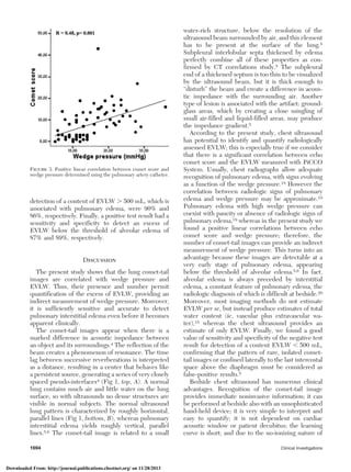
A significant positive linear correlation was found
between echo comet score and radiologic score
(r ϭ 0.60, p Ͻ 0.0001), and no significant difference
in the mean difference between these two scores was
observed (bias, 4.7; 95% limits of agreement, Ϫ 9.9
to 19.3). There was a significant positive linear
correlation between echo comet score and EVLW
(r ϭ 0.42, p ϭ 0.001) [Fig 2].
Comparison Between Chest Ultrasound and
Hemodynamic Parameters
Positive linear correlations were found between
echo comet score and wedge pressure (r ϭ 0.48,
p Ͻ 0.0001) and systolic pulmonary pressure
(r ϭ 0.53, p ϭ 0.007) determined using pulmonary
artery catheterization (Fig 3). No significant correla-
tions between echo comet score and CO and cardiac
index were observed.
Positive vs Negative Comet Test Results
Thirty-two examination results were considered
positive and 28 were negative. When we compared
the group of test results considered positive vs
negative, a significant difference in mean EVLW was
found (742 Ϯ 277 mL vs 392 Ϯ 92 mL, p ϭ 0.0001).
The mean content of EVLW in negative test result
was below the normal limit of EVLW (Ͻ 500 mL).
The sensitivity and specificity of the negative test
result for detection of a content of EVLW Ͻ 500 mL
were 90% and 89% respectively, whereas the sensi-
tivity and specificity of the positive test result for
Table 2—Radiologic Scoring of EVLW
Variables Score*
Hilar vessels
Enlarged 1 2 3
Increased in density 2 4 6
Blurred 3 6 9
Kerley lines
A 4 8
B 4 8
C 4 8
Micronoduli 4 8
Widening of interlobar fissures 4 8 12
Peribronchial and perivascular cuffs 4 8 12
Extensive perihilar haze 4 8 12
Subpleural effusion 5 10
Diffuse increase in density 5 10 15
*The score assigned to each variable depends on the severity of
involvement: ie, Hilary vessels enlarged: 1, normal mild enlarge-
ment; 2, moderate enlargement; 3, severe enlargement.
Figure 2. Significant positive linear correlation between comet
score and EVLW determined with the indicator thermodilution
method (PiCCO System).
www.chestjournal.org CHEST / 127 / 5 / MAY, 2005 1693
Downloaded From: http://journal.publications.chestnet.org/ on 11/28/2013
5. detection of a content of EVLW Ͼ 500 mL, which is
associated with pulmonary edema, were 90% and
86%, respectively. Finally, a positive test result had a
sensitivity and specificity to detect an excess of
EVLW below the threshold of alveolar edema of
87% and 89%, respectively.
Discussion
The present study shows that the lung comet-tail
images are correlated with wedge pressure and
EVLW. Thus, their presence and number permit
quantification of the excess of EVLW, providing an
indirect measurement of wedge pressure. Moreover,
it is sufficiently sensitive and accurate to detect
pulmonary interstitial edema even before it becomes
apparent clinically.
The comet-tail images appear when there is a
marked difference in acoustic impedance between
an object and its surroundings.4 The reflection of the
beam creates a phenomenon of resonance. The time
lag between successive reverberations is interpreted
as a distance, resulting in a center that behaves like
a persistent source, generating a series of very closely
spaced pseudo-interfaces4 (Fig 1, top, A). A normal
lung contains much air and little water on the lung
surface, so with ultrasounds no dense structures are
visible in normal subjects. The normal ultrasound
lung pattern is characterized by roughly horizontal,
parallel lines (Fig 1, bottom, B), whereas pulmonary
interstitial edema yields roughly vertical, parallel
lines.5,6 The comet-tail image is related to a small
water-rich structure, below the resolution of the
ultrasound beam surrounded by air, and this element
has to be present at the surface of the lung.5
Subpleural interlobular septa thickened by edema
perfectly combine all of these properties as con-
firmed by CT correlations study.5 The subpleural
end of a thickened septum is too thin to be visualized
by the ultrasound beam, but it is thick enough to
“disturb” the beam and create a difference in acous-
tic impedance with the surrounding air. Another
type of lesion is associated with the artifact: ground-
glass areas, which by creating a close mingling of
small air-filled and liquid-filled areas, may produce
the impedance gradient.5
According to the present study, chest ultrasound
has potential to identify and quantify radiologically
assessed EVLW; this is especially true if we consider
that there is a significant correlation between echo
comet score and the EVLW measured with PiCCO
System. Usually, chest radiographs allow adequate
recognition of pulmonary edema, with signs evolving
as a function of the wedge pressure.18 However the
correlation between radiologic signs of pulmonary
edema and wedge pressure may be approximate.19
Pulmonary edema with high wedge pressure can
coexist with paucity or absence of radiologic signs of
pulmonary edema,19 whereas in the present study we
found a positive linear correlations between echo
comet score and wedge pressure; therefore, the
number of comet-tail images can provide an indirect
measurement of wedge pressure. This turns into an
advantage because these images are detectable at a
very early stage of pulmonary edema, appearing
below the threshold of alveolar edema.5,6 In fact,
alveolar edema is always preceded by interstitial
edema, a constant feature of pulmonary edema, the
radiologic diagnosis of which is difficult at bedside.20
Moreover, most imaging methods do not estimate
EVLW per se, but instead produce estimates of total
water content (ie, vascular plus extravascular wa-
ter),15 whereas the chest ultrasound provides an
estimate of only EVLW. Finally, we found a good
value of sensitivity and specificity of the negative test
result for detection of a content EVLW Ͻ 500 mL,
confirming that the pattern of rare, isolated comet-
tail images or confined laterally to the last intercostal
space above the diaphragm must be considered as
false-positive results.5
Bedside chest ultrasound has numerous clinical
advantages. Recognition of the comet-tail image
provides immediate noninvasive information; it can
be performed at bedside also with an unsophisticated
hand-held device; it is very simple to interpret and
easy to quantify; it is not dependent on cardiac
acoustic window or patient decubitus; the learning
curve is short; and due to the no-ionizing nature of
Figure 3. Positive linear correlation between comet score and
wedge pressure determined using the pulmonary artery catheter.
1694 Clinical Investigations
Downloaded From: http://journal.publications.chestnet.org/ on 11/28/2013
6. the examination, it is useful in following up the
patient over time and tailoring therapy.6 Further-
more, the possibility of obtaining information on
EVLW with a very simple, bedside technology fur-
ther expands the possibility of a diagnosis based on
ultrasound for assessing both cardiac function and
pulmonary congestion, which are the two fundamen-
tal parameters needed for primary diagnosis, serial
follow-up, and therapy tailoring in heart failure
patients.21
The present study has its limitations. We excluded
patients with lung diseases. In this way, lung pathol-
ogy may have been underrepresented in our series,
reducing the possibility of false-positive results.5,6
Ultrasound detection of interstitial syndrome does
not necessarily imply a cardiogenic origin: pneumo-
nia, ARDS, chronic interstitial lung diseases, or
third-space syndrome after cardiopulmonary bypass,
as in our patients, can give comet-tail images.22
However, the aim of the present study was to
validate the presence and the number of these
images with a quantitative measurement of EVLW
independently of the generating cause.
In conclusion, the analysis of the presence and the
number of “sonographic Kerley lines” allowed us to
detect and quantify pulmonary edema. The possibil-
ity of obtaining information on EVLW with simplic-
ity and high feasibility with bedside technology
makes the ultrasound an attractive and easy to use
diagnostic tool at the bedside for assessing cardiac
function and pulmonary congestion.
References
1 Halperin B, Feeley T, Mihm F, et al. Evaluation of the
portable chest roentgenogram for quantitating extravascular
lung water in critically ill adults. Chest 1985; 88:649–652
2 Eisenberg PR, Hansbrough JR, Anderson D, et al. A prospec-
tive study of lung water measurements during patient man-
agement in an intensive care unit. Am Rev Respir Dis 1987;
136:662–668
3 Targhetta R, Chavagneaux R, Bourgeois JM, et al. Sono-
graphic approach to diagnosing pulmonary consolidation. J
Ultrasound Med 1992; 11:667–672
4 Ziskin MC, Thickman DI, Goldenberg NJ, et al. The comet
tail artifact. J Ultrasound Med 1982; 1:1–7
5 Lichtenstein D, Me´zie´re G, Biderman P, et al. The Comet-
tail artifact: an ultrasound sign of alveolar-interstitial syn-
drome. Am J Respir Crit Care Med 1997; 156:1640–1646
6 Lichtenstein D, Me´zie´re G. A lung ultrasound sign allowing
bedside distinction between pulmonary edema and COPD:
the comet-tail artifact. Intensive Care Med 1998; 24:133–
1334
7 Sivak ED, Wiedemann HP. Clinical measurement of ex-
travascular lung water. Crit Care Clin 1986; 2:511–526
8 Effros RM. Lung water measurements with the mean transit
time approach. J Appl Physiol 1985; 59:673–683
9 Jambrik Z, Monti S, Coppola V, et al. Usefulness of ultra-
sound lung comets as a nonradiologic sign of extravascular
lung water. Am J Cardiol 2004; 93:1265–1270
10 Milne EN, Pistolesi M, Miniati M, et al. The radiologic
distinction of cardiogenic and noncardiogenic edema. AJR
Am J Roentgenol 1985; 144:879–894
11 Miniati M, Pistolesi M, Milne EN, et al. Detection of lung
edema. Crit Care Med 1987; 15:1146–1155
12 Pistolesi M, Miniati M, Milne EN, et al. Measurement of
extravascular lung water. Intensive Care World 1991; 8:16–21
13 Lewis FR, Elings VB, Sturm JA. Bedside measurement of
lung water. J Surg Res 1979; 27:250–261
14 Sibbald WJ, Warshawski FJ, Short AK, et al. Clinical studies
of measuring extravascular lung water by the thermal dye
technique in critically ill patients. Chest 1983; 83:725–731
15 Lange NR, Schuster DP. The measurement of lung water.
Crit Care 1999; 3: R19–R24
16 Bongard FS, Matthay M, Mackersie RC, et al. Morphologic
and physiologic correlates of increased extravascular lung
water. Surgery 1984; 96:395–403
17 Bland JM, Altman DG. Statistical methods for assessing
agreement between two methods of clinical measurements.
Lancet 1986; 1:307–310
18 Givertz MM, Colucci WS, Braunwald E. Clinical aspects of
heart failure: high-output failure, pulmonary edema. In:
Braunwald E, Zipes DS, Libby P, eds. Heart disease. 6th ed.
Philadelphia, PA: WB Saunders, 2001; 545–546
19 Chakko S, Woska D, Martinez H, et al. Clinical, radiographic
and hemodynamic correlations in congestive heart failure:
conflicting results may lead to inappropriate care. Am J Med
1991; 90:353–359
20 Staub NC. Pulmonary edema. Physiol Rev 1974; 54:678–811
21 Remme WJ, Swedberg K. European Society of Cardiology.
Comprehensive guidelines for the diagnosis and treatment of
chronic heart failure. Task Force for the Diagnosis and
Treatment of Chronic Heart Failure of the European Society
of Cardiology. Eur J Heart Fail 2002; 4:11–22
22 Weiss YG, Merin G, Koganov E, et al. Postcardiopulmonary
bypass hypoxemia: a prospective study of incidence, risk
factor and clinical significance. J Cardiothorac Vasc Anesth
2000; 14:506–513
www.chestjournal.org CHEST / 127 / 5 / MAY, 2005 1695
Downloaded From: http://journal.publications.chestnet.org/ on 11/28/2013