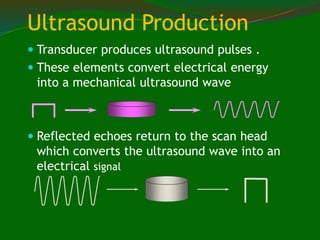
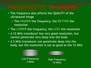
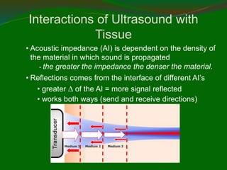
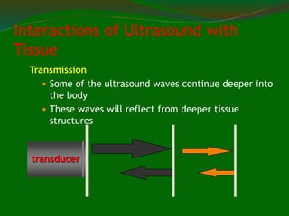
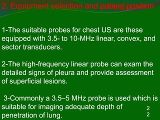
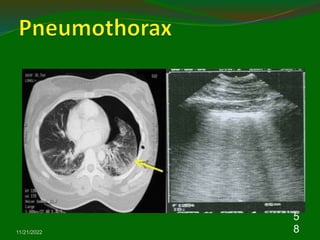
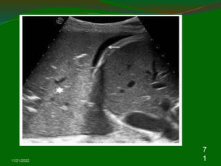
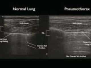
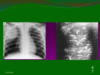
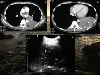
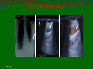
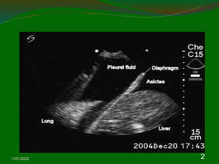
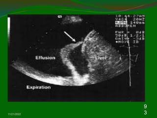
Dr-Mousa El-shamly
11/21/2022 3
1. Introduction
Ultrasound (US) has been proved to be valuable for the evaluation
of a wide variety of chest diseases,
The advantages of US
1- It is a relatively inexpensive
2-widely available, mobile form of multi-planar
imaging and free from ionizing.
3-Chest US can guides a variety of diagnostic
and therapeutic procedures.
4-Chest US is a useful tool in the diagnosis of
pleural diseases and peripheral pulmonary
lesions.
5- Moreover, endobronchial ultrasound (EBUS) is
the real advance in recent chest medicine.
Ultrasound Physics
Sound is a mechanical, longitudinal
wave that travels in a straight line
Sound requires a medium through which
to travel
Ultrasound is a mechanical, longitudinal
wave with a frequency exceeding the
upper limit of human hearing, which is
20,000 Hz or 20 kHz.
 Medical Ultrasound 2MHz to 16MHz
 1MHz=1million Hertz
ULTRASOUND – How is it produced?
Produced by passing an electrical current through a
piezoelectrical (material that expands and
contracts with current) crystal
Ultrasound Production
 Transducer produces ultrasound pulses .
 These elements convert electrical energy
into a mechanical ultrasound wave
 Reflected echoes return to the scan head
which converts the ultrasound wave into an
electrical signal
Frequency vs. Resolution
 The frequency also affects the QUALITY of the
ultrasound image
 The HIGHER the frequency, the BETTER the
resolution
 The LOWER the frequency, the LESS the resolution
 A 12 MHz transducer has very good resolution, but
cannot penetrate very deep into the body
 A 3 MHz transducer can penetrate deep into the
body, but the resolution is not as good as the 12 MHz
Low Frequency
3 MHz
High Frequency
12 MHz
Image Formation
Electrical signal produces ‘dots’ on the
screen
 Brightness of the dots is proportional to
the strength of the returning echoes
 Location of the dots is determined by
travel time. The velocity in tissue is
assumed constant at 1540 m/sec
Distance = Velocity
Time
• Acoustic impedance (AI) is dependent on the density of
the material in which sound is propagated
- the greater the impedance the denser the material.
• Reflections comes from the interface of different AI’s
• greater  of the AI = more signal reflected
• works both ways (send and receive directions)
Medium 1 Medium 2 Medium 3
Transducer
Interactions of Ultrasound with
Tissue
Interactions of Ultrasound
with Tissue
Reflection
Refraction
Transmission
Attenuation
Transmission
 Some of the ultrasound waves continue deeper into
the body
 These waves will reflect from deeper tissue
structures
transducer
Interactions of Ultrasound with
Tissue
Refraction
Incident
reflective
refraction
Angle of incidence = angle of reflection
Scattered
echoes
Attenuation
 Defined - the deeper the wave travels in the
body, the weaker it becomes -3 processes:
reflection, absorption, refraction
 Air (lung)> bone > muscle > soft tissue >blood >
water
Interactions of Ultrasound with
Tissue
Refraction
Incident
reflective
refraction
Angle of incidence = angle of reflection
Scattered
echoes
Reflected Echo’s
 Strong Reflections = White dots
Diaphragm, tendons, bone
‘Hyperechoic’
Weaker Reflections =
Grey dots
 Most solid organs,
 thick fluid – ‘isoechoic’
Reflected Echo’s
Reflected Echo’s
 No Reflections = Black dots
 Fluid within a cyst, urine, blood
‘Hypoechoic’ or echofree
What determines how far ultrasound waves can
travel?
 The FREQUENCY of the transducer
 The HIGHER the frequency, the LESS it
can penetrate
 The LOWER the frequency, the DEEPER it
can penetrate
 Attenuation is directly related to
frequency
Liver metastases
www.upei.ca/~vetrad
Attenuation
 Absorption = energy is captured by the
tissue then converted to heat
 Reflection = occurs at interfaces between
tissues of different acoustic properties
 Scattering = beam hits irregular interface –
beam gets scattered
www.upei.ca/~vetrad
Ultrasound Terminology
 Never use dense, opaque, lucent
 Anechoic
 No returning echoes= black (acellular
fluid)
 Echogenic
 Regarding fluid--some shade of grey d/t
returning echoes
 Relative terms
 Comparison to normal echogenicity of the
same organ or other structure
 Hypoechoic, isoechoic, hyperechoic
 Spleen should be hyperechoic to liver
2
2
2. Equipment selection and patient position
1-The suitable probes for chest US are these
equipped with 3.5- to 10-MHz linear, convex, and
sector transducers.
2-The high-frequency linear probe can exam the
detailed signs of pleura and provide assessment
of superficial lesions.
3-Commonly a 3.5–5 MHz probe is used which is
suitable for imaging adequate depth of
penetration of lung.
11/21/2022
2
3
4- During chest US examinations, patients
can be in the seated or supine position .
5-The probe is moved along the
intercostals space to avoid interference by
ribs or sternum. The transducer can be
moved longitudinally or horizontally in the
chest wall.
2
5
11/21/2022
2
6
3. Chest wall: Muscle layers, bone, and
pleura
Because air cannot be visualized by US, the
normal lung parenchyma cannot be detected by
US theoretically.
The image of chest US in chest wall including
muscle, fascia, bone, and pleura .
The soft tissue echogenicity with multiple layers
means muscles and fascia.
The normal ribs appear hyperechoic surfaces
with prominent acoustic shadows beneath the
ribs.
11/21/2022
2
7
Approximal 0.5 cm below the ribs shadows,
the visceral and parietal pleura appear as an
enchogenic bright line.
During respiratory movement, the two pleural
lines glide with each other which is referred to
as the “Gliding sign”. Therefore, the “Gliding
sign” means normal parietal and visceral
pleura slide over each other during respiration
and the loss of “Gliding sign” can be seen in
pneumothorax or diffuse pleural thickening
11/21/2022
2
8
Lungs –normal static findings
Normal lung considered “invisible” to
ultrasonographer Artefact scan be used to infer
normality or abnormality
A lines horizontal reverberation artifacts from
pleural line the only finding in 2/3 of normal lung
US
B lines vertical narrow bands from pleural line to
edge of screen obliterate the A line
Multiple B lines = Ultrasound Lung Rockets =
Abnormal lung has characteristics that are
clinically useful
NORMAL LUNG
A lines = default normal
 Horizontal echo
reflection at exact
multiples of
intervals from
surface to bright
reflector.
 Dry lung
 Decay with depth
 Obliterated by B
pleura A
A
A
A
A
A
B lines = fluid in alveolus or
interstitium
 Originates from
pleural line
 Reaches base of
screen OR
ALMOST
 MORE THAN 2 at
once is abnormal
EXCEPT in lung base
Remember
as ‘Kerley
Bs’
Not exactly
the same.
RI
B
RI
B
B B B B
B
B Lines = Crackles
Confluent B lines = Bad Bad
 ‘White’ or ‘shining’
lung
 Means increased
severity
 Probably indicates
thicker fluid in
alveoli eg protein or
inflammatory cells
 % space / 10
C = Pleural line abnormalities
Acute
inflammation
Old fibrosis.
Indicates
abnormal
interstitium
RI
B
C
C can be grossly abnormalIf CCF is
clinically
likely,
need to
cross
check with
heart or
IVC view.
RI
B
B x 3 x 2 x 2 = CCF
Makes assumption that ‘globally’ wet
lungs are most likely to be CCF
12
11/21/2022
3
9
Lungs –normal dynamic findings
Pleural sliding (lung sliding sign) Pleural line “shimmers”
with respiration Presence of lung sliding rules out
pneumothorax Lung sliding greatest in lower thorax
(greatest expansion) Absence of lung sliding has a
number of causes
1- Pneumothorax
2-Apnoea
3- Pleural adhesions
4-Mainstem bronchial intubation or occlusion
5-Critical parenchymal lung disease e.g. ARDS,
contusion
11/21/2022
4
0
11/21/2022
4
1
11/21/2022
4
2
4. Pleural disease
4.1. Pleural effusion
For the purpose of investigation pleural fluid can
be divided into three broad categories according
to etiology: infective, malignant and
miscellaneous.
The infective etiologies result in either a para-
pneumonic effusion or an empyema.
Malignant effusions are due to primary or
secondary thoracic disease, which may be
pulmonary or pleural.
11/21/2022
4
3
The miscellaneous causes of pleural fluid
include sterile benign effusions,
haemothoraces and chylothoraces.
Pleural effusions are differentiated into
transudates or exudates on the basis of
biochemical analysis of the aspirated fluid.
The erect chest radiograph is the most
common first line radiological investigation
PLEURAL EFFUSIONS
VERY BIG MEDIUM
11/21/2022
4
5
11/21/2022
4
6
Whereas approximately 500 ml of pleural fluid is
required before an effusion can be identified
clinically, as little 200 ml will blunt the
costophrenic angle.
US in either a standing or sitting position not only
is able to detect smaller volumes of pleural fluid
than the erect frontal chest radiograph but it also
gives useful information about the nature of the
effusion.
The pleural effusion images in ultrasound
appearances are characterized by an echofree
space between the visceral and parietal pleura
.
11/21/2022
4
7
The classifications of the volume of pleural
effusion known currently are minimal, small,
moderate, and massive.
The minimal effusion indicate the echo- free
space is seen within the costophrenic angle;
small effusion indicates the space is greater than
costphrenic angle but still within a one-probe
range; moderate effusion indicates the space is
greater than one-probe range but within a two-
probe; and massive effusion indicates the space
is bigger than two-probe range.
11/21/2022
4
8
Pleural effusion.
11/21/2022
11/21/2022
5
0
4.2. Pleural effusion echogenicity
. Four basic ultrasounds patterns of internal
echogenicity of pleural effusion were identified and they
can be subclassified as anechoic, complex
nonseptated, complex septated, and homogenously
echogenic
Anechoic effusion is defined as echo-free spaces
between visceral and parietal pleura.
Complex nonseptated effusion is defined as
heterogenous echogenic materials inside the pleural
effusions.
Complex septataed effusion is defined as fibrin strands
or septa floating inside the pleural effusions.
Homogenously enchogenic effusion is defined as
echogenic spots density evenly distributed within the
11/21/2022
5
1
A purely anechoic collection is found in exudates
and transudates with equal frequency.
However, internal echoes in the form of
septations or focal areas of debris are due
invariably to exudates. US presentations in
transudative pleural effusions are not always in
an anechoic pattern.
Tran‐ sudative pleural effusions may have a
complex nonseptated pattern or an anechoic
pattern
Pleural thickening.
11/21/2022
5
2
11/21/2022
5
3
There was no transudative pleural effusion with complex
septated or homogenously echogenic pattern .
The applications of sonographic appearances in
effusions of febrile patients in the intensive care unit
(ICU) can determine the necessity of thoracentesis in
high risk patients with effusion in ICU
This study reported that complex nonseptated and
relatively hyperechoic, complex septated and
homogenously echogenic pleural effusion patterns might
predict the possibility of empyema in febrile patients in
the ICU.
The sonographic septation in lymphocyte-rich exudative
pleural effusions can help us differentiate tuberculosis
pleurisy from malignant pleural effusion
11/21/2022
5
4
5. Pneumothorax
Chest US may be help in the diagnosis of
pneumothoraces.
Normal parietal and visceral pleura slide over each other
during respiration and a pneumothorax is suspected
when this ‘Gliding sign’ is absent in chest US
A recently published systemic review, chest US had a
sensitivity of 90.0% and a specificity of 98.2% .
The confirmation of lung gliding has a 100% negative
predictive value for the absence of pneumothorax .
The use of M-mode can also objectify the presence or
absence of lung gliding
11/21/2022
5
5
Pneumothorax
Normal Lung
Pneumothorax
M-mode for sliding - normal
straight
speckled
11/21/2022
5
8
Pneumothorax
11/21/2022
5
9
11/21/2022
6
0
5.1. Pulmonary lesions
The normal aerated lung is difficult to image
because the dramatic change in acoustic impe‐
dance between chest wall and lung results in
specular reflection of ultrasound waves at the
pleura. However, consolidated lung has a tissue
density and echo-texture similar to liver,
analogous to pathological hepatisation. This
removes the change in acoustic impedance at the
pleural interface, and ultrasound waves pass
directly into the affected lung
11/21/2022
6
1
When patient with lobar or segmental
pneumonia and the lesion is adjacent to pleura
or in the pleural effusion, the pneumonia may be
detected by chest US. A marked consolidation
with air-bronchogram and treelike ramifications
is easily seen Within the consolidated area,
hyperechoic (white) foci may be visible, again
representing a change in acoustic impedance,
but this time at the tissue interface between solid
lung and air-filled bronchi. Subpleural nodule
also can be seen in chest US
Pneumonia complicated by
abscess
11/21/2022
6
2
11/21/2022
6
3
11/21/2022
6
4
Chest US with color Doppler is a powerful tool for
differentiating the peripheral air-fluid abscess from
empyema .
The empyema can be detected by chest US with an
image of a hypoechoic lesion with complex-septated
effusions, passive atelectasis, width uniformity, and
smooth luminal and outer margins.
Color Doppler ultra‐ sound could not identify vessel
signals in pericavitary atelectasis.
The lung abscess in the US image reveals hypoechoic
lesion with typical pulmonary consolidation, irregular wall
width, and irregular luminal and outer margins.
Color Doppler ultrasound could identify vessel signals in
pericavitary consolidation
11/21/2022
6
5
.2. Alveolar interstitial syndrome
Alveolar interstitial syndrome constitutes a group
of diseases that is caused by an increase in lung
fluid and/or a reduction in its air content.
The result of this thickening of the interlobular
septa causes a particular artifact that is seen
arising from pleura line.
The ultra‐ sound appearance of alveolar
interstitial syndrome is a vertical artifact, called B-
line. Presence of the comet-tail artifact allowed
diagnosis of alveolar-interstitial syndrome by
chest ultrasound
11/21/2022
6
6
The major causes of alveolar interstitial
syndrome include pulmonary edema, acute
respiratory distress syndrome (ARDS), and
interstitial fibrosis. In the advanced study,
application of chest ultrasound in the patients
with respiratory failure, chest ultra‐ sounds can
help the clinician make a rapid diagnosis in
patients with acute respiratory failure according
the BLUE protocol
Acute Pulmonary oedema
11/21/2022
6
7
Acute pulmonary oedema
11/21/2022
6
8
11/21/2022
6
9
Consolidation
 Irregular hypoechic area of varying size and
shape.
 Echotexure can appear homogenous or
inhomogenous.
 The most common sonographic feature is the
air bronchogram which is characterized by lens
shaped internal echoes within hypodene area
or echogenic lines.
11/21/2022
7
0
11/21/2022
7
1
11/21/2022
7
2
11/21/2022
7
3
COLLASPE OF LUNG
11/21/2022
7
4
Peripheral bronchial carcinoma
 Can help to define better necrotic areas that
are depicted as anehoic region inside the
tumour.
 The infiltrative growth of solid tissue
without regard to anatomical structures is
characteristic of malignancy.
11/21/2022
7
5
Lung ca infiltrating chest wall.
11/21/2022
7
6
Pulmonary metastasis
11/21/2022
7
7
Pheripheral bronchial ca.
11/21/2022
7
8
11/21/2022
7
9
6. Ultrasound-guided intervention
6.1. Diagnostic thoracentasis
The aim of the initial assessment of pleural fluid is to identify
malignancy or infection and starts with a visual inspection of the
aspirate prior to laboratory analysis.
Therefore diagnostic thoracentesis is mandatory. When
performed with image guidance thoracentesis provides clinically
useful information in more than 90% of cases.
If the pleural effusion spans three intercostal spaces and its
presence can be confirmed clinically by percussion then aspiration
can be performed safely at the bedside without the aid of image
guidance. When the effusion is small,
loculated or associated with underlying pulmonary collapse this
becomes more difficult and hazardous. In all these situations US
examination is required to confirm the presence of fluid and to
guide thoracentesi
11/21/2022
8
0
6.4. Endobronchial Ultrasound (EBUS)
Endobronchial ultrasound (EBUS) technology is a relatively new
bronchoscopic method of visualizing the tracheobronchial tree, the
surrounding pulmonary parenchyma, and the mediastinal
structures, with a particular role in lung cancer diagnosis, staging,
and treatment .
There are 2 types of probes used in EBUS: the peripheral or radial
probe (RP) and the linear or convex probe (CP) EBUS, which
have technical differences and distinct diagnostic abilities. Both
are used for EBUS-guided biopsies and transbronchial needle
aspirations (TBNA), which increases the diagnostic yield over
conventional bronchoscopic techniques, thus providing advanced
information on staging, diagnosis, and treatment. The 20-MHz
RP- EBUS is positioned inside a water-inflatable balloon and is
inserted through the working channel of the bronchoscope.
Acute pulmonary oedema
11/21/2022
8
1
Pneumothorax
11/21/2022
8
2
11/21/2022
8
3
11/21/2022
8
4
Consolidation
 Irregular hypoechic area of varying size and
shape.
 Echotexure can appear homogenous or
inhomogenous.
 The most common sonographic feature is the
air bronchogram which is characterized by lens
shaped internal echoes within hypodene area
or echogenic lines.
11/21/2022
8
5
11/21/2022
8
6
11/21/2022
8
7
11/21/2022
8
8
Summarizing sonographic
finding in consolidation
 Liver like in early stage
 Air bronchogram
 Fluid bronchogram
 Blurred and serrated margin
 Reberberation echoes in the margin
 Hypoechoic abscess formation
11/21/2022
8
9
COLLASPE OF LUNG
11/21/2022
9
0
Pleural effusion.
11/21/2022
11/21/2022
9
2
11/21/2022
9
3
Pleural thickening.
11/21/2022
9
4
Lung abscess
 They typically appear as round or oval
anechoic lesions
 Margin may be smooth and echodense
 Microabscess are often visible as anechoic
areas within the pneumonic consolidation.
11/21/2022
9
5
Peripheral bronchial carcinoma
 Can help to define better necrotic areas that
are depicted as anehoic region inside the
tumour.
 The infiltrative growth of solid tissue
without regard to anatomical structures is
characteristic of malignancy.
11/21/2022
9
6
Pheripheral bronchial ca.
11/21/2022
9
7
Lung ca infiltrating chest wall.
11/21/2022
9
8
11/21/2022
9
9
Pulmonary metastasis
11/21/2022
1
0
0
Summary of pulmonary ca
 Hypoechoic, inhomogenous.
 Rounded, nearly rounded.
 Sharp,serrated margins
 Infiltration of chest wall
 Irregular vascularization
11/21/2022
1
0
1
Limitation of lung ultrasound
 Require formal training ,knowledge and skills.
 Obese patients are frequently difficult to examine.
 The presence of subcutaneous emphysema.
 Lung ultrasound cannot detect lung overinflation.
11/21/2022
1
0
2
Conclusion
 Easy to learn and apply
 It can be used quickly to rule out any
significant pneumothorax in critically ill
patient.
 More sensitive than cxr in pleural effusion or
pleural pathology.
 The routine use of lung ultrasound appears
attractive alternative to bedside chest
radiography
11/21/2022
1
0
3
Cp5

chestultrasou.ppt

  • 2.
  • 3.
    11/21/2022 3 1. Introduction Ultrasound(US) has been proved to be valuable for the evaluation of a wide variety of chest diseases, The advantages of US 1- It is a relatively inexpensive 2-widely available, mobile form of multi-planar imaging and free from ionizing. 3-Chest US can guides a variety of diagnostic and therapeutic procedures. 4-Chest US is a useful tool in the diagnosis of pleural diseases and peripheral pulmonary lesions. 5- Moreover, endobronchial ultrasound (EBUS) is the real advance in recent chest medicine.
  • 4.
    Ultrasound Physics Sound isa mechanical, longitudinal wave that travels in a straight line Sound requires a medium through which to travel Ultrasound is a mechanical, longitudinal wave with a frequency exceeding the upper limit of human hearing, which is 20,000 Hz or 20 kHz.  Medical Ultrasound 2MHz to 16MHz  1MHz=1million Hertz
  • 5.
    ULTRASOUND – Howis it produced? Produced by passing an electrical current through a piezoelectrical (material that expands and contracts with current) crystal
  • 6.
    Ultrasound Production  Transducerproduces ultrasound pulses .  These elements convert electrical energy into a mechanical ultrasound wave  Reflected echoes return to the scan head which converts the ultrasound wave into an electrical signal
  • 7.
    Frequency vs. Resolution The frequency also affects the QUALITY of the ultrasound image  The HIGHER the frequency, the BETTER the resolution  The LOWER the frequency, the LESS the resolution  A 12 MHz transducer has very good resolution, but cannot penetrate very deep into the body  A 3 MHz transducer can penetrate deep into the body, but the resolution is not as good as the 12 MHz Low Frequency 3 MHz High Frequency 12 MHz
  • 8.
    Image Formation Electrical signalproduces ‘dots’ on the screen  Brightness of the dots is proportional to the strength of the returning echoes  Location of the dots is determined by travel time. The velocity in tissue is assumed constant at 1540 m/sec Distance = Velocity Time
  • 9.
    • Acoustic impedance(AI) is dependent on the density of the material in which sound is propagated - the greater the impedance the denser the material. • Reflections comes from the interface of different AI’s • greater  of the AI = more signal reflected • works both ways (send and receive directions) Medium 1 Medium 2 Medium 3 Transducer Interactions of Ultrasound with Tissue
  • 10.
    Interactions of Ultrasound withTissue Reflection Refraction Transmission Attenuation
  • 11.
    Transmission  Some ofthe ultrasound waves continue deeper into the body  These waves will reflect from deeper tissue structures transducer Interactions of Ultrasound with Tissue
  • 12.
  • 13.
    Attenuation  Defined -the deeper the wave travels in the body, the weaker it becomes -3 processes: reflection, absorption, refraction  Air (lung)> bone > muscle > soft tissue >blood > water Interactions of Ultrasound with Tissue
  • 14.
  • 15.
    Reflected Echo’s  StrongReflections = White dots Diaphragm, tendons, bone ‘Hyperechoic’
  • 16.
    Weaker Reflections = Greydots  Most solid organs,  thick fluid – ‘isoechoic’ Reflected Echo’s
  • 17.
    Reflected Echo’s  NoReflections = Black dots  Fluid within a cyst, urine, blood ‘Hypoechoic’ or echofree
  • 18.
    What determines howfar ultrasound waves can travel?  The FREQUENCY of the transducer  The HIGHER the frequency, the LESS it can penetrate  The LOWER the frequency, the DEEPER it can penetrate  Attenuation is directly related to frequency
  • 19.
  • 20.
    www.upei.ca/~vetrad Attenuation  Absorption =energy is captured by the tissue then converted to heat  Reflection = occurs at interfaces between tissues of different acoustic properties  Scattering = beam hits irregular interface – beam gets scattered
  • 21.
    www.upei.ca/~vetrad Ultrasound Terminology  Neveruse dense, opaque, lucent  Anechoic  No returning echoes= black (acellular fluid)  Echogenic  Regarding fluid--some shade of grey d/t returning echoes  Relative terms  Comparison to normal echogenicity of the same organ or other structure  Hypoechoic, isoechoic, hyperechoic  Spleen should be hyperechoic to liver
  • 22.
    2 2 2. Equipment selectionand patient position 1-The suitable probes for chest US are these equipped with 3.5- to 10-MHz linear, convex, and sector transducers. 2-The high-frequency linear probe can exam the detailed signs of pleura and provide assessment of superficial lesions. 3-Commonly a 3.5–5 MHz probe is used which is suitable for imaging adequate depth of penetration of lung.
  • 23.
    11/21/2022 2 3 4- During chestUS examinations, patients can be in the seated or supine position . 5-The probe is moved along the intercostals space to avoid interference by ribs or sternum. The transducer can be moved longitudinally or horizontally in the chest wall.
  • 25.
  • 26.
    11/21/2022 2 6 3. Chest wall:Muscle layers, bone, and pleura Because air cannot be visualized by US, the normal lung parenchyma cannot be detected by US theoretically. The image of chest US in chest wall including muscle, fascia, bone, and pleura . The soft tissue echogenicity with multiple layers means muscles and fascia. The normal ribs appear hyperechoic surfaces with prominent acoustic shadows beneath the ribs.
  • 27.
    11/21/2022 2 7 Approximal 0.5 cmbelow the ribs shadows, the visceral and parietal pleura appear as an enchogenic bright line. During respiratory movement, the two pleural lines glide with each other which is referred to as the “Gliding sign”. Therefore, the “Gliding sign” means normal parietal and visceral pleura slide over each other during respiration and the loss of “Gliding sign” can be seen in pneumothorax or diffuse pleural thickening
  • 28.
    11/21/2022 2 8 Lungs –normal staticfindings Normal lung considered “invisible” to ultrasonographer Artefact scan be used to infer normality or abnormality A lines horizontal reverberation artifacts from pleural line the only finding in 2/3 of normal lung US B lines vertical narrow bands from pleural line to edge of screen obliterate the A line Multiple B lines = Ultrasound Lung Rockets = Abnormal lung has characteristics that are clinically useful
  • 29.
  • 31.
    A lines =default normal  Horizontal echo reflection at exact multiples of intervals from surface to bright reflector.  Dry lung  Decay with depth  Obliterated by B pleura A A A A A A
  • 33.
    B lines =fluid in alveolus or interstitium  Originates from pleural line  Reaches base of screen OR ALMOST  MORE THAN 2 at once is abnormal EXCEPT in lung base Remember as ‘Kerley Bs’ Not exactly the same. RI B RI B B B B B B
  • 34.
    B Lines =Crackles
  • 35.
    Confluent B lines= Bad Bad  ‘White’ or ‘shining’ lung  Means increased severity  Probably indicates thicker fluid in alveoli eg protein or inflammatory cells  % space / 10
  • 36.
    C = Pleuralline abnormalities Acute inflammation Old fibrosis. Indicates abnormal interstitium RI B C
  • 37.
    C can begrossly abnormalIf CCF is clinically likely, need to cross check with heart or IVC view. RI B
  • 38.
    B x 3x 2 x 2 = CCF Makes assumption that ‘globally’ wet lungs are most likely to be CCF 12
  • 39.
    11/21/2022 3 9 Lungs –normal dynamicfindings Pleural sliding (lung sliding sign) Pleural line “shimmers” with respiration Presence of lung sliding rules out pneumothorax Lung sliding greatest in lower thorax (greatest expansion) Absence of lung sliding has a number of causes 1- Pneumothorax 2-Apnoea 3- Pleural adhesions 4-Mainstem bronchial intubation or occlusion 5-Critical parenchymal lung disease e.g. ARDS, contusion
  • 40.
  • 41.
  • 42.
    11/21/2022 4 2 4. Pleural disease 4.1.Pleural effusion For the purpose of investigation pleural fluid can be divided into three broad categories according to etiology: infective, malignant and miscellaneous. The infective etiologies result in either a para- pneumonic effusion or an empyema. Malignant effusions are due to primary or secondary thoracic disease, which may be pulmonary or pleural.
  • 43.
    11/21/2022 4 3 The miscellaneous causesof pleural fluid include sterile benign effusions, haemothoraces and chylothoraces. Pleural effusions are differentiated into transudates or exudates on the basis of biochemical analysis of the aspirated fluid. The erect chest radiograph is the most common first line radiological investigation
  • 44.
  • 45.
  • 46.
    11/21/2022 4 6 Whereas approximately 500ml of pleural fluid is required before an effusion can be identified clinically, as little 200 ml will blunt the costophrenic angle. US in either a standing or sitting position not only is able to detect smaller volumes of pleural fluid than the erect frontal chest radiograph but it also gives useful information about the nature of the effusion. The pleural effusion images in ultrasound appearances are characterized by an echofree space between the visceral and parietal pleura .
  • 47.
    11/21/2022 4 7 The classifications ofthe volume of pleural effusion known currently are minimal, small, moderate, and massive. The minimal effusion indicate the echo- free space is seen within the costophrenic angle; small effusion indicates the space is greater than costphrenic angle but still within a one-probe range; moderate effusion indicates the space is greater than one-probe range but within a two- probe; and massive effusion indicates the space is bigger than two-probe range.
  • 48.
  • 49.
  • 50.
    11/21/2022 5 0 4.2. Pleural effusionechogenicity . Four basic ultrasounds patterns of internal echogenicity of pleural effusion were identified and they can be subclassified as anechoic, complex nonseptated, complex septated, and homogenously echogenic Anechoic effusion is defined as echo-free spaces between visceral and parietal pleura. Complex nonseptated effusion is defined as heterogenous echogenic materials inside the pleural effusions. Complex septataed effusion is defined as fibrin strands or septa floating inside the pleural effusions. Homogenously enchogenic effusion is defined as echogenic spots density evenly distributed within the
  • 51.
    11/21/2022 5 1 A purely anechoiccollection is found in exudates and transudates with equal frequency. However, internal echoes in the form of septations or focal areas of debris are due invariably to exudates. US presentations in transudative pleural effusions are not always in an anechoic pattern. Tran‐ sudative pleural effusions may have a complex nonseptated pattern or an anechoic pattern
  • 52.
  • 53.
    11/21/2022 5 3 There was notransudative pleural effusion with complex septated or homogenously echogenic pattern . The applications of sonographic appearances in effusions of febrile patients in the intensive care unit (ICU) can determine the necessity of thoracentesis in high risk patients with effusion in ICU This study reported that complex nonseptated and relatively hyperechoic, complex septated and homogenously echogenic pleural effusion patterns might predict the possibility of empyema in febrile patients in the ICU. The sonographic septation in lymphocyte-rich exudative pleural effusions can help us differentiate tuberculosis pleurisy from malignant pleural effusion
  • 54.
    11/21/2022 5 4 5. Pneumothorax Chest USmay be help in the diagnosis of pneumothoraces. Normal parietal and visceral pleura slide over each other during respiration and a pneumothorax is suspected when this ‘Gliding sign’ is absent in chest US A recently published systemic review, chest US had a sensitivity of 90.0% and a specificity of 98.2% . The confirmation of lung gliding has a 100% negative predictive value for the absence of pneumothorax . The use of M-mode can also objectify the presence or absence of lung gliding
  • 55.
  • 56.
  • 57.
    M-mode for sliding- normal straight speckled
  • 58.
  • 59.
  • 60.
    11/21/2022 6 0 5.1. Pulmonary lesions Thenormal aerated lung is difficult to image because the dramatic change in acoustic impe‐ dance between chest wall and lung results in specular reflection of ultrasound waves at the pleura. However, consolidated lung has a tissue density and echo-texture similar to liver, analogous to pathological hepatisation. This removes the change in acoustic impedance at the pleural interface, and ultrasound waves pass directly into the affected lung
  • 61.
    11/21/2022 6 1 When patient withlobar or segmental pneumonia and the lesion is adjacent to pleura or in the pleural effusion, the pneumonia may be detected by chest US. A marked consolidation with air-bronchogram and treelike ramifications is easily seen Within the consolidated area, hyperechoic (white) foci may be visible, again representing a change in acoustic impedance, but this time at the tissue interface between solid lung and air-filled bronchi. Subpleural nodule also can be seen in chest US
  • 62.
  • 63.
  • 64.
    11/21/2022 6 4 Chest US withcolor Doppler is a powerful tool for differentiating the peripheral air-fluid abscess from empyema . The empyema can be detected by chest US with an image of a hypoechoic lesion with complex-septated effusions, passive atelectasis, width uniformity, and smooth luminal and outer margins. Color Doppler ultra‐ sound could not identify vessel signals in pericavitary atelectasis. The lung abscess in the US image reveals hypoechoic lesion with typical pulmonary consolidation, irregular wall width, and irregular luminal and outer margins. Color Doppler ultrasound could identify vessel signals in pericavitary consolidation
  • 65.
    11/21/2022 6 5 .2. Alveolar interstitialsyndrome Alveolar interstitial syndrome constitutes a group of diseases that is caused by an increase in lung fluid and/or a reduction in its air content. The result of this thickening of the interlobular septa causes a particular artifact that is seen arising from pleura line. The ultra‐ sound appearance of alveolar interstitial syndrome is a vertical artifact, called B- line. Presence of the comet-tail artifact allowed diagnosis of alveolar-interstitial syndrome by chest ultrasound
  • 66.
    11/21/2022 6 6 The major causesof alveolar interstitial syndrome include pulmonary edema, acute respiratory distress syndrome (ARDS), and interstitial fibrosis. In the advanced study, application of chest ultrasound in the patients with respiratory failure, chest ultra‐ sounds can help the clinician make a rapid diagnosis in patients with acute respiratory failure according the BLUE protocol
  • 67.
  • 68.
  • 69.
  • 70.
    Consolidation  Irregular hypoechicarea of varying size and shape.  Echotexure can appear homogenous or inhomogenous.  The most common sonographic feature is the air bronchogram which is characterized by lens shaped internal echoes within hypodene area or echogenic lines. 11/21/2022 7 0
  • 71.
  • 72.
  • 73.
  • 74.
  • 75.
    Peripheral bronchial carcinoma Can help to define better necrotic areas that are depicted as anehoic region inside the tumour.  The infiltrative growth of solid tissue without regard to anatomical structures is characteristic of malignancy. 11/21/2022 7 5
  • 76.
    Lung ca infiltratingchest wall. 11/21/2022 7 6
  • 77.
  • 78.
  • 79.
    11/21/2022 7 9 6. Ultrasound-guided intervention 6.1.Diagnostic thoracentasis The aim of the initial assessment of pleural fluid is to identify malignancy or infection and starts with a visual inspection of the aspirate prior to laboratory analysis. Therefore diagnostic thoracentesis is mandatory. When performed with image guidance thoracentesis provides clinically useful information in more than 90% of cases. If the pleural effusion spans three intercostal spaces and its presence can be confirmed clinically by percussion then aspiration can be performed safely at the bedside without the aid of image guidance. When the effusion is small, loculated or associated with underlying pulmonary collapse this becomes more difficult and hazardous. In all these situations US examination is required to confirm the presence of fluid and to guide thoracentesi
  • 80.
    11/21/2022 8 0 6.4. Endobronchial Ultrasound(EBUS) Endobronchial ultrasound (EBUS) technology is a relatively new bronchoscopic method of visualizing the tracheobronchial tree, the surrounding pulmonary parenchyma, and the mediastinal structures, with a particular role in lung cancer diagnosis, staging, and treatment . There are 2 types of probes used in EBUS: the peripheral or radial probe (RP) and the linear or convex probe (CP) EBUS, which have technical differences and distinct diagnostic abilities. Both are used for EBUS-guided biopsies and transbronchial needle aspirations (TBNA), which increases the diagnostic yield over conventional bronchoscopic techniques, thus providing advanced information on staging, diagnosis, and treatment. The 20-MHz RP- EBUS is positioned inside a water-inflatable balloon and is inserted through the working channel of the bronchoscope.
  • 81.
  • 82.
  • 83.
  • 84.
  • 85.
    Consolidation  Irregular hypoechicarea of varying size and shape.  Echotexure can appear homogenous or inhomogenous.  The most common sonographic feature is the air bronchogram which is characterized by lens shaped internal echoes within hypodene area or echogenic lines. 11/21/2022 8 5
  • 86.
  • 87.
  • 88.
  • 89.
    Summarizing sonographic finding inconsolidation  Liver like in early stage  Air bronchogram  Fluid bronchogram  Blurred and serrated margin  Reberberation echoes in the margin  Hypoechoic abscess formation 11/21/2022 8 9
  • 90.
  • 91.
  • 92.
  • 93.
  • 94.
  • 95.
    Lung abscess  Theytypically appear as round or oval anechoic lesions  Margin may be smooth and echodense  Microabscess are often visible as anechoic areas within the pneumonic consolidation. 11/21/2022 9 5
  • 96.
    Peripheral bronchial carcinoma Can help to define better necrotic areas that are depicted as anehoic region inside the tumour.  The infiltrative growth of solid tissue without regard to anatomical structures is characteristic of malignancy. 11/21/2022 9 6
  • 97.
  • 98.
    Lung ca infiltratingchest wall. 11/21/2022 9 8
  • 99.
  • 100.
  • 101.
    Summary of pulmonaryca  Hypoechoic, inhomogenous.  Rounded, nearly rounded.  Sharp,serrated margins  Infiltration of chest wall  Irregular vascularization 11/21/2022 1 0 1
  • 102.
    Limitation of lungultrasound  Require formal training ,knowledge and skills.  Obese patients are frequently difficult to examine.  The presence of subcutaneous emphysema.  Lung ultrasound cannot detect lung overinflation. 11/21/2022 1 0 2
  • 103.
    Conclusion  Easy tolearn and apply  It can be used quickly to rule out any significant pneumothorax in critically ill patient.  More sensitive than cxr in pleural effusion or pleural pathology.  The routine use of lung ultrasound appears attractive alternative to bedside chest radiography 11/21/2022 1 0 3
  • 104.