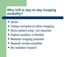
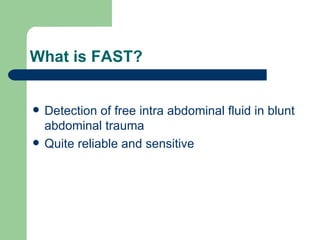
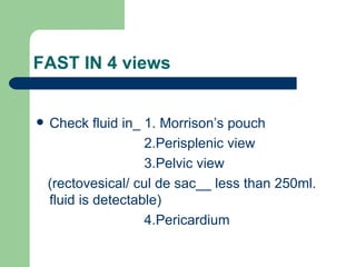
Ultra sound imaging A routine imaging  modality
Why U/S is day to day imaging modality? Quick Cheap compare to other imaging Strict patient prep. not required Patient position is flexible Bedside imaging possible Repeat/ review possible No radiation hazard
Ultrasound in emergency It is focused in YES or NO question eg.  Is there ruptured entopic? Is there cardiac tamponade? Is there abdominal aortic aneurysm? etc. etc.
Indication in emergency Trauma Cardiac Bleeding in pregnancy Acute abdominal pain Torsion of testis
Trauma Focussed abdominal sonography for trauma……….FAST
What is FAST? Detection of free intra abdominal fluid in blunt abdominal trauma Quite reliable and sensitive
FAST IN 4 views Check fluid in_ 1. Morrison’s pouch 2.Perisplenic view 3.Pelvic view  (rectovesical/ cul de sac__ less than 250ml. fluid is detectable) 4.Pericardium
Cardiac To detect cardiac activity if pulse less electrical activity To detect pericardial effusion if yes, Is patient in cardiac tamponade? ( thumping on right cardiac chamber)
Bleeding in pregnancy Whatever is the gestational period It could be_  ectopic pregnancy threatened abortion placenta praevia any other
Acute abdominal colic/pain Few  eg._  acute cholelithiasis  acute choledocholithiasis  acute pancreatitis acute nephrolithiasis acute torsion of ovary ruptured aortic aneurysm
Torsion of testes Use of Doppler to assess vascular supply
U/S is not diagnostic for Intestinal obstruction Perforation Plain X-ray supine/ erect or lateral decubitus is the first line of imaging modality
Indications other than emergency Abdominal Cardiac Vascular Pelvic
Contd. Eye Neck Breast Neonatal head/ brain Any other in consultation with clinician
Abdominal problems Ascitis_  to know the underlying cause hepatic - chr. liver disease renal  - renal failure cardiac - cardiac failure extra pulmonary tubercular malignancy
Contd. Jaundice_  Is it?  extrahepatic or intrahepatic  and then  to see the cause for it
Mass in abdomen To assess the size, shape and texture Origin of mass Extent of mass Adjoining vessels/ viscera Associated lesion( ascitis/ pl.effusion/ PE)
Fever of unknown origion Sub clinical/ occult malignancy Abdominal tuberculosis HIV ( immunosuppression) related complication eg. abscess
Cardiac problem All cardiac lesion (except conduction defect or arrhythmia)  like_  cardio-megaly on X-ray chest valvular lesion congenital defects
Vascular problem Any pulsatile swelling-  aneurysm Arterial thrombus Deep vein thrombosis Varicosity Peripheral vessel disease( limited help)
Pelvic problems Gynaecological_  infertility bleeding disorder mass in pelvic cavity pelvic pain lost IUCD
Trans/endo vaginal scanning Pregnancy less than 6 wks. Ectopic pregnancy  Post menopausal bleeding Follicular study
Obstetric ultrasound To ascertain pregnancy_ size/ gestational age site( IU/ ectopic) viability( cardiac activity ) number position/ lie
Contd. Placental localization Amniotic fluid ( normal AFI- 10)  Umbilical cord Any congenital anomaly
Doppler U/S in Obs. To assess IUGR ( though the specificity is low ) Fetal distress Commonly umbilical, middle cerebral, uterine artery are examined for systolic/ diastolic peak to assess RI/ PI
Eye problem Proptosis Trauma/ foreign body Retinal detachment/ tear/ haemorrhage Vitrous haemorrhage/ collapse
Contd. Ocular mass Cataract Retrobulbar mass Optic nerve( gross lesion )  Whenever funduscopy is inconclusive
Neck problem Enlarged thyroid Submandibular swelling Cervical lymphadenopathy Carotid artery plaque/ thrombosis(doppler)
Breast problem Enlarged breast eg. Fibrocystic disease Mass in breast Bleeding from nipple U/S guided FNAC or cyst aspiration
Neonatal head/ brain Enlarged head- ? hydrocephalus Neonatal fits- hypoxia haemorrhage Congenital anomaly Birth trauma- ? subdural hematoma
Last but not least Any problem in consultation with clinician  where ultrasound can help directly or by way of exclusion.
Eur.J.emergency.Med 2009 Jul4 Epub Imaging studies performed by technologist and then reviewed by radiologist with no patient contact are better quality studies. This para digm of imaging misses the point of clinicians performed U/S Clinician performed sonography in emergency has better accuracy
Request from clinicians  Accurate clinical picture is really helpful for correct  interpretation of U/S image
Recent advances in U/S imaging Despite developments other imaging techniques, the role of U/S continues to expand. Its unrivalled ability to show events in the body in real time with continuing technological advances will

Ultra sound imaging general presentation

  • 1.
    Ultra sound imagingA routine imaging modality
  • 2.
    Why U/S isday to day imaging modality? Quick Cheap compare to other imaging Strict patient prep. not required Patient position is flexible Bedside imaging possible Repeat/ review possible No radiation hazard
  • 3.
    Ultrasound in emergencyIt is focused in YES or NO question eg. Is there ruptured entopic? Is there cardiac tamponade? Is there abdominal aortic aneurysm? etc. etc.
  • 4.
    Indication in emergencyTrauma Cardiac Bleeding in pregnancy Acute abdominal pain Torsion of testis
  • 5.
    Trauma Focussed abdominalsonography for trauma……….FAST
  • 6.
    What is FAST?Detection of free intra abdominal fluid in blunt abdominal trauma Quite reliable and sensitive
  • 7.
    FAST IN 4views Check fluid in_ 1. Morrison’s pouch 2.Perisplenic view 3.Pelvic view (rectovesical/ cul de sac__ less than 250ml. fluid is detectable) 4.Pericardium
  • 8.
    Cardiac To detectcardiac activity if pulse less electrical activity To detect pericardial effusion if yes, Is patient in cardiac tamponade? ( thumping on right cardiac chamber)
  • 9.
    Bleeding in pregnancyWhatever is the gestational period It could be_ ectopic pregnancy threatened abortion placenta praevia any other
  • 10.
    Acute abdominal colic/painFew eg._ acute cholelithiasis acute choledocholithiasis acute pancreatitis acute nephrolithiasis acute torsion of ovary ruptured aortic aneurysm
  • 11.
    Torsion of testesUse of Doppler to assess vascular supply
  • 12.
    U/S is notdiagnostic for Intestinal obstruction Perforation Plain X-ray supine/ erect or lateral decubitus is the first line of imaging modality
  • 13.
    Indications other thanemergency Abdominal Cardiac Vascular Pelvic
  • 14.
    Contd. Eye NeckBreast Neonatal head/ brain Any other in consultation with clinician
  • 15.
    Abdominal problems Ascitis_ to know the underlying cause hepatic - chr. liver disease renal - renal failure cardiac - cardiac failure extra pulmonary tubercular malignancy
  • 16.
    Contd. Jaundice_ Is it? extrahepatic or intrahepatic and then to see the cause for it
  • 17.
    Mass in abdomenTo assess the size, shape and texture Origin of mass Extent of mass Adjoining vessels/ viscera Associated lesion( ascitis/ pl.effusion/ PE)
  • 18.
    Fever of unknownorigion Sub clinical/ occult malignancy Abdominal tuberculosis HIV ( immunosuppression) related complication eg. abscess
  • 19.
    Cardiac problem Allcardiac lesion (except conduction defect or arrhythmia) like_ cardio-megaly on X-ray chest valvular lesion congenital defects
  • 20.
    Vascular problem Anypulsatile swelling- aneurysm Arterial thrombus Deep vein thrombosis Varicosity Peripheral vessel disease( limited help)
  • 21.
    Pelvic problems Gynaecological_ infertility bleeding disorder mass in pelvic cavity pelvic pain lost IUCD
  • 22.
    Trans/endo vaginal scanningPregnancy less than 6 wks. Ectopic pregnancy Post menopausal bleeding Follicular study
  • 23.
    Obstetric ultrasound Toascertain pregnancy_ size/ gestational age site( IU/ ectopic) viability( cardiac activity ) number position/ lie
  • 24.
    Contd. Placental localizationAmniotic fluid ( normal AFI- 10) Umbilical cord Any congenital anomaly
  • 25.
    Doppler U/S inObs. To assess IUGR ( though the specificity is low ) Fetal distress Commonly umbilical, middle cerebral, uterine artery are examined for systolic/ diastolic peak to assess RI/ PI
  • 26.
    Eye problem ProptosisTrauma/ foreign body Retinal detachment/ tear/ haemorrhage Vitrous haemorrhage/ collapse
  • 27.
    Contd. Ocular massCataract Retrobulbar mass Optic nerve( gross lesion ) Whenever funduscopy is inconclusive
  • 28.
    Neck problem Enlargedthyroid Submandibular swelling Cervical lymphadenopathy Carotid artery plaque/ thrombosis(doppler)
  • 29.
    Breast problem Enlargedbreast eg. Fibrocystic disease Mass in breast Bleeding from nipple U/S guided FNAC or cyst aspiration
  • 30.
    Neonatal head/ brainEnlarged head- ? hydrocephalus Neonatal fits- hypoxia haemorrhage Congenital anomaly Birth trauma- ? subdural hematoma
  • 31.
    Last but notleast Any problem in consultation with clinician where ultrasound can help directly or by way of exclusion.
  • 32.
    Eur.J.emergency.Med 2009 Jul4Epub Imaging studies performed by technologist and then reviewed by radiologist with no patient contact are better quality studies. This para digm of imaging misses the point of clinicians performed U/S Clinician performed sonography in emergency has better accuracy
  • 33.
    Request from clinicians Accurate clinical picture is really helpful for correct interpretation of U/S image
  • 34.
    Recent advances inU/S imaging Despite developments other imaging techniques, the role of U/S continues to expand. Its unrivalled ability to show events in the body in real time with continuing technological advances will